. 2023 Sep 5;25(9):1672-1685.
doi: 10.1093/neuonc/noad074.

Prognostic evaluation of re-resection for recurrent glioblastoma using the novel RANO classification for extent of resection: A report of the RANO resect group

Affiliations

Prognostic evaluation of re-resection for recurrent glioblastoma using the novel RANO classification for extent of resection: A report of the RANO resect group

Philipp Karschnia et al. Neuro Oncol. .

Abstract

Background: The value of re-resection in recurrent glioblastoma remains controversial as a randomized trial that specifies intentional incomplete resection cannot be justified ethically. Here, we aimed to (1) explore the prognostic role of extent of re-resection using the previously proposed Response Assessment in Neuro-Oncology (RANO) classification (based upon residual contrast-enhancing (CE) and non-CE tumor), and to (2) define factors consolidating the surgical effects on outcome.

Methods: The RANO resect group retrospectively compiled an 8-center cohort of patients with first recurrence from previously resected glioblastomas. The associations of re-resection and other clinical factors with outcome were analyzed. Propensity score-matched analyses were constructed to minimize confounding effects when comparing the different RANO classes.

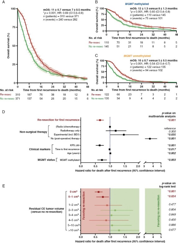
Results: We studied 681 patients with first recurrence of Isocitrate Dehydrogenase (IDH) wild-type glioblastomas, including 310 patients who underwent re-resection. Re-resection was associated with prolonged survival even when stratifying for molecular and clinical confounders on multivariate analysis; ≤1 cm3 residual CE tumor was associated with longer survival than non-surgical management. Accordingly, "maximal resection" (class 2) had superior survival compared to "submaximal resection" (class 3). Administration of (radio-)chemotherapy in the absence of postoperative deficits augmented the survival associations of smaller residual CE tumors. Conversely, "supramaximal resection" of non-CE tumor (class 1) was not associated with prolonged survival but was frequently accompanied by postoperative deficits. The prognostic role of residual CE tumor was confirmed in propensity score analyses.

Conclusions: The RANO resect classification serves to stratify patients with re-resection of glioblastoma. Complete resection according to RANO resect classes 1 and 2 is prognostic.

Keywords: classification; extent of resection; glioblastoma recurrence; outcome; surgical re-resection.


Conflict of interest statement

MW - Research grants: Quercis, Versameb. Honoraria or advisory board participation and consulting: Bayer, CureVac, Medac, Novartis, Orbus, Philogen. M.vdB. - Consultant: Celgene, BMS, Agios, Boehringer, Abbvie, Bayer, Carthera, Nerviano, Genenta. N. Ta-Research grant: Medtronic. Founder: BrainDynamics; Advisory Board: Nervonik, BrainGrade. RR - Honoraria, advisory board, and consulting: UCB, Bayer, Novocure, Genenta, Servier. MAV - Indirect equity and patent royalty interests: Infuseon Therapeutics. Honoraria: Chimerix, Midatech. Research grants: DeNovo Pharma, Oncosynergy, Infuseon, Chimerix. JCT -Research grants: Novocure, Munich Surgical Imaging. Advisory board: AAA Novartis. Royalties: Springer Publisher. PK, AD, JSY, STJ, N. Te, LH, TS, CYM, FB, LN, RAM, AFH, JB, SHJ, AMM, LB, OS, SJG, SMC, MSB, and YE - None.

Figures

Figure 1.
Figure 1.
Baseline characteristics of patients with first recurrence of a previously resected glioblastoma. (A) Schematic representation of the 8 neuro-oncological centers participating in the study. (B, C) Kaplan–Meier estimates of overall survival after first recurrence for the entire study cohort (B; n = 681) and for patients with MGMT promotor status available (C; n = 512). Points indicate deceased or censored patients, light shading indicates standard error of the mean (SEM). (D, E) Distribution of residual tumor following first resection (measured according to the RANO classification) and MGMT promotor methylation status across patients with re-resection (D; n = 310) and patients managed without re-resection (E; n = 371) at first recurrence. (F, G) Therapeutic approaches following diagnosis and first recurrence among patients with re-resection (F; n = 310) and patients managed without re-resection (G; n = 371) at first recurrence. Nodes of the Sankey plots represent time points in the disease course (new diagnosis, first recurrence, second recurrence); and time to first progression is indicated. Therapeutic approaches are color-coded and arc thickness corresponds to patient numbers.
Figure 2.
Figure 2.
Prognostic role of re-resection compared to non-surgically managed patients. (A–C): Kaplan–Meier estimates of overall survival after first recurrence for the entire study cohort (A; n = 681) and for patients with MGMT promotor status being methylated (B; n = 255) or unmethylated (C; n = 257). Patients were stratified according to whether re-resection was provided at first recurrence. Points indicate deceased or censored patients, light shading indicates SEM. (D) Multivariate analysis using a Cox proportional hazard regression model estimating the hazard ratio for death after first recurrence. All included variables were of significance on univariate analysis. BEV: Bevacizumab. Hazard ratio ± 95% confidence interval. (E) Univariate analysis using log-rank tests comparing patients with different amounts of residual contrast-enhancing (CE) tumor volumes following re-resection to non-surgically managed patients. Note that an association favoring re-resection was only observed for residual CE tumor volumes of ≤1 cm3. Hazard ratio ± 95% confidence interval.
Figure 3.
Figure 3.
Implications of the stratification per RANO classification by residual contrast-enhancing (CE) and non-CE tumor volumes on outcome. (A) Axial brain MRI with contrast-enhanced T1- (left on each panel) and T2-weighted (right on each panel) sequences demonstrating a left temporo-mesial glioblastoma. The CE (yellow) and non-CE tumor (blue) is delineated. On post-operative imaging, complete CE resection becomes apparent. Note the residual non-CE tumor being surrounded by edema. (B) Stratification of all patients undergoing re-resection with pre- and post-operative volumetrics available for review (n = 307) according to the previously proposed RANO classification. (C) Kaplan–Meier estimates of overall survival after first recurrence for patients stratified to RANO class 3 (n = 90) compared to non-surgically managed patients (n = 371). No differences were observed. (D) Hazard ratios for death after first recurrence calculated for each individual residual CE tumor volume among patients undergoing re-resection (n = 307). An exponential hazard increase can be seen for higher residual CE volumes. (E) Kaplan–Meier estimates of overall survival after first recurrence for patients stratified to RANO class 1/2 (corresponding to ≤1 cm3 residual CE tumor; n = 217) compared to RANO class 3 (n = 90). A significant survival difference is calculated. (F) Hazard ratios for death after first recurrence calculated for each individual residual non-CE tumor volume among patients without any residual CE tumor (n = 155). Only a minimal hazard ratio increase can be seen for large residual non-CE volumes. (G) Kaplan–Meier estimates of overall survival after first recurrence for patients stratified to RANO class 1 (corresponding to ≤5 cm3 residual non-CE tumor; n = 60) compared to RANO class 2 (n = 157). No benefit of non-CE tumor resection was detected. Points indicate deceased or censored patients, light shading indicates SEM.
Figure 4.
Figure 4.
Associations of medical therapies on outcome following re-resection. (A) Overview on the non-surgical approaches provided after re-resection (n = 301). (B) Kaplan–Meier estimates of overall survival after first recurrence for patients with re-resection stratified for (radio-)chemotherapy ((R)CTx; n = 193), radiotherapy (XRT; n = 38), and re-resection (or experimental) therapy only (n = 70). Points indicate deceased or censored patients, light shading indicates SEM. (C) Contingency table stratifying patients to residual contrast-enhancing (CE) tumor (x-axis; yellow: RANO class 1/2, pink: RANO class 3) and further medical management (y-axis). Each dot represents one individual patient, the median survival of the respective patient subgroup is indicated, and the rate of new post-operative deficits is indicated by a pie chart next to each patient subgroup. Darker colors in each pie charts indicate individuals with post-operative deficits. (D) Distribution of age (left panel), post-operative Karnofsky performance status (KPS; second to left), tumor localization (second to right panel), and MGMT promotor methylation status (right panel) across the different therapy subgroups. Asterisks indicate P ≤ .05 and n.s. indicates “not significant” when all 3 groups were tested together using a Kruskal–Wallis test (for continuous data) or a χ2-test (for categorical variables). Mean ± SEM for continuous data.
Figure 5.
Figure 5.
Prognostic confirmation of the RANO classification using propensity score-matched analyses. (A) Schematic representation of the principals of propensity-scored based matching. Nearest neighbor matching for multiple covariates results in 2 comparable cohorts who only differ for the variable of interest. (B–E) Kernel density estimates before and after propensity score-based matching (B, D) and Kaplan–Meier estimates for survival after first recurrence (C, E). Patients stratified to RANO class 1/2 (corresponding to ≤1 cm3 residual contrast-enhancing (CE) tumor; n = 215) were matched to controls selected from RANO class 3 (n = 215) (B, C); and patients stratified to RANO class 1 (corresponding to ≤5 cm3 residual non-CE tumor; n = 60) were matched to controls selected from RANO class 2 (n = 60) (D, E). Note that survival differences were only observed for the comparison based on CE tumor, but not for non-CE tumor. Points indicate deceased or censored patients, light shading indicates SEM. (F) Proposed strategy for surgical decision-making in patients with recurrent glioblastoma. Importantly, the optimal therapy beyond surgery warrants prospective evaluation as the current study is only hypothesis-generating in this regard.

References

    1. Weller M, van den Bent M, Preusser M, et al. . EANO guidelines on the diagnosis and treatment of diffuse gliomas of adulthood. Nat Rev Clin Oncol. 2020:33293629. - PMC - PubMed
    1. Wen PY, Weller M, Lee EQ, et al. . Glioblastoma in adults: A Society for Neuro-Oncology (SNO) and European Society of Neuro-Oncology (EANO) consensus review on current management and future directions. Neuro Oncol. 2020;22(8):1073–1113. - PMC - PubMed
    1. Gorlia T, Stupp R, Brandes AA, et al. . New prognostic factors and calculators for outcome prediction in patients with recurrent glioblastoma: A pooled analysis of EORTC Brain Tumour Group phase I and II clinical trials. Eur J Cancer. 2012;48(8):1176–1184. - PubMed
    1. Tsien CI, Pugh SL, Dicker AP, et al. . NRG oncology/RTOG1205: A randomized phase II trial of concurrent bevacizumab and reirradiation versus bevacizumab alone as treatment for recurrent glioblastoma. J Clin Oncol. 2022;36260832:Jco2200164. - PMC - PubMed
    1. Wick W, Gorlia T, Bendszus M, et al. . Lomustine and bevacizumab in progressive glioblastoma. N Engl J Med. 2017;377(20):1954–1963. - PubMed