

7T amygdala and hippocampus subfields in volumetry-based associations with memory: A 3-year follow-up study of early Alzheimer's disease
- PMID: 37253284
- PMCID: PMC10236463
- DOI: 10.1016/j.nicl.2023.103439
7T amygdala and hippocampus subfields in volumetry-based associations with memory: A 3-year follow-up study of early Alzheimer's disease
Abstract
Introduction: The hippocampus is the most prominent single region of interest (ROI) for the diagnosis and prediction of Alzheimer's disease (AD). However, its suitability in the earliest stages of cognitive decline, i.e., subjective cognitive decline (SCD), remains uncertain which warrants the pursuit of alternative or complementary regions. The amygdala might be a promising candidate, given its implication in memory as well as other psychiatric disorders, e.g. depression and anxiety, which are prevalent in SCD. In this 7 tesla (T) magnetic resonance imaging (MRI) study, we aimed to compare the contribution of volumetric measurements of the hippocampus, the amygdala, and their respective subfields, for early diagnosis and prediction in an AD-related study population.
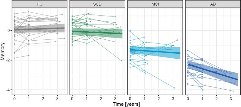
Methods: Participants from a longitudinal study were grouped into SCD (n = 29), mild cognitive impairment (MCI, n = 23), AD (n = 22) and healthy control (HC, n = 31). All participants underwent 7T MRI at baseline and extensive neuropsychological testing at up to three visits (baseline n = 105, 1-year n = 78, 3-year n = 39). Analysis of covariance (ANCOVA) was used to assess group differences of baseline volumes of the amygdala and the hippocampus and their subfields. Linear mixed models were used to estimate the effects of baseline volumes on yearly changes of a z-scaled memory score. All models were adjusted to age, sex and education.
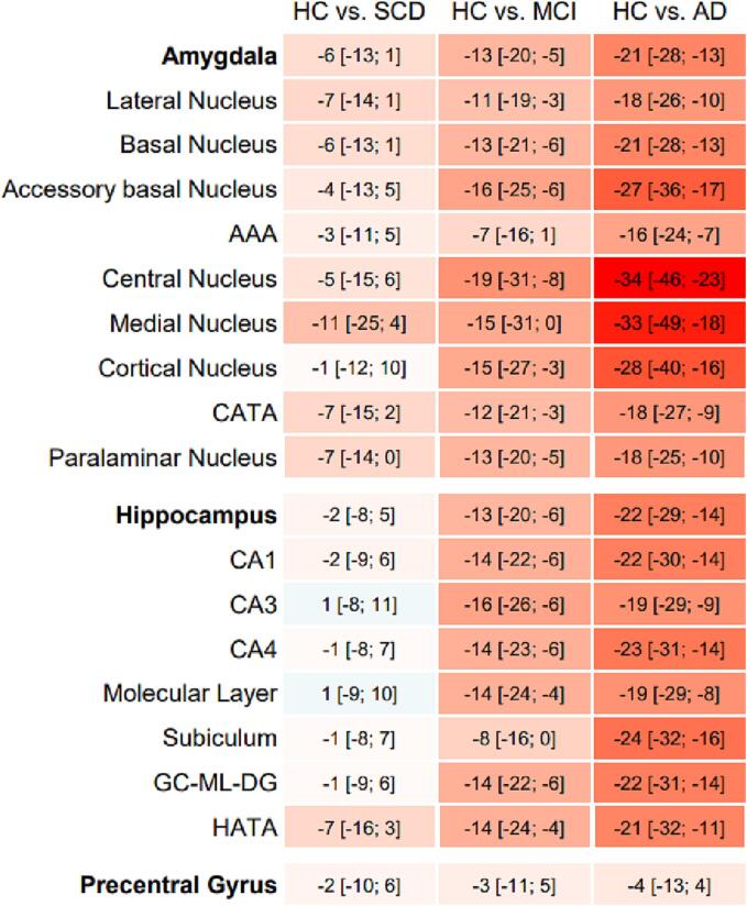
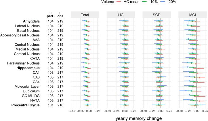
Results: Compared to the HC group, individuals with SCD showed smaller amygdala ROI volumes (range across subfields -11% to -1%), but not hippocampus ROI volumes (-2% to 1%) except for the hippocampus-amygdala-transition-area (-7%). However, cross-sectional associations between baseline memory and volumes were smaller for amygdala ROIs (std. ß [95% CI] ranging between 0.16 [0.08; 0.25] and 0.46 [0.31; 0.60]) than hippocampus ROIs (between 0.32 [0.19; 0.44] and 0.53 [0.40; 0.67]). Further, the association of baseline volumes with yearly memory change in the HC and SCD groups was similarly weak for amygdala ROIs and hippocampus ROIs. In the MCI group, volumes of amygdala ROIs were associated with a relevant yearly memory decline [95% CI] ranging between -0.12 [-0.24; 0.00] and -0.26 [-0.42; -0.09] for individuals with 20% smaller volumes than the HC group. However, effects were stronger for hippocampus ROIs with a corresponding yearly memory decline ranging between -0.21 [-0.35; -0.07] and -0.31 [-0.50; -0.13].
Conclusion: Volumes of amygdala ROIs, as determined by 7T MRI, might contribute to objectively and non-invasively identify patients with SCD, and thus aid early diagnosis and treatment of individuals at risk to develop dementia due to AD, however associations with other psychiatric disorders should be evaluated in further studies. The amygdala's value in the prediction of longitudinal memory changes in the SCD group remains questionable. Primarily in patients with MCI, memory decline over 3 years appears to be more strongly associated with volumes of hippocampus ROIs than amygdala ROIs.
Keywords: 7T MRI; Alzheimer’s disease; Amygdala; Hippocampus; Memory; SCD.
Copyright © 2023 The Author(s). Published by Elsevier Inc. All rights reserved.
Conflict of interest statement
Declaration of Competing Interest The authors declare that they have no known competing financial interests or personal relationships that could have appeared to influence the work reported in this paper.
Figures
References
-
- Basso M., Yang J., Warren L., MacAvoy M.G., Varma P., Bronen R.A., van Dyck C.H. Volumetry of amygdala and hippocampus and memory performance in Alzheimer's disease. Psychiatry Res. 2006;146(3):251–261. - PubMed
-
- Bates D., Mächler M., Bolker B., Walker S. Fitting Linear Mixed-Effects Models Using lme4. J. Stat. Softw. 2015;67:1–48.
-
- Brady D.R., Mufson E.J. Amygdaloid pathology in Alzheimer's disease: Qualitative and quantitative analysis. Dementia. 1990;1:5–17.
-
- de Flores R., La Joie R., Chetelat G. Structural imaging of hippocampal subfields in healthy aging and Alzheimer's disease. Neuroscience. 2015;309:29–50. - PubMed
-
- Dell'Orco, A., 2022. 0rC0/NeuroMet_MP2rage_pypes: v0.1.1-beta. Zenodo.
Publication types
MeSH terms
LinkOut - more resources
Full Text Sources
Medical
Research Materials
