. 2023 May 30;13(1):8728.
doi: 10.1038/s41598-023-35558-x.

Microfibril-associated protein 5 and the regulation of skin scar formation

Affiliations

Microfibril-associated protein 5 and the regulation of skin scar formation

Chen Han et al. Sci Rep. .

Abstract

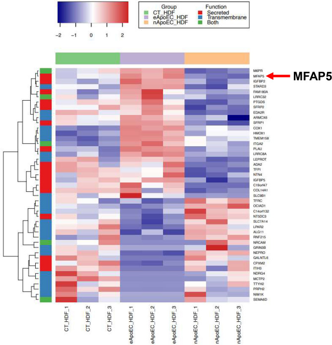
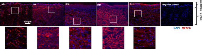
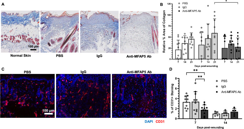
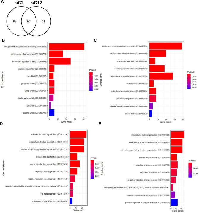
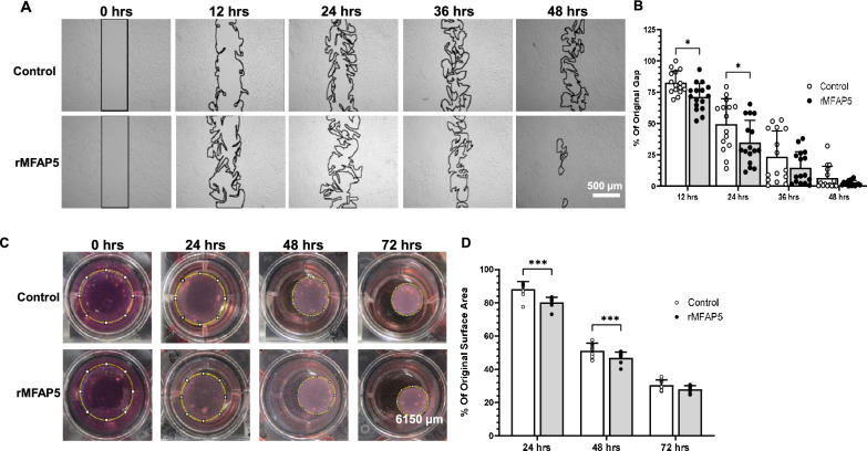
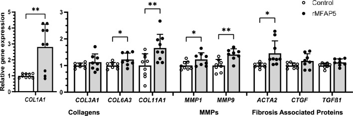
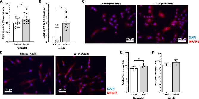
Many factors regulate scar formation, which yields a modified extracellular matrix (ECM). Among ECM components, microfibril-associated proteins have been minimally explored in the context of skin wound repair. Microfibril-associated protein 5 (MFAP5), a small 25 kD serine and threonine rich microfibril-associated protein, influences microfibril function and modulates major extracellular signaling pathways. Though known to be associated with fibrosis and angiogenesis in certain pathologies, MFAP5's role in wound healing is unknown. Using a murine model of skin wound repair, we found that MFAP5 is significantly expressed during the proliferative and remodeling phases of healing. Analysis of existing single-cell RNA-sequencing data from mouse skin wounds identified two fibroblast subpopulations as the main expressors of MFAP5 during wound healing. Furthermore, neutralization of MFAP5 in healing mouse wounds decreased collagen deposition and refined angiogenesis without altering wound closure. In vitro, recombinant MFAP5 significantly enhanced dermal fibroblast migration, collagen contractility, and expression of pro-fibrotic genes. Additionally, TGF-ß1 increased MFAP5 expression and production in dermal fibroblasts. Our findings suggest that MFAP5 regulates fibroblast function and influences scar formation in healing wounds. Our work demonstrates a previously undescribed role for MFAP5 and suggests that microfibril-associated proteins may be significant modulators of wound healing outcomes and scarring.


Conflict of interest statement

The authors declare no competing interests.

Figures

Figure 1
Figure 1
MFAP5 is upregulated in phagocytic fibroblasts. Heatmap of differentially expressed genes between phagocytic and non-phagocytic fibroblasts incubated with GFP-HDMECs (padj ≤ 0.05) that were identified as putative secreted proteins (red), transmembrane proteins (blue) or both (green). Genes were hierarchically clustered using the complete agglomeration method. Each row represents a gene, and each column represents an individual sample in the following groups: control fibroblasts (CT_HDF), fibroblasts phagocytosing apoptotic endothelial cells (eApoEC_HDF), and non-phagocytosing fibroblasts exposed to apoptotic endothelial cells (nApoEC_HDF). N = 3 for each group. The data used was log2 transformed count data. Adjusted p-values were calculated with a Wald test.
Figure 2
Figure 2
Mfap5 expression is upregulated during the proliferative and remodeling phases of wound healing. RT-PCR of full thickness 3-mm (A) and 5-mm (B) dorsal mouse wound tissue over the time course of healing. Mice used for this experiment were all female. Mfap5 expression was normalized to Gapdh expression and is expressed as 2−ΔΔCT. Bars indicate mean ± SD. N = 3–6 with each dot representing a biological replicate that consists of 3 technical replicates at each time point. Day 0 post-wounding samples are normal skin. One-way ANOVA with two-stage linear step-up procedure of Benjamini, Krieger, and Yekutieli post-hoc testing (vs 0), * = p < 0.05, ** = p < 0.01, *** = p < 0.001.
Figure 3
Figure 3
MFAP5 is predominantly expressed extracellularly in unwounded normal skin and in dermal wounds. Representative photomicrographs of fluorescence microscopy images of MFAP5 (red) immunohistochemical staining of normal skin (NS) and 3-mm full thickness dorsal mouse wounds from female mice at 7, 10, 14, and 21 days post-wounding. Nuclei in blue (DAPI). Magnified portions of each original photomicrograph are depicted by the white box and shown below the original.
Figure 4
Figure 4
Mouse wounds treated with anti-MFAP5 antibodies exhibit decreased collagen deposition and angiogenesis. Collagen content and angiogenesis in full thickness 5-mm dorsal mouse wounds treated with PBS, IgG, or anti-MFAP5 antibody was assessed by Masson's trichrome and immunofluorescence staining, respectively. (A) Representative photomicrographs of 5-mm full thickness dorsal mouse wounds at 21 days post-wounding. (B) Quantitative collagen deposition in mouse wounds expressed as percent area-stained blue at days 7, 14, and 21 post-wounding, relative to normal skin (NS). N = 9–10 in each group. (C) Representative photomicrographs of fluorescence microscopy images of CD31 (red) staining in tissue from 7 days post-wounding. Nuclei in blue (DAPI). (D) Vessel density in tissue at days 7 and 14 post-wounding following treatments expressed as the percent CD31 positive area in wounds relative to NS. N = 5–10 in each group. Bars on all graphs indicate mean ± SD. * = p < 0.05, ** = p < 0.01. Two-way ANOVA with two-stage linear step-up procedure of Benjamini, Krieger, and Yekutieli post-hoc testing (vs. PBS or IgG).
Figure 5
Figure 5
Top 10 cellular components (CC) and biological processes (BP) gene ontology (GO) terms of wound fibroblasts that differentially express Mfap5 in vivo identify functions likely related to ECM synthesis and organization. (A) Venn diagram comparing the number of distinct and shared differentially expressed genes for sC2 and sC12. Top 10 significantly enriched (padj < 0.05) GO terms for CC of wound fibroblast subpopulation sC2 (B) and sC12 (C). Top 10 significantly enriched (padj < 0.05) GO terms for BP of wound fibroblast subpopulation sC2 (D) and sC12 (E). Adjusted p-values for GO terms were determined by Kolmogorov–Smirnov testing.
Figure 6
Figure 6
Exogenous MFAP5 treatment increases fibroblast migration and collagen gel contraction in vitro. (A) Representative photos of the fibroblast cell migration assay in media supplemented with or without 200 ng/mL recombinant MFAP5 (rMFAP5). Areas not covered by cells are outlined by a black line. (B) Rate of cell migration, expressed as a percentage of the original uncovered area. N = 15–16. (C) Representative photos of the collagen gel contraction assay for dermal fibroblasts cultured in media with or without 200 ng/mL rMFAP5. Gel area is depicted by a yellow line. Scale bar = 6150 μm. (D) Rate of gel contraction, expressed as a percentage of the original gel surface area. N = 9. * = p < 0.05, ** = p < 0.01, *** = p < 0.0001. Two-way ANOVA with two-stage linear step-up procedure of Benjamini, Krieger, and Yekutieli post-hoc testing (vs. Control).
Figure 7
Figure 7
Relative gene expression levels of pro-fibrotic genes and ECM proteins in dermal fibroblasts treated with exogenous MFAP5 in vitro. RT-PCR performed on fibroblasts cultured with or without 200 ng/mL recombinant MFAP5 (rMFAP5) for 24 h. Gene expression levels were normalized to GAPDH and expressed as 2−ΔΔCT. Bars indicate mean ± SD. N = 8–9, with each dot representing a biological replicate that consists of 3 technical replicates. * = p < 0.05, ** = p < 0.01. Two-tailed unpaired t-test with Welch’s correction.
Figure 8
Figure 8
TGF-ß1 increases relative MFAP5 gene and protein expression in neonatal and adult dermal fibroblasts in vitro. RT-PCR performed on neonatal (A) and adult (B) fibroblasts treated with or without 10 ng/mL TGF-ß1 for 36 h. MFAP5 expression was normalized to GAPDH expression and is expressed as 2−ΔΔCT. N = 6–9, with each dot representing a biological replicate that consists of 3 technical replicates. Representative images of fluorescence microscopy of MFAP5 (red) in TGFß-1 treated or control neonatal (C) and adult (D) fibroblasts. Nuclei in blue (DAPI). Relative fluorescence of MFAP5 in TGF-ß1 treated or control neonatal (E) and adult (F) fibroblasts. N = 3–5. Bars on all graphs indicate mean ± SD. * = p < 0.05. Two-tailed unpaired t-test with Welch’s correction was used for (A, B, E, and F).

References

    1. Mascharak S, et al. Preventing Engrailed-1 activation in fibroblasts yields wound regeneration without scarring. Science. 2021 doi: 10.1126/science.aba2374. - DOI - PMC - PubMed
    1. Guerrero-Juarez CF, et al. Single-cell analysis reveals fibroblast heterogeneity and myeloid-derived adipocyte progenitors in murine skin wounds. Nat. Commun. 2019;10:650. doi: 10.1038/s41467-018-08247-x. - DOI - PMC - PubMed
    1. Maurer B, et al. Vascular endothelial growth factor aggravates fibrosis and vasculopathy in experimental models of systemic sclerosis. Ann. Rheum. Dis. 2014;73:1880–1887. doi: 10.1136/annrheumdis-2013-203535. - DOI - PubMed
    1. DiPietro LA. Angiogenesis and wound repair: When enough is enough. J. Leukoc. Biol. 2016;100:979–984. doi: 10.1189/jlb.4MR0316-102R. - DOI - PMC - PubMed
    1. Park S, Kim JW, Kim JH, Lim CW, Kim B. Differential roles of angiogenesis in the induction of fibrogenesis and the resolution of fibrosis in liver. Biol. Pharm. Bull. 2015;38:980–985. doi: 10.1248/bpb.b15-00325. - DOI - PubMed

Publication types

Substances