. 2023 May 30;13(1):8743.
doi: 10.1038/s41598-023-35070-2.

Correlating the differences in the receptor binding domain of SARS-CoV-2 spike variants on their interactions with human ACE2 receptor

Affiliations

Correlating the differences in the receptor binding domain of SARS-CoV-2 spike variants on their interactions with human ACE2 receptor

Gokulnath Mahalingam et al. Sci Rep. .

Abstract

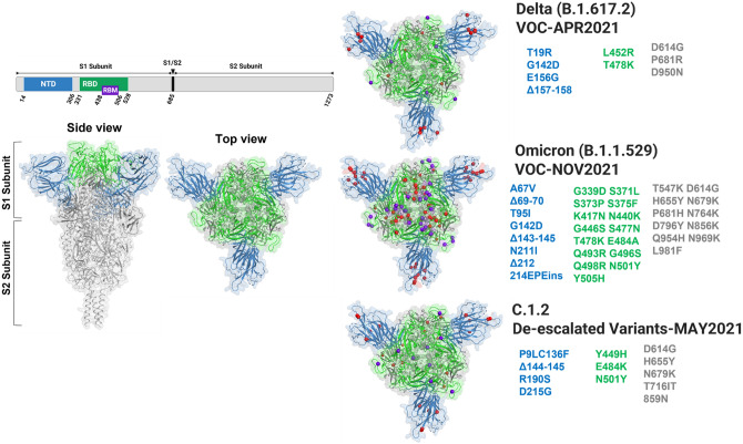
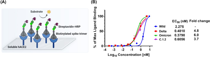
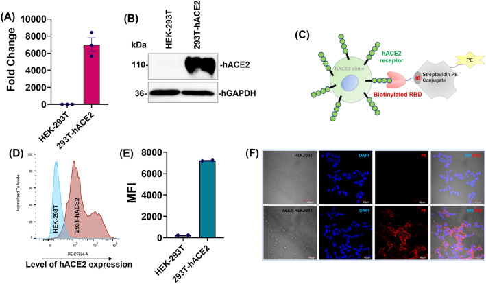
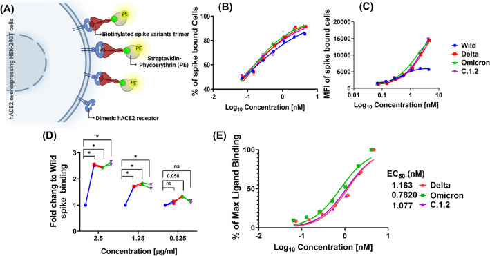
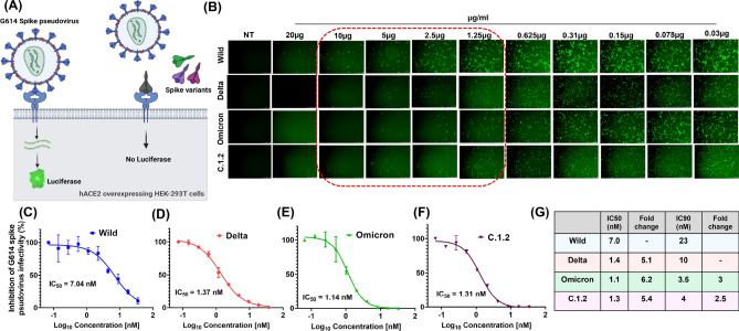
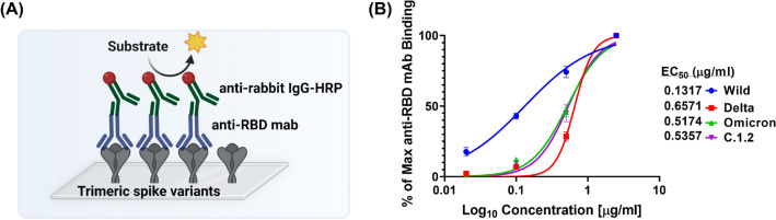
Spike glycoprotein of SARS-CoV-2 variants plays a critical role in infection and transmission through its interaction with human angiotensin converting enzyme 2 (hACE2) receptors. Prior findings using molecular docking and biomolecular studies reported varied findings on the difference in the interactions among the spike variants with the hACE2 receptors. Hence, it is a prerequisite to understand these interactions in a more precise manner. To this end, firstly, we performed ELISA with trimeric spike glycoproteins of SARS-CoV-2 variants including Wuhan Hu-1(Wild), Delta, C.1.2 and Omicron. Further, to study the interactions in a more specific manner by mimicking the natural infection, we developed hACE2 receptors expressing HEK-293T cell line, evaluated their binding efficiencies and competitive binding of spike variants with D614G spike pseudotyped virus. In line with the existing findings, we observed that Omicron had higher binding efficiency compared to Delta in both ELISA and Cellular models. Intriguingly, we found that cellular models could differentiate the subtle differences between the closely related C.1.2 and Delta in their binding to hACE2 receptors. Our study using the cellular model provides a precise method to evaluate the binding interactions between spike sub-lineages to hACE2 receptors.


Conflict of interest statement

The authors declare no competing interests.

Figures

Figure 1
Figure 1
3D crystal structure of ancestral SARS-CoV-2 spike trimer (PDB ID 6XR8) was generated, highlighting the mutational landscape by purple balls in RBM domain, red balls in NTD (blue), RBD (green) and other (gray) regions of Delta, Omicron and C.1.2 spike trimers using PyMOL software.
Figure 2
Figure 2
Concentration − response curves of trimeric SARS-CoV-2 spike variants, binding to hACE2 receptor protein. Schematic representation of non-competitive ELISA assay: plate coated with soluble hACE2 receptors and titrated with varying concentrations of biotinylated trimeric spike variants. The level of binding was quantified using streptavidin-HRP conjugate (A). The binding curves and EC50’s of each spike variants was quantified using ELISA by titrating of biotinylated spike variants to soluble hACE2 coated on the plate and the amount of spike trimer binding were estimated using streptavidin-HRP conjugate (B) (Mean ± SEM, N = 2).
Figure 3
Figure 3
Generation and characterisation of hACE2 receptor stably expressing HEK-293T cell line. The VSV-G lentiviral particles were produced with pLenti-hACE2-P2A-PuroR plasmid and transduced into HEK-293T cells. The stable expressing hACE2 HEK-293T cell line was selected with puromycin. The expression level of hACE2 in 293T-hACE2 cells were analysed by qPCR (A) and western blotting (with 20 µg of protein lysates) techniques (B). The functional characterization of 293T-hACE2 stable cells were done by RBD-Biotin surface staining. Graphical representation of RBD-biotin surface staining principle (C). After RBD-Biotin surface staining, the ancestral SARS-CoV-2 RBD binding hACE2 level on surface of 293T-hACE2 cells were quantified by flow cytometry (D, E) (MFI-mean florescent intensity) and RBD-hACE2 interaction was visualised by confocal microscopy (F).
Figure 4
Figure 4
Potency of spike variants affinity to dimeric hACE2 receptors present on surface of the cells. Schematic representation of in vitro flow cytometry binding assay to estimate binding affinity of spike variants (A). The 293T-hACE2 cells were surface stained with biotinylated trimeric spike variants at different concentration. Percentage (B) and level (MFI) (C) of spike trimer bound to cells was quantified using streptavidin-PE conjugate in flow cytometry. The fold change of Delta, Omicron and C.1.2 binding (MFI) related to Wild was calculated at indicated concentration (D). Percentage of maximum spike binding was calculated from MFI, plotted against concentration and a non-linear curve fitting was used to estimate the EC50 of spike variants (E).
Figure 5
Figure 5
In vitro competitive pseudovirus assay for evaluating the binding affinities of spike variants. Schematic representation of in vitro competitive pseudovirus assay (A). The 293T-hACE2 cells was incubated with D614G spike pseudovirus (expressing luciferase and ZsGreen protein) and different concentration of spike variants. After 60–72 h, infectivity of D614G spike pseudovirus was visualized by expression of ZsGreen protein in fluorescent microscopy at different spike variant treatment (B). The percentage of D614G spike pseudovirus infectivity was quantified by internalized pseudovirus luciferase activity and inhibitory concentration 50 (IC50) of Wild (C) Delta (D) Omicron (E) C.1.2 (F) spike proteins were quantified using non-linear curve fitting models. Table of IC50 and IC90 values of each spike variants (G).
Figure 6
Figure 6
Concentration − response curves of anti-RBD antibody binding to trimeric SARS-CoV-2 spike variants. Schematic representation of ELISA assay to quantify the binding of anti-RBD antibody against trimeric spike variants (A). EC50 was calculated by titrating of anti-RBD mAbs against trimeric spike variants and level of anti-RBD antibody bound to each spike variants were quantified using anti-rabbit IgG-HRP conjugate (B).

References

    1. Luo R, Delaunay-Moisan A, Timmis K, Danchin A. SARS-CoV-2 biology and variants: Anticipation of viral evolution and what needs to be done. Environ. Microbiol. 2021;23:2339–2363. doi: 10.1111/1462-2920.15487. - DOI - PMC - PubMed
    1. Jackson CB, Farzan M, Chen B, Choe H. Mechanisms of SARS-CoV-2 entry into cells. Nat. Rev. Mol. Cell Biol. 2022;23:3–20. doi: 10.1038/s41580-021-00418-x. - DOI - PMC - PubMed
    1. Kumar A, Parashar R, Kumar S, Faiq MA, Kumari C, Kulandhasamy M, Narayan RK, Jha RK, Singh HN, Prasoon P, et al. Emerging SARS-CoV-2 variants can potentially break set epidemiological barriers in COVID-19. J. Med. Virol. 2022;94:1300–1314. doi: 10.1002/jmv.27467. - DOI - PMC - PubMed
    1. Korber B, Fischer WM, Gnanakaran S, Yoon H, Theiler J, Abfalterer W, Hengartner N, Giorgi EE, Bhattacharya T, Foley B, et al. Tracking changes in SARS-CoV-2 spike: Evidence that D614G increases infectivity of the COVID-19 virus. Cell. 2020;182:812–827 e819. doi: 10.1016/j.cell.2020.06.043. - DOI - PMC - PubMed
    1. Yurkovetskiy L, Wang X, Pascal KE, Tomkins-Tinch C, Nyalile TP, Wang Y, Baum A, Diehl WE, Dauphin A, Carbone C, et al. Structural and functional analysis of the D614G SARS-CoV-2 spike protein variant. Cell. 2020;183:739–751 e738. doi: 10.1016/j.cell.2020.09.032. - DOI - PMC - PubMed

Publication types

Substances

Supplementary concepts