Multicenter Study
. 2023 May 30;13(1):8747.
doi: 10.1038/s41598-023-35759-4.

Three-month outcomes of faricimab loading therapy for wet age-related macular degeneration in Japan

Affiliations
Multicenter Study

Three-month outcomes of faricimab loading therapy for wet age-related macular degeneration in Japan

Ryo Mukai et al. Sci Rep. .

Abstract

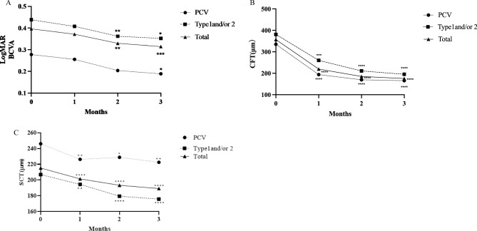
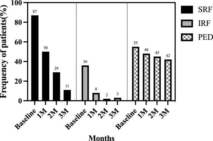
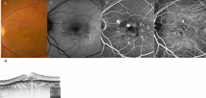
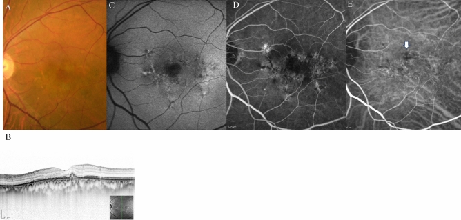
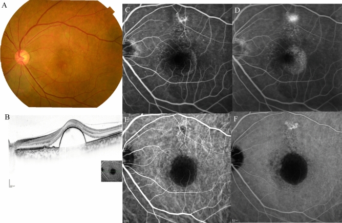
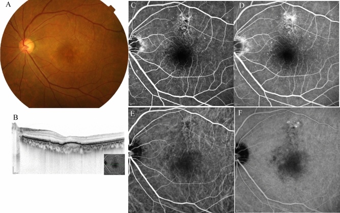
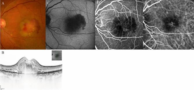
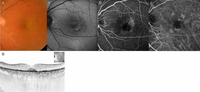
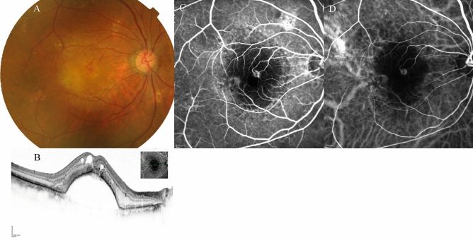
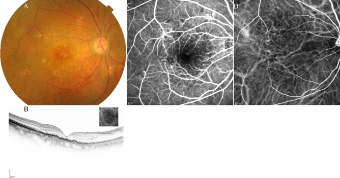
This multicenter study aimed to assess the short-term effectiveness and safety of faricimab in treatment-naïve patients with wet age-related macular degeneration (wAMD) in Japan. We retrospectively reviewed 63 eyes of 61 patients with wAMD, including types 1, 2, and 3 macular neovascularization as well as polypoidal choroidal vasculopathy (PCV). Patients received three consecutive monthly intravitreal injections of faricimab as loading therapy. Over these 3 months, visual acuity improved gradually compared to baseline. Moreover, the central foveal thickness decreased significantly at 1, 2, and 3 months compared to baseline (p < 0.0001). At 3 months after initiation of faricimab therapy, a dry macula (defined as absence of intraretinal or subretinal fluid) was achieved in 82% of the eyes. Complete regression of polypoidal lesions was observed in 52% of eyes with PCV. Subfoveal choroidal thickness also decreased significantly at 1, 2, and 3 months compared to baseline (p < 0.0001). Although retinal pigment epithelium tears developed in two eyes, there were no other ocular or systemic complications observed during the 3 months of loading therapy. In conclusion, loading therapy using faricimab resulted in improved visual acuity and retinal morphology in Japanese patients with wAMD without particular safety issues.


Conflict of interest statement

The authors declare no competing interests.

Figures

Figure 1
Figure 1
(A) Changes in best-corrected visual acuity in 62 eyes treated with 3 monthly faricimab injections. *p < 0.05, **p < 0.01, ***p < 0.001. (B) Changes in average central foveal thickness (CFT) in 62 eyes treated with 3 monthly faricimab injections. ***p < 0.001, ****p < 0.0001. (C) Changes in average subfoveal choroidal thickness (SCT) in 62 eyes treated with 3 monthly faricimab injections. *p < 0.05, **p < 0.01, ****p < 0.0001.
Figure 2
Figure 2
Presence of subretinal fluid (SRF), intraretinal fluid (IRF) and pigment epithelial detachment (PED) at baseline, 1, 2, and 3 months after initiation of faricimab.
Figure 3
Figure 3
Case 1 A 75-year-old male patient with PCV at baseline. (A) Fundus photography revealed pigment epithelial detachment (PED) with fibrin at the fovea. (B) Optical coherence tomography (OCT) revealed PED with fibrin and subretinal fluid. (C) Fundus autofluorescence imaging revealed a blockage of the PED. (D) Late-phase fluorescein angiography revealed leakage from polypoidal lesions at the macula. (E) Middle-phase indocyanine green angiography identified polypoidal lesions at the fovea (arrowhead) with a branching neovascular network.
Figure 4
Figure 4
The same 75-year-old male patient as shown in Fig. 1, here at 3 months after loading therapy with faricimab. (A) Fundus photography shows partial regression of pigment epithelial detachment (PED) as well as complete fibrin absorption at the fovea. (B) Optical coherence tomography (OCT) revealed regressed PED and absorption of serous retinal detachment. (C) Fundus autofluorescence imaging revealed no enlargement of the patchy atrophy. (D) Late-phase fluorescein angiography detected no leakage from a polypoidal lesion at the fovea. (E) Middle-phase indocyanine green angiography identified no polypoidal lesions at the fovea.
Figure 5
Figure 5
Case 2 A 63-year-old male patient with type 1 macular neovascularisation (MNV) at baseline. (A) Fundus photography shows a pigment epithelial detachment (PED) with subretinal fluid (SRF) at the fovea. (B) Optical coherence tomography (OCT) revealed a PED with SRF and a shallow PED at the nasal side of the large PED. (C, D) Early and late-phase fluorescein angiography detected a type 1 MNV. (E, F) Early and late-phase indocyanine green angiography identified abnormal MNV at the nasal edge of the large PED.
Figure 6
Figure 6
The same 63-year-old male patient as shown in Fig. 6, here at 3 months after initiation of faricimab therapy. (A) Fundus photography shows absorption of the subretinal fluid (SRF), and the pigment epithelial detachment (PED) was well absorbed. (B) Optical coherence tomography (OCT) revealed absorption of SRF, and a drastic decrease in the height of PED. (C, D) Early and late-phase fluorescein angiography detected no leakage from MNV. (E) Early and late-phase indocyanine green angiography identified no blockade of the large PED.
Figure 7
Figure 7
A 68-year-old male patient with type 2 macular neovascularisation (MNV) at baseline. (A) Fundus photography shows yellowish MNV surrounded by retinal haemorrhage with fibrin at the fovea. (B) Optical coherence tomography (OCT) revealed a subretinal mass surrounded by haemorrhage and fibrin with subretinal fluid. (C) Fundus autofluorescence imaging revealed a hemispherical blockade due to the subretinal haemorrhage. (D) Middle-phase fluorescein angiography detected MNV; however, haemorrhage caused a blockade. (E) Middle-phase indocyanine green angiography identified abnormal choroidal neovascularisation at the fovea.
Figure 8
Figure 8
The same 68-year-old male patient as shown in Fig. 7, here at 3 months after initiation of faricimab therapy. (A) Fundus photography shows absorption of the haemorrhage, fibrin, and yellowish mass. (B) Optical coherence tomography (OCT) revealed absorption of fibrin and haemorrhage, serous retinal detachment, and a small mass retained in the subretinal region. (C) Fundus autofluorescence imaging revealed very slight fine granular atrophy. (D) Late-phase fluorescein angiography detected no leakage from MNV. (E) Middle-phase indocyanine green angiography identified no abnormal vasculature.
Figure 9
Figure 9
Case 4 A 78-year-old female patient with type 3 macular neovascularisation (MNV) at baseline. (A) Fundus photography revealed a pigment epithelial detachment (PED) with intra retinal fluid (IRF) and intraretinal haemorrhage at the fovea. (B) Optical coherence tomography (OCT) revealed the large PED with IRF. (C) Early-phase fluorescein angiography detected dye leakage from a type 3 MNV. (D) Early-phase indocyanine green angiography also identified the type 3 neovascularisation.
Figure 10
Figure 10
The same 78-year-old female patient as shown in Fig. 9, here at 3 months after initiation of faricimab therapy. (A) Fundus photography shows absorption of the intraretinal fluid (IRF) and disappearance of intra retinal haemorrhage. The pigment epithelial detachment (PED) was absorbed. (B) Optical coherence tomography (OCT) revealed absorption of the IRF, and the PED was almost completely regressed. (C, D) Early-phase fluorescein angiography detected no leakage from the type 3 macular neovascularisation (MNV). (E) Early-phase indocyanine green angiography identified no type 3 MNV.
Figure 11
Figure 11
A case of retinal pigment epithelial (RPE) tear in 77-year-old male patient with polypoidal choroidal vasculopathy. (A) Fundus photography revealed a haemorrhagic pigment epithelial detachment (PED) with subretinal fluid (SRF) and subretinal haemorrhage at the fovea. (B) Fundus autofluorescence (FAF) revealed no RPE tear at baseline (C) Optical coherence tomography (OCT) revealed the large haemorrhagic PED with SRF. (D) Fundus photography at 1 months after the third injection of faricimab revealed that RPE tear developed at the inferior of the PED. (E) FAF identified hemispherical RPE defect at the inferior part of the PED. (F) OCT revealed RPE defect at the inferior part of the pre-existing PED.SRF was completely absorbed. Regardless of the RPE tear development, visual acuity improved from 20/125 to 20/40.

References

    1. Klein R, Klein BEK, Cruickshanks KJ. The prevalence of age-related maculopathy by geographic region and ethnicity. Prog. Retin. Eye Res. 1999;18:371–389. doi: 10.1016/S1350-9462(98)00025-1. - DOI - PubMed
    1. Klaver CC, et al. Incidence and progression rates of age-related maculopathy: The Rotterdam Study. Invest Ophthalmol. Vis. Sci. 2001;42:2237–2241. - PubMed
    1. Kawasaki R, et al. The prevalence of age-related macular degeneration in Asians: A systematic review and meta-analysis. Ophthalmology. 2010;117:921–927. doi: 10.1016/j.ophtha.2009.10.007. - DOI - PubMed
    1. Regula JT, et al. Targeting key angiogenic pathways with a bispecific CrossMAb optimized for neovascular eye diseases. EMBO Mol. Med. 2016;8:1265–1288. doi: 10.15252/emmm.201505889. - DOI - PMC - PubMed
    1. Joussen AM, et al. Angiopoietin/Tie2 signalling and its role in retinal and choroidal vascular diseases: A review of preclinical data. Eye. 2021;35:1305–1316. doi: 10.1038/s41433-020-01377-x. - DOI - PMC - PubMed

Publication types

MeSH terms