. 2024 Feb;52(1):59-71.
doi: 10.1007/s15010-023-02048-1. Epub 2023 May 30.

Detection of bornavirus-reactive antibodies and BoDV-1 RNA only in encephalitis patients from virus endemic areas: a comparative serological and molecular sensitivity, specificity, predictive value, and disease duration correlation study

Affiliations

Detection of bornavirus-reactive antibodies and BoDV-1 RNA only in encephalitis patients from virus endemic areas: a comparative serological and molecular sensitivity, specificity, predictive value, and disease duration correlation study

Petra Allartz et al. Infection. 2024 Feb.

Abstract

Purpose: Human Borna disease virus (BoDV-1) encephalitis is an emerging disease in Germany. This study investigates the spectrum of human BoDV-1 infection, characterizes anti-BoDV-1-antibodies and kinetics, and compares laboratory test performances.

Methods: Three hundred four encephalitis cases, 308 nation-wide neuropsychiatric conditions, 127 well-defined psychiatric cases from Borna disease-endemic areas, and 20 persons with contact to BoDV-1 encephalitis patients or animals were tested for BoDV-1 infections by serology and PCR.

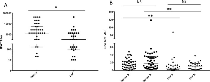
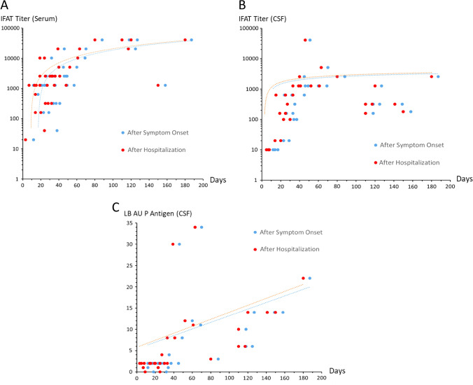
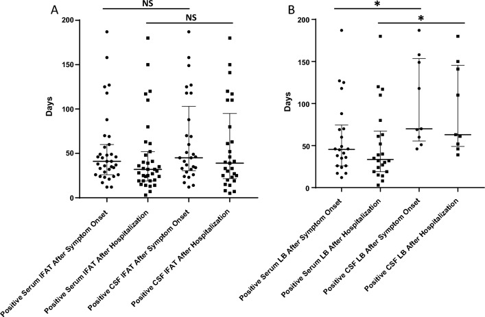
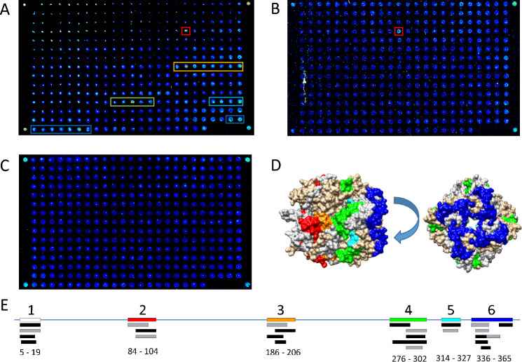
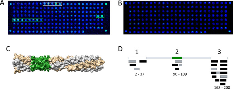
Results: BoDV-1 infections were only found in encephalitis patients with residence in, or recent travel to, virus-endemic areas. Antibodies were detected as early as 12 days after symptom onset. Serum antibody levels correlated with disease duration. Serology was ordered after 50% of the disease duration had elapsed, reflecting low awareness. BoDV-1-antibodies were of IgG1 subclass, and the epitope on BoDV-1 antigens was determined. Specificity of the indirect immunofluorescence antibody test (IFAT) and lineblot (LB) from serum and cerebrospinal fluid (CSF), as well as PCR testing from CSF, was 100%. Sensitivity, depending on first or all samples, reached 75-86% in serum and 92-94% in CSF for the IFAT, and 33-57% in serum and 18-24% in CSF for the LB. Sensitivity for PCR in CSF was 25-67%. Positive predictive values were 100% each, while negative predictive values were 99% (IFAT), 91-97% (LB), and 90% (PCR).

Conclusions: There is no hint that BoDV-1 causes other diseases than encephalitis in humans. Awareness has to be increased in virus-endemic areas. Tests are robust but lack sensitivity. Detection of IgG1 against specific peptides may facilitate diagnosis. Screening of healthy individuals is likely not beneficial.

Keywords: Bornavirus; IgG1; PCR; Peptide; Serology.


Conflict of interest statement

No conflict of interest concerning this publication has to be disclosed by the authors.

Figures

Fig. 1
Fig. 1
Disease duration in confirmed BoDV-1 encephalitis cases, time point of taking diagnostic samples, and time point of diagnosis over years. A Duration of BoDV-1 encephalitis in the patient cohort. Median length of illness was 46 days. The two patients with the longest disease duration were severely immunosuppressed as therapeutic approach for BoDV-1 encephalitis. Median with interquartile range is shown. B Time point of taking diagnostic samples. Median duration before a sample was taken after symptom onset was 25 days, corresponding to median day 14 after hospital admission. Median with interquartile range is shown. C Diagnosis at days after hospitalization. A weak tendency to request bornavirus serology earlier each year is seen over time (linear regression; R2=0.045)
Fig. 2
Fig. 2
Height of antibody levels in BoDV-1 patients. A Immunofluorescence antibody test (IFAT) results for serum and cerebrospinal fluid (CSF) of all BoDV-1 patients tested in this study. Serum titers are significantly higher than CSF titers (*p < 0.05; Mann–Whitney test). Median with interquartile range is shown. B Line blot antibody test results for serum and CSF of all BoDV-1 patients. Serum antibodies against individual antigens are significantly higher than respective CSF antibody levels (**p < 0.005; Mann–Whitney test (respective one-on-one comparison)). Median with interquartile range is shown. AU arbitrary units; NS not significant
Fig. 3
Fig. 3
Height of antibody levels in relation to disease duration. A Immunofluorescence antibody test (IFAT) results for serum of all BoDV-1 patients tested in this study over time. Correlation of titers with time after symptom onset is 0.67 and after hospitalization 0.72. Logarithmic scale. B IFAT results for cerebrospinal fluid (CSF) of all BoDV-1 patients tested in this study over time. Correlation of titers with time after symptom onset is 0.62 and after hospitalization 0.63. Logarithmic scale. C Line blot (LB) arbitrary units (AU) of antibodies against BoDV-1 P antigen in CSF of all BoDV-1 patients tested in this study. Correlation of AU with time after symptom onset and after hospitalization is 0.78 each. Linear scale
Fig. 4
Fig. 4
Presence of antibodies at time points of disease. A Positive immunofluorescence antibody test (IFAT) result for serum and cerebrospinal fluid (CSF) of all BoDV-1 patients tested in this study at given time points in days after symptom onset and hospital admission. Antibodies in serum were non-significantly earlier positive than in CSF. Median with interquartile range is shown. B Positive line blot (LB) results for serum and CSF of all BoDV-1 patients tested in this study at given time points in days after symptom onset and hospital admission. Antibodies in serum were significantly earlier positive than in CSF (*p < 0.05). Median with interquartile range is shown
Fig. 5
Fig. 5
BoDV-1 N epitope mapping. A Serological fingerprint of a serum sample on the peptide microarray, represented as a 16-bit pseudocolor image obtained from the 650 nm channel. The detected antibody targets are designated by colored boxes if they were detected in two independent array experiments. B CSF serological fingerprint from same patient as in A showed a strong single spot bound antibody, binding to the same region as in A (red box). C Serum sample without observable signals. D Mapping of antibody target sequences onto the structure of the N protein (1N93). The colors correspond to peptide binding regions as found in A. E Overlay of antibody target sequences on the N protein revealing six immunogenic sites. The amino acid numbers are given below. Colors of the regions correspond to those in A and D. Region 1 is not resolved in the crystal structure (white box). Black boxes designate antibodies found in serum, gray boxes indicate antibodies from CSF samples
Fig. 6
Fig. 6
BoDV-1 P epitope mapping. A Serological fingerprint of a CSF sample on the peptide microarray, represented as a 16-bit pseudocolor image obtained from the 650 nm channel. B Serum sample without detectable signals. C Mapping of antibody target sequences onto the structure of the P protein (8B8A), color correspond to AA sequence found in A. D Overlay of antibody target sequences on the P protein revealing three immunogenic sites. The amino acid numbers are given below. The colors of the regions correspond to those in A and C. Regions 1 and 3 are not resolved in the crystal structure (white boxes). Black boxes represent antibodies found in serum, gray boxes indicate antibodies from CSF samples
Fig. 7
Fig. 7
Distribution of BoDV-1 qPCR cycle threshold (ct) values and viral copy numbers in cerebrospinal fluid. A ct values with 36.2 in median, ranging from 29.3 to 37.9 are displayed. The two samples with the lowest ct values were from patients with the second shortest duration of disease when the material was taken and with the lowest antibody levels in CSF. Median with interquartile range is shown. B BoDV-1 copy numbers with 3086/mL in median, ranging from 55 to 477,844/mL are demonstrated. Median with interquartile range is shown

References

    1. Schlottau K, Forth L, Angstwurm K, Höper D, Zecher D, Liesche F, Hoffmann B, Kegel V, Seehofer D, Platen S, Salzberger B, Liebert UG, Niller HH, Schmidt B, Matiasek K, Riemenschneider MJ, Brochhausen C, Banas B, Renders L, Moog P, Wunderlich S, Seifert CL, Barreiros A, Rahmel A, Weiss J, Tappe D, Herden C, Schmidt-Chanasit J, Schwemmle M, Rubbenstroth D, Schlegel J, Pietsch C, Hoffmann D, Jantsch J, Beer M. Fatal encephalitic borna disease virus 1 in solid-organ transplant recipients. N Engl J Med. 2018;379:1377–1379. doi: 10.1056/NEJMc1803115. - DOI - PubMed
    1. Korn K, Coras R, Bobinger T, Herzog SM, Lücking H, Stöhr R, Huttner HB, Hartmann A, Ensser A. Fatal encephalitis associated with borna disease virus 1. N Engl J Med. 2018;379:1375–1377. doi: 10.1056/NEJMc1800724. - DOI - PubMed
    1. Dürrwald R, Kolodziejek J, Weissenböck H, Nowotny N. The bicolored white-toothed shrew Crocidura leucodon (HERMANN 1780) is an indigenous host of mammalian Borna disease virus. PLoS ONE. 2014;9:e93659. doi: 10.1371/journal.pone.0093659. - DOI - PMC - PubMed
    1. Nobach D, Bourg M, Herzog S, Lange-Herbst H, Encarnação JA, Eickmann M, Herden C. Shedding of infectious borna disease virus-1 in living bicolored white-toothed shrews. PLoS ONE. 2015;10:e0137018. doi: 10.1371/journal.pone.0137018. - DOI - PMC - PubMed
    1. Weissenböck H, Bagó Z, Kolodziejek J, Hager B, Palmetzhofer G, Dürrwald R, Nowotny N. Infections of horses and shrews with bornaviruses in upper Austria: a novel endemic area of Borna disease. Emerg Microbes Infect. 2017;6:e52. doi: 10.1038/emi.2017.36. - DOI - PMC - PubMed