. 2023 May 30;13(1):8773.
doi: 10.1038/s41598-023-35511-y.

Identification and localization of polar tube proteins in the extruded polar tube of the microsporidian Anncaliia algerae

Affiliations

Identification and localization of polar tube proteins in the extruded polar tube of the microsporidian Anncaliia algerae

Maurine Fayet et al. Sci Rep. .

Abstract

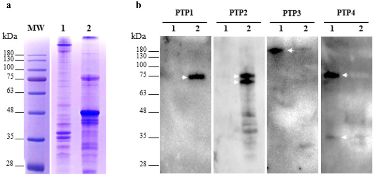
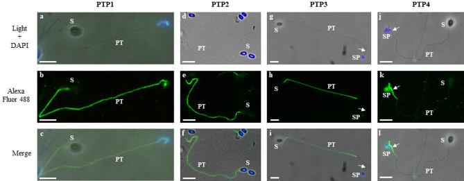
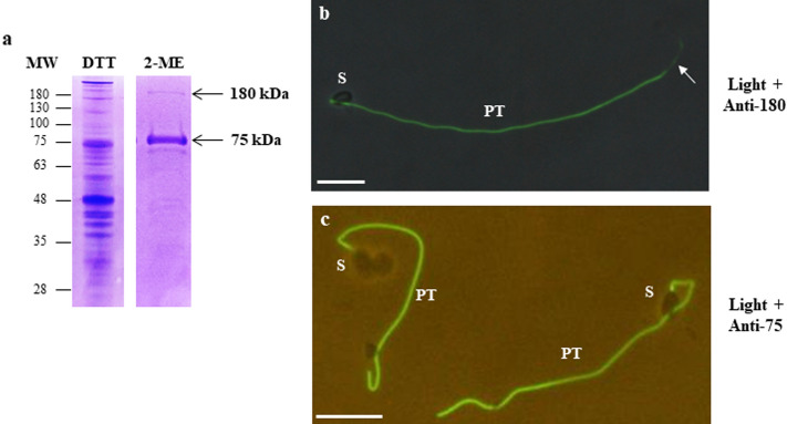
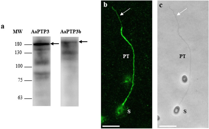
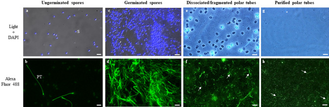
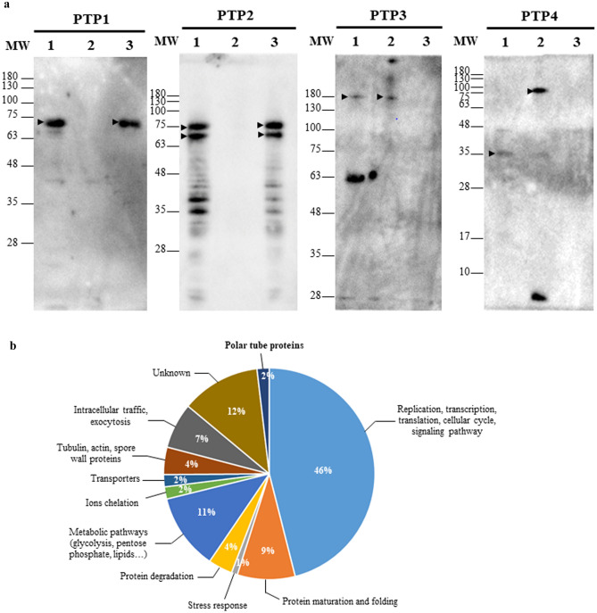
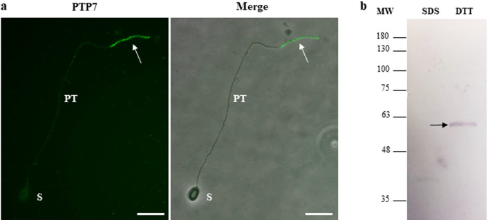
Microsporidia are obligate intracellular parasites able to infect a wide range of hosts from invertebrates to vertebrates. The success of their invasion process is based on an original organelle, the polar tube, which is suddenly extruded from the spore to inoculate the sporoplasm into the host cytoplasm. The polar tube is mainly composed of proteins named polar tube proteins (PTPs). A comparative analysis allowed us to identify genes coding for 5 PTPs (PTP1 to PTP5) in the genome of the microsporidian Anncaliia algerae. While PTP1 and PTP2 are found on the whole polar tube, PTP3 is present in a large part of the extruded polar tube except at its end-terminal part. On the contrary, PTP4 is specifically detected at the end-terminal part of the polar tube. To complete PTPs repertoire, sequential sporal protein extractions were done with high concentration of reducing agents. In addition, a method to purify polar tubes was developed. Mass spectrometry analysis conducted on both samples led to the identification of a PTP3-like protein (PTP3b), and a new PTP (PTP7) only found at the extremity of the polar tube. The specific localization of PTPs asks the question of their roles in cell invasion processes used by A. algerae.


Conflict of interest statement

The authors declare no competing interests.

Figures

Figure 1
Figure 1
Immunodetection of PTPs in A. algerae sporal protein extracts. (a) Coomassie blue stained 10% SDS-PAGE of proteins extracted from A. algerae spores. The first extraction was done in 2.5% SDS (lane 1). Then, the insoluble proteins were solubilized in a buffer containing 100 mM DTT (lane 2). (b) Western Blot analysis using antisera raised against PTP1, PTP2, PTP3 and PTP4 on SDS (lane 1) and DTT extracts (lane 2). PTP1 and PTP2 are only soluble in presence of reducing agent (arrows). In contrast, PTP3 and PTP4 are detected in the SDS extract (arrows). All antisera were used at a 1:200 dilution.
Figure 2
Figure 2
Indirect immunofluorescence assay using antisera raised against recombinant PTPs revealing the specific location of each PTP in A. algerae extruded polar tubes. Germinated spores are visualized in phase contrast (a,d,g,j), and after anti-PTP1 (b), anti-PTP2 (e), anti-PTP3 (h) and anti-PTP4 (k) labelling. Merged pictures were generated with GIMP 2.10.8 software (c,f,i,l). Antibodies against PTP1 and PTP2 proteins labelled the entire polar tube. In contrast, antibodies against the PTP3 protein labelled a large part of the polar tube except the extremity, whereas anti-PTP4 antibodies exclusively labelled the extremity of the polar tube (arrows). All antisera were used at a 1:100 dilution. Secondary antibody was Alexa 488-conjugated goat anti-mouse (or anti-rabbit for PTP1) IgG. At least 10 different fields were observed which corresponds to ~ 50 to 130 spores with their polar tube extruded. See Supplementary Fig. S2 for additional pictures illustrating the different localizations of PTPs. PT polar tube, S spore, SP sporoplasm. Scale bar: 10 µm.
Figure 3
Figure 3
Antibodies produced against two A. algerae protein bands soluble in high concentration of 2-mercaptoethanol specifically labelled the extruded polar tube. (a) SDS-PAGE (10%) of sporal protein extracts obtained first in a buffer containing 100 mM DTT. Then, the insoluble fraction was incubated in a 50% 2-mercaptoethanol (2-ME) solution. Two protein bands running at 75 and 180 kDa (arrows) were selected in the 2-ME extract to produce mouse antibodies and were analysed in mass spectrometry (see Table S2). (b,c) IFA with mouse antisera raised against 180-kDa (b) and 75-kDa (c) protein bands. Both antisera labelled the entire polar tube of germinated A. algerae spores but the signal was less intensive at the end of the tube with the anti-180 kDa antiserum (arrow). Mouse antisera were used at a 1:100 dilution. Secondary antibody was Alexa 488-conjugated goat anti-mouse IgG. Merged pictures were generated with GIMP 2.10.8 software. PT polar tube; S spore; Scale bar: 10 µm.
Figure 4
Figure 4
Antibodies produced against the recombinant PTP3b specifically labelled the extruded polar tube. (a) Immunoblotting with anti-PTP3 and anti-PTP3b against A. algerae total sporal protein extract (10% SDS-PAGE). PTP3b is detected at a higher molecular weight than PTP3 (arrows). The other lower detected-bands probably correspond to protein degradation. (b) IFA with anti-PTP3b antibodies. The entire extruded polar tube is stained but with a less intensive signal at the end-terminal part of the tube (white arrow). (c) phase contrast. At least 10 different fields were observed which corresponds to ~ 50 to 130 spores with their polar tube extruded. See Supplementary Fig. S2 for additional pictures illustrating the different localizations of PTPs. PT polar tube; S spore, Scale bar: 10 µm.
Figure 5
Figure 5
Purification of A. algerae polar tubes. 109 spores of A. algerae were incubated in PBS (a,b) or in a solution containing 0.2 M NaHCO3-Na2CO3 at pH 9.5 for 2 h at 20 °C to stimulate the germination (c,d). Polar tubes were then dissociated from the germinated spores and fragmented (e,f). Finally, fragmented polar tubes were purified by 3 successive centrifugations (g,h). At each step, biological material was collected and analysed by DAPI staining (a,c,e,g) and by IFA (b,d,f,h) using mouse polyclonal antibodies raised against the polar tube (anti-PTP3 antibodies). Fragmented and purified polar tubes are indicated by arrows. In the final purified polar tubes fraction, some fragmented polar tubes are labelled with DAPI (g, *). PT polar tube, S spore. Scale bars: 10 µm.
Figure 6
Figure 6
Analysis of purified polar tube fraction. (a) Immunodetection of PTPs in protein extracts from A. algerae purified polar tubes. Anti-PTP polyclonal antibodies were applied to 3 different protein extracts from purified polar tubes. The total extract (lane 1) contains proteins soluble in 2.5% SDS and 100 mM DTT. The two other extracts correspond to a differential protein extraction. Proteins from purified polar tubes were first solubilized with 2.5% SDS (lane 2), then insoluble proteins were extracted in a buffer containing 100 mM DTT (lane 3). Both PTP1 and PTP2 were only soluble in presence of reducing agents (lane 3, arrows). In contrast, anti-PTP3 and anti-PTP4 antisera detected protein bands in the SDS extract without DTT (lane 2). For PTP3, an additional band around 63 kDa was also stained in the total extract. For PTP4, the SDS extract revealed three bands of 75, 35 and < 10 kDa (lane 2). All antisera were used at a 1:200 dilution. (b) Functional classification and distribution of the 214 proteins identified by mass spectrometry in the purified polar tube fraction using InterProScan prediction (see Table S3).
Figure 7
Figure 7
Localization of the newly identified PTP7 at the terminal end of the extruded polar tube. (a) IFA with anti-PTP7 mouse antiserum (dilution 1:100) revealing a specific labelling of the terminal end of the A. algerae extruded polar tube (arrow). Secondary antibodies were Alexa 488-conjugated goat anti-mouse IgG. At least 10 different fields were observed which corresponds to ~ 50 to 130 spores with their polar tube extruded. See Supplementary Fig. S2 for additional pictures illustrating the different localizations of PTP7. PT polar tube; S spore; Scale bar: 10 µm. (b) Western Blot analysis on sporal proteins solubilized first in presence of 2.5% SDS (SDS extract) and then in a 100 mM DTT- containing solution (DTT extract). The antiserum reacted with a unique A. algerae sporal protein band at around 60 kDa in the DTT extract (arrow).

References

    1. Bojko J, et al. Microsporidia: A new taxonomic, evolutionary, and ecological synthesis. Trends Parasitol. 2022;38:642–659. doi: 10.1016/j.pt.2022.05.007. - DOI - PubMed
    1. Stentiford GD, et al. Microsporidia—Emergent pathogens in the global food chain. Trends Parasitol. 2016;32:336–348. doi: 10.1016/j.pt.2015.12.004. - DOI - PMC - PubMed
    1. Han B, Pan G, Weiss LM. Microsporidiosis in humans. Clin. Microbiol. Rev. 2021;34:e0001020. doi: 10.1128/CMR.00010-20. - DOI - PMC - PubMed
    1. Ruan Y, et al. The largest meta-analysis on the global prevalence of microsporidia in mammals, avian and water provides insights into the epidemic features of these ubiquitous pathogens. Parasites Vectors. 2021;14:186. doi: 10.1186/s13071-021-04700-x. - DOI - PMC - PubMed
    1. Seatamanoch N, Kongdachalert S, Sunantaraporn S, Siriyasatien P, Brownell N. Microsporidia, a highly adaptive organism and its host expansion to humans. Front. Cell Infect. Microbiol. 2022;12:924007. doi: 10.3389/fcimb.2022.924007. - DOI - PMC - PubMed

Publication types

Substances

Supplementary concepts