

A vanished gastric gastrointestinal stromal tumor
- PMID: 37254018
- PMCID: PMC10229511
- DOI: 10.1186/s40792-023-01674-z
A vanished gastric gastrointestinal stromal tumor
Abstract
Background: Local resection is the standard treatment for gastrointestinal stromal tumors (GISTs). Laparoscopic and endoscopic cooperative surgery (LECS) is a minimally invasive surgery used to resect GISTs. Herein, we report an extremely rare case of a gastric GIST that grossly vanished during LECS.
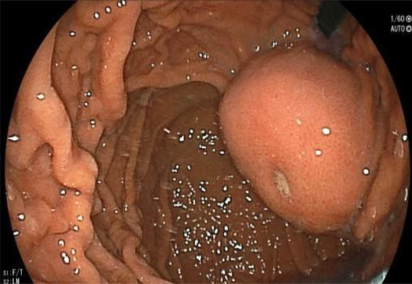
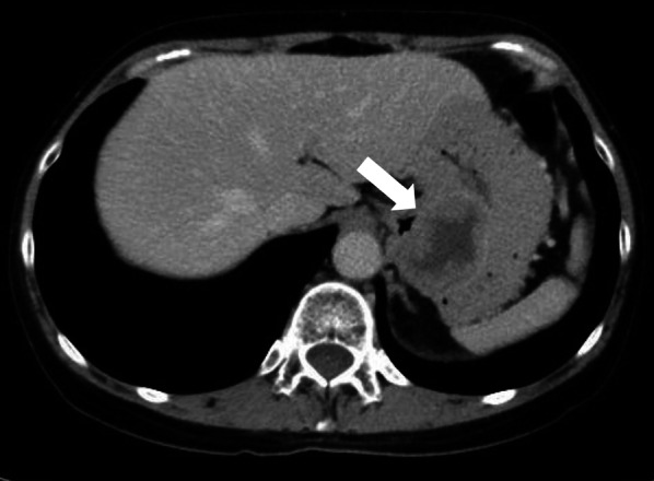
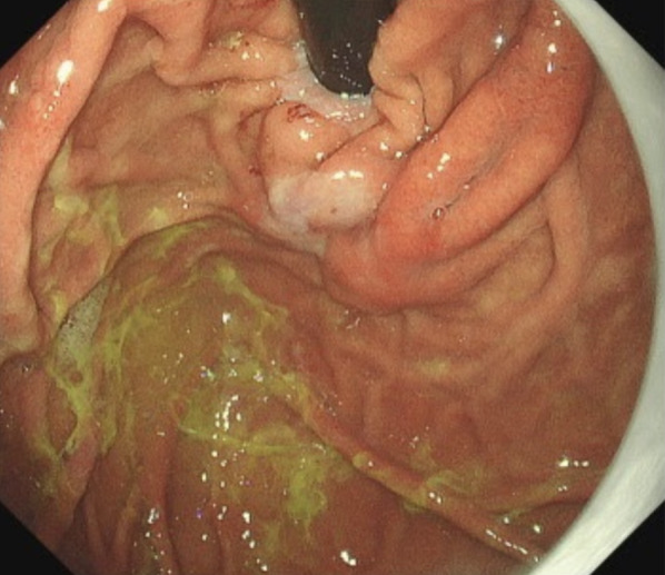
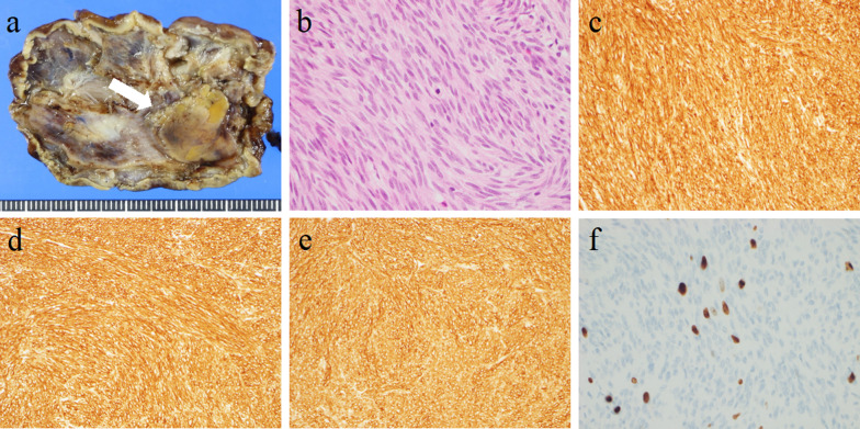
Case presentation: A 50-year-old Japanese female was referred to our hospital after an abnormality was detected during an esophagogastroduodenoscopy (EGD) at her annual health checkup. Based on EGD, endoscopic ultrasound (EUS), and computer tomography (CT) findings, the patient was diagnosed with a 50-mm submucosal tumor (SMT) with intraluminal growth on the anterior wall of the lesser curvature of the upper body of the stomach. We routinely use LECS to treat the intraluminal growth type of GISTs. During the intraoperative endoscopy, the intraluminal submucosal tumor, which was detected preoperatively, had vanished. A red-white scar was observed in the regressed tumor region. LECS was performed by resecting at a distance away from the scar tissue and closing the gastric wall with intracavitary sutures. In the evaluation from the tumor section view of the original resected specimen, a 22 × 14 × 8 mm lobular neoplasm was observed that was predominantly located in the gastric submucosa to the muscularis propia. Pathological findings confirmed the diagnosis of GIST with intermediate risk indicated by the Fletcher classification. The patient continued postoperative adjuvant chemotherapy with imatinib and no recurrence was detected over 12 months after surgery.
Conclusion: LECS was performed on the vanished gastric GIST, providing the best surgical treatment and leading to an accurate diagnosis and optimal postoperative care.
Keywords: Gastrointestinal stroma tumor (GIST); Laparoscopy and endoscopy cooperative surgery (LECS); Spontaneous regression; Vanished.
© 2023. The Author(s).
Conflict of interest statement
The authors declare that they have no competing interests.
Figures


References
-
- Stewart FW. Experiences in spontaneous regression of neoplastic disease in man. Tex Rep Biol Med. 1952;10(1):239–253. - PubMed
-
- Chodorowski Z, Anand JS, Wiśniewski M, Madaliński M, Wierzba K, Wiśniewski J. Spontaneous regression of cancer–review of cases from 1988 to 2006. Przegl Lek. 2007;64(4–5):380–382. - PubMed
LinkOut - more resources
Full Text Sources
