. 2023 May 30;23(1):188.
doi: 10.1186/s12890-023-02474-9.

ILC2 regulates hyperoxia-induced lung injury via an enhanced Th17 cell response in the BPD mouse model

Affiliations

ILC2 regulates hyperoxia-induced lung injury via an enhanced Th17 cell response in the BPD mouse model

Yue Zhu et al. BMC Pulm Med. .

Abstract

Backgroud: Recent research has focused on the role of immune cells and immune responses in the pathogenesis of bronchopulmonary dysplasia (BPD), but the exact mechanisms have not yet been elucidated. Previously, the key roles of type 2 innate lymphoid cells (ILC2) in the lung immune network of BPD were explored. Here, we investigated the role Th17 cell response in hyperoxia-induced lung injury of BPD, as well as the relationship between ILC2 and Th17 cell response.

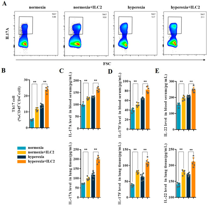
Methods: A hyperoxia-induced BPD mouse model was constructed and the pathologic changes of lung tissues were evaluated by Hematoxylin-Eosin staining. Flow cytometry analysis was conducted to determine the levels of Th17 cell, ILC2 and IL-6+ILC2. The expression levels of IL-6, IL-17 A, IL-17 F, and IL-22 in the blood serum and lung tissues of BPD mice were measured by ELISA. To further confirm the relationship between ILC2 and Th17 cell differentiation, ILC2 depletion was performed in BPD mice. Furthermore, we used immunomagnetic beads to enrich ILC2 and then flow-sorted mouse lung CD45+Lin-CD90.2+Sca-1+ILC2. The sorted ILC2s were injected into BPD mice via tail vein. Following ILC2 adoptive transfusion, the changes of Th17 cell response and lung injury were detected in BPD mice.

Results: The expression levels of Th17 cells and Th17 cell-related cytokines, including IL-17 A, IL-17 F, and IL-22, were significantly increased in BPD mice. Concurrently, there was a significant increase in the amount of ILC2 and IL-6+ILC2 during hyperoxia-induced lung injury, which was consistent with the trend for Th17 cell response. Compared to the control BPD group, ILC2 depletion was found to partially abolish the Th17 cell response and had protective effects against lung injury after hyperoxia. Furthermore, the adoptive transfer of ILC2 enhanced the Th17 cell response and aggravated lung injury in BPD mice.

Conclusions: This study found that ILC2 regulates hyperoxia-induced lung injury by targeting the Th17 cell response in BPD, which shows a novel strategy for BPD immunotherapy.

Keywords: Bronchopulmonary dysplasia; Lung injury; Th17 cell response; Type 2 innate lymphoid cells.


Conflict of interest statement

The authors declare no competing interests.

Figures

Fig. 1
Fig. 1
Hyperoxia induced lung injury in BPD mice. A C57BL/6 newborn mice were treated with normoxia or hyperoxia exposure from birth. Neonatal mice from the different groups were sacrificed at day 14. B Representative H&E staining of the lungs from the neonatal mice in the normoxia and hyperoxia groups. C, D lung injury was assessed by RAC and lung injure score. E Representative PAS staining of the lungs from the neonatal mice in the normoxia and hyperoxia groups. Data presented are the mean ± SD (n = 6); ** P < 0.05 vs. normoxia group
Fig. 2
Fig. 2
Hyperoxia induced Th17 cell response in BPD mice. A Representative FACS analysis showing the gating strategy used to identify CD45+CD4+IL-17 A+Th17 in lung of BPD mice. B Representative results of Th17 cells detected by flow cytometry in lung tissues from the normoxia and hyperoxia groups. C Percentage of Th17 cells detected by flow cytometry in lung tissues from the normoxia and hyperoxia groups. D–F IL-17 A, IL-17 F, and IL-22 levels in the blood serum and lung tissues were assessed in the normoxia and hyperoxia groups. Data presented are the mean ± SD (n = 6); ** P < 0.05 vs. normoxia group
Fig. 3
Fig. 3
Hyperoxia induced ILC2 and IL-6+ILC2 in BPD mice. A Representative FACS analysis showing the gating strategy used to identify CD45+Lin-CD90.2+Sca-1+ILC2 in lung of BPD mice. B, C Representative results and percentage of ILC2 detected by flow cytometry in lung tissues from the normoxia and hyperoxia groups. D Representative Wright-Giemsa staining of sorted ILC2. E, F Representative results and percentage of IL-6+ILC2 (gate: CD45+Lin-CD90.2+Sca-1+) detected by flow cytometry in normoxia and hyperoxia groups. G RT-qPCR analysis of IL-6 mRNA levels in ILC2 of normoxia and hyperoxia groups. H ELISA analysis of IL-6 expression in the blood serum and lung tissues from the normoxia and hyperoxia groups. Data represented as mean ± SD (n = 6); ** P < 0.05 vs. normoxia group
Fig. 4
Fig. 4
ILC2 depletion inhibited Th17 cell response. A Newborn mice were treated with anti-CD90.2 antibody or control IgG twice during hyperoxia exposure. Neonatal mice in different groups were sacrificed at day 14. B Representative results and the percentage of ILC2 detected by flow cytometry in the lungs of the hyperoxia, hyperoxia + control IgG, and hyperoxia + anti-CD90.2 groups. C Representative results and percentage of Th17 cells detected by flow cytometry in lung samples of the hyperoxia, hyperoxia + control IgG, and hyperoxia + anti-CD90.2 groups. D–G IL-6, IL-17 A, IL-17 F, and IL-22 levels in the blood serum and lung tissues were assessed in the hyperoxia, hyperoxia + control IgG, and hyperoxia + anti-CD90.2 groups. Data presented are the mean ± SD (n = 6); ** P < 0.05 vs. hyperoxia group
Fig. 5
Fig. 5
ILC2 depletion inhibited hyperoxia-induced lung injury in BPD mice. A Representative H&E staining of the lung samples from the neonatal mice in the hyperoxia, hyperoxia + control IgG, and hyperoxia + anti-CD90.2 groups. B, C lung injury was assessed by RAC and lung injure score. D Representative PAS staining of the lungs from the neonatal mice in the hyperoxia, hyperoxia + control IgG, and hyperoxia + anti-CD90.2 groups. Data presented are the mean ± SD (n = 6); ** P < 0.05 vs. hyperoxia group
Fig. 6
Fig. 6
ILC2 adoptive transfusion in neonatal mice. A Neonatal mice were treated with an adoptive transfusion at day 7 during hyperoxia exposure. Neonatal mice in different groups were sacrificed at day 14. B In vivo imaging of Cy7-stained ILC2 in BPD mice; upper panel: whole-body imaging at 0, 6, and 30 h; lower panel: ex vivo imaging of the heart, lung, liver, spleen, kidney, and stomach at 30 h. C, D Representative results and percentage of ILC2 detected by flow cytometry in the lung tissues from the different groups. E IL-6 levels in the blood serum and lung tissues were assessed in different groups. Data presented are the mean ± SD (n = 6); ** P < 0.05
Fig. 7
Fig. 7
ILC2 adoptive transfusion enhanced the Th17 cell response in neonatal mice. A, B Representative results and the percentage of Th17 cells detected by flow cytometry in the lung tissues from the different groups. C–E IL-17 A, IL-17 F, and IL-22 levels in the blood serum and lung tissues were assessed in the different groups. Data presented are the mean ± SD (n = 6); ** P < 0.05
Fig. 8
Fig. 8
ILC2 adoptive transfusion enhanced lung injury in neonatal mice. A Representative H&E staining of the neonatal lung tissues from the mice in the different groups. B, C lung injury was assessed by RAC and lung injure score. D Representative PAS staining of the lungs from the neonatal mice in the different groups. Data presented are the mean ± SD (n = 6); ** P < 0.05
Fig. 9
Fig. 9
Schematic representation of the mechanism of ILC2 on regulating hyperoxia-induced lung injury by targeting Th17 cell response in BPD mice model

References

    1. Thébaud B, Goss KN, Laughon M, Whitsett JA, Abman SH, Steinhorn RH, Aschner JL, Davis PG, McGrath-Morrow SA, Soll RF, et al. Bronchopulmonary dysplasia. Nat Rev Dis Primers. 2019;5(1):78. doi: 10.1038/s41572-019-0127-7. - DOI - PMC - PubMed
    1. Hwang JS, Rehan VK. Recent advances in Bronchopulmonary Dysplasia: pathophysiology, Prevention, and treatment. Lung. 2018;196(2):129–38. doi: 10.1007/s00408-018-0084-z. - DOI - PMC - PubMed
    1. Northway WH, Jr, Rosan RC, Porter DY. Pulmonary disease following respirator therapy of hyaline-membrane disease. Bronchopulmonary dysplasia. N Engl J Med. 1967;276(7):357–68. doi: 10.1056/NEJM196702162760701. - DOI - PubMed
    1. Gibbs K, Jensen EA, Alexiou S, Munson D, Zhang H. Ventilation strategies in severe bronchopulmonary dysplasia. Neoreviews. 2020;21(4):e226–37. doi: 10.1542/neo.21-4-e226. - DOI - PubMed
    1. Giusto K, Wanczyk H, Jensen T, Finck C. Hyperoxia-induced bronchopulmonary dysplasia: better models for better therapies. Dis Model Mech. 2021;14(2):047753. doi: 10.1242/dmm.047753. - DOI - PMC - PubMed