. 2023 May 30;23(1):242.
doi: 10.1186/s12886-023-02993-3.

Geographic Atrophy after Reabsorption of Pigment Epithelial Detachment (GARPED) study

Affiliations

Geographic Atrophy after Reabsorption of Pigment Epithelial Detachment (GARPED) study

Enoch T Peng et al. BMC Ophthalmol. .

Abstract

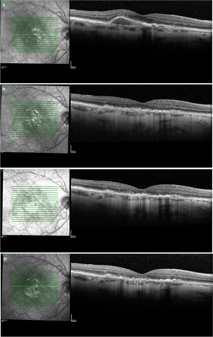
Background: To describe the occurrence, rate of geographic atrophy (GA) expansion, and changes in visual acuity (VA) after reabsorption of subfoveal pigment epithelial detachments (PED).

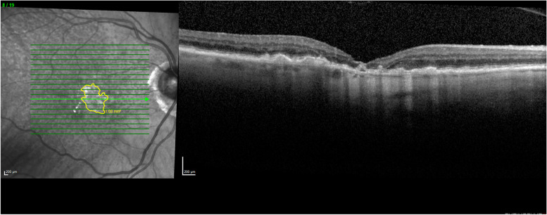
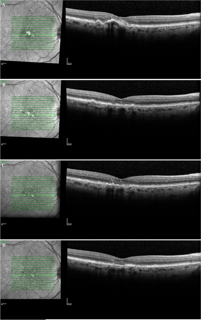
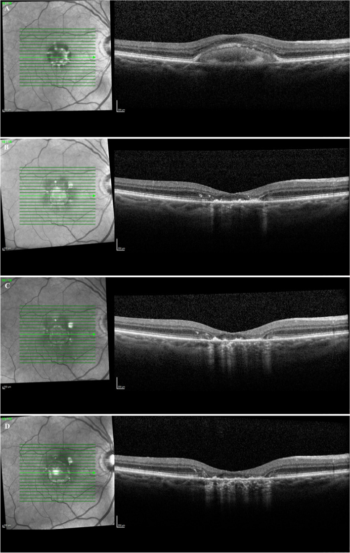
Methods: Included patients had reabsorption of a PED followed by GA. Patients underwent clinical examination with SD-OCT. Images were classified by size with grading occurring post reabsorption. VA was recorded pre-reabsorption, post-reabsorption, and over time.

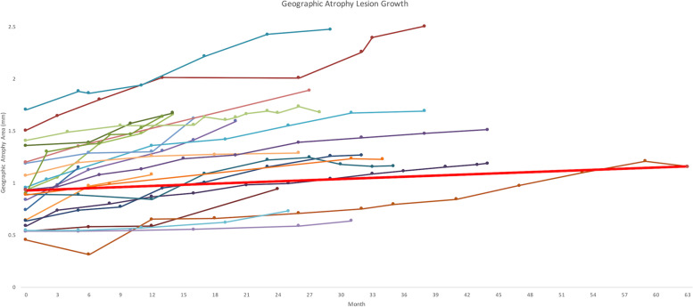
Results: The average age of the cohort, consisting of 22 eyes from 19 participants, was 86.9 years at reabsorption. Prior to reabsorption, the VA was 20/80 and then declined to 20/200 (p = 0.001) with an average follow-up time of 30.2 months. There was no significant VA change after the initial loss with reabsorption. The average initial lesion size of GA was 0.987 mm2 with an average growth rate of 0.274 mm/year.

Conclusions: This study longitudinally examined GA growth rate in patients with reabsorbed PEDs. These patients started with a drusenoid or serous PED, had a dramatic reduction in vision and GA that occurred in place of the PED. These GA lesions have a slower growth rate and a smaller area of onset compared to rates previously reported in the literature. They do not show significant VA change after reabsorption. As we have entered the era of GA therapy, these patients may not benefit from current treatments.

Keywords: AMD; Age-related macular degeneration; Geographic atrophy; Pigment epithelial detachment.


Conflict of interest statement

No conflicting relationship exists for any author.

Figures

Fig. 1
Fig. 1
Geographic atrophy lesion expansion following RPED
Fig. 2
Fig. 2
Optical Coherence Tomography of fovea with GA delineation
Fig. 3
Fig. 3
Optical Coherence Tomography of fovea A) Drusenoid PED (20/80); B) Post Drusenoid PED reabsorption 20/200; C) GA growth 6 months post reabsorption (20/100); D) GA growth 12 months post reabsorption (20/100)
Fig. 4
Fig. 4
Optical Coherence Tomography of fovea A) Serous PED (20/40); B) Post Serous PED reabsorption (20/400); C) GA growth 7 months post reabsorption (20/400); D) GA growth 9 months post reabsorption (CF)
Fig. 5
Fig. 5
Optical Coherence Tomography of fovea A) Serous PED (20/40); B) Post Serous PED (20/100); C) GA growth 8 months post reabsorption (20/200); D) GA growth 12 months post reabsorption (20/200)

Similar articles

Cited by

References

    1. Fleckenstein M, Mitchell P, Freund KB, et al. The progression of geographic atrophy secondary to age-related macular degeneration. Ophthalmology. 2018;125(3):369–390. doi: 10.1016/j.ophtha.2017.08.038. - DOI - PubMed
    1. Schmitz-Valckenberg S, Sahel JA, Danis R, et al. Natural history of geographic atrophy progression secondary to age-related macular degeneration (Geographic Atrophy Progression Study) Ophthalmology. 2016;123(2):361–368. doi: 10.1016/j.ophtha.2015.09.036. - DOI - PubMed
    1. Friedman DS, O’Colmain BJ, Muñoz B, et al. Prevalence of age-related macular degeneration in the United States. Arch Ophthalmol Chic Ill 1960. 2004;122(4):564–572. doi: 10.1001/archopht.122.4.564. - DOI - PubMed
    1. Yu JJ, Agrón E, Clemons TE, et al. Natural history of drusenoid pigment epithelial detachment associated with age-related macular degeneration: Age-Related Eye Disease Study 2 Report No. 17. Ophthalmology. 2019;126(2):261–273. doi: 10.1016/j.ophtha.2018.08.017. - DOI - PMC - PubMed
    1. Green WR, Enger C. Age-related macular degeneration histopathologic studies: the 1992 Lorenz E. Zimmerman Lecture. 1992. Retina Phila Pa. 2005;25(5 Suppl):1519–1535. doi: 10.1097/00006982-200507001-00015. - DOI - PubMed

MeSH terms