. 2023 May 30;25(1):59.
doi: 10.1186/s13058-023-01662-z.

Breast metastatic tumors in lung can be substituted by lung-derived malignant cells transformed by alternative splicing H19 lncRNA

Affiliations

Breast metastatic tumors in lung can be substituted by lung-derived malignant cells transformed by alternative splicing H19 lncRNA

Jin Biao Xu et al. Breast Cancer Res. .

Abstract

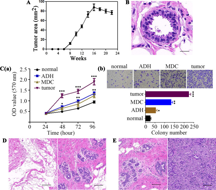
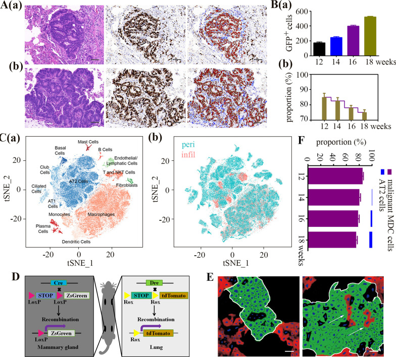
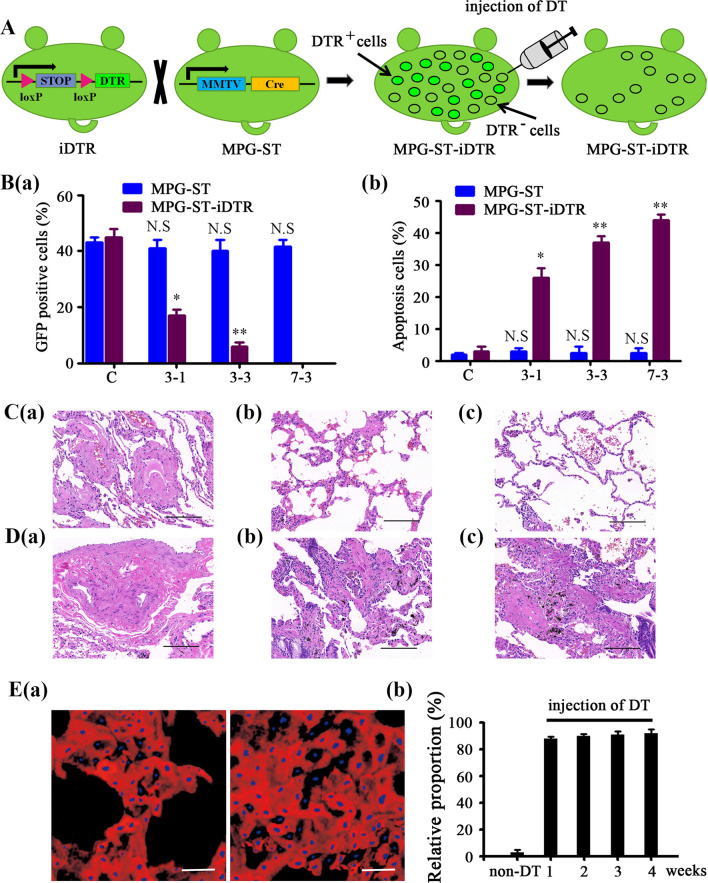
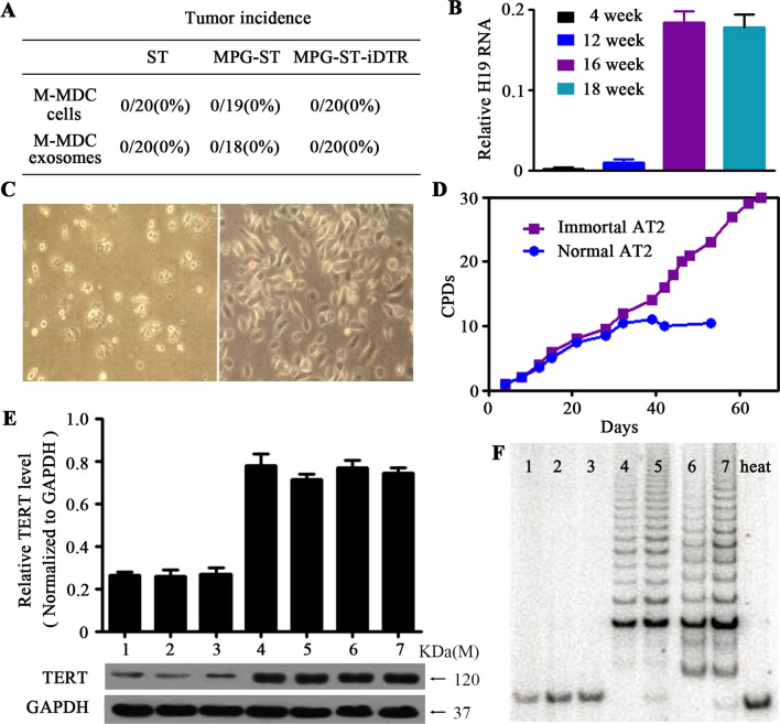
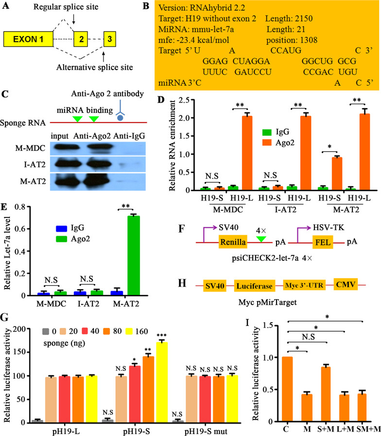
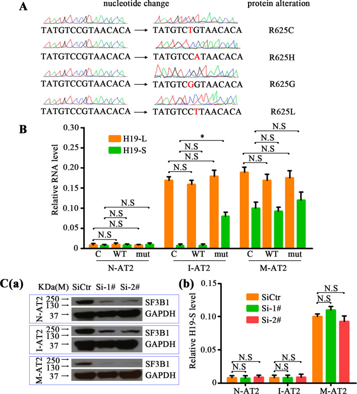
Metastasis accounts for most cancer-associated deaths; yet, this complex process remains poorly understood, particularly the relationship between distant metastasis and primary site-derived cells. Here, we modified the classical MMTV-PyMT breast carcinoma model to trace the fate of mammary-derived carcinoma cells. We show that within the lung, when the metastatic breast carcinoma cells are conditionally depleted, transformed lung epithelial cells generate new metastases. Metastatic breast carcinoma cells transmit H19 long noncoding (lnc) RNA to lung epithelial cells through exosomes. SF3B1 bearing mutations at arginine-625 alternatively splices H19 lncRNA in lung epithelial cells, which selectively acts like a molecular sponge to sequester let-7a and induces Myc upregulation. Under the conditional elimination of primary site-derived breast carcinoma cells, lung malignant cells expressing the mutated SF3B1 splice variant dominate the newly created tumors. Our study suggests that these new carcinoma cells originating from within the colonized organ can replace the primary site-derived malignant cells whenever their expansion is abrogated using an inducible diphtheria toxin receptor in our designed system. These findings should call for a better understanding of metastatic tumors with the specific origin during cancer metastasis.


Conflict of interest statement

The authors declare no competing interests.

Figures

Fig. 1
Fig. 1
Early dissemination of mammary cells from the MMTV–PyMT transgenic model of breast cancer. A Increase in tumor area in MMTV–PyMT-Green (MPG) transgenic female mice (number of mice = 5) over time. Data are expressed as mean ± standard deviation. B The mammary gland and mammary gland-derived cells (MDCs) in MPG mice at 4 weeks (number of mice = 5). Histological sections of mammary glands showing that lateral buds display the morphology of atypical ductal hyperplasia (ADH). Scale bar, 50 μm. C Proliferation and invasive capacities in GFP-positive cells isolated from ADH areas (ADH) or lung tissues (MDC) of 4-week-old MPG mice, normal cells (normal) digested from wild-type syngeneic mice and GFP-positive cells isolated from the breast tumor (tumor) of 12-week-old MPG mice, as assessed by MTT (a) and in vitro invasion assay (b), respectively. Data were quantified and presented as the mean ± SD of triplicate experiments. *P < 0.05, **P < 0.01, ***P < 0.001by Student’s t-test. D GFP-positive cells isolated from ADH areas of 4-week-old MPG mice (donor mice = 8) were transplanted subcutaneously (left panel) or orthotopically (right panel) into syngeneic wild-type (WT) recipient mice (1 million cells/mice, recipient mice = 40). Histopathology of transplanted mammary cells at 18 weeks after transplantation. Scale bar, 200 μm. E FACS-sorted early MDC cells (E-MDCs) from the lung of MPG mice (donor mice = 20) at 4 weeks were transplanted into the mammary gland of syngeneic WT recipient animals (1 million cells/mice, recipient mice = 20). Histopathology of the mammary gland at 4 weeks (left panel) or 18 weeks (right panel) after transplantation. n = number of donor or recipient mice. Scale bar, 200 μm
Fig. 2
Fig. 2
Metastasis expansion is not dependent on malignant MDCs. A H&E staining (left panel) or GFP immunohistochemistry (middle panel) of lung metastases forming in MMTV–PyMT-Green (MPG) mice at 12 (a) or 16 weeks (b). Blue, DAPI staining. To quantify the proportion of GFP-positive cells (right panel), the layers of images were decomposed by Image J software and restained using chromophore fluorescence for GFP expression (red) and DAPI (blue). Scale bar, 50 μm. B The absolute numbers (a) and proportion (b, number of GFP-positive cells divided by the total number of cells) of malignant mammary gland-derived cells (MDCs) in lung metastases of MPG mice (7 metastases/group, n = 5) at different ages (from 12 to 18 weeks) was quantified. n = number of groups. Data are expressed as mean ± standard deviation. C Single-cell RNA-Seq was performed on single-cell suspensions generated from the infiltrating cells (excluding T lymphocytes) or peritumoral cells of the lung metastatic tissues of 16-week-old MPG mice. a Cells were clustered using a graph-based shared nearest neighbor clustering approach and visualized using a t-distributed Stochastic Neighbor Embedding (tSNE) plot. b Cells on the tSNE plot were colored as originating either from the infiltrating cells (Infil) or peritumoral cells (Peri). D Schema of the genesis of MMTV–PyMT-Green/Sftpc-Tomato mice (MPG-ST) mice. E Reconstructed confocal images of the lung sections collected from MPG-ST mice at 12 (left panel) and 16 (right panel) weeks. The white frames showed metastases, and the arrows showed interspersed red fluorescent cells. Blue, DAPI; Green, malignant MDCs; Red, AT2 cells; Scale bar, 20 μm. F The proportion of malignant MDCs and AT2 cells in lung metastases of MPG-ST mice at different ages was quantified. Data are expressed as mean ± standard deviation
Fig. 3
Fig. 3
The newly generated growth of distant metastasis is not dependent on malignant MDCs. A Scheme of the Cre-inducible diphtheria toxin receptor (DTR) transgenic mice (iDTR) system. Crossing the iDTR strain with the MMTV–PyMT-Green/ Sftpc-tdTomato (MPG-ST) strain renders Cre-induced GFP-positive cells sensitive to DT. B Four-week-old MPG-ST and MPG-ST-iDTR mice (n = 6) were injected with 100 ng DT once daily for 3 consecutive days (3-1), 3 times a day for 3 consecutive days (3-3), or 3 times a day for 7 consecutive days (7-3). Mammary glands were analyzed 36 h later. The percentage of GFP-positive cells (a) or apoptotic cells stained with Annexin V (b) in different conditions are indicated. Mice without treatment by DT were used as control (C). Data are Mean ± s.d., n = 3 independent experiments per condition. N.S, no significance; *P < 0.05, **P < 0.01 by Student’s t test. MPG-ST-iDTR mice at 12 weeks (C) or 16 weeks (D) were injected with 100 ng diphtheria toxin (DT) every 8 h for 7 consecutive days. Lung metastases were detected by H&E staining (ac; Scale bar, 200 μm). Representative images of lung sections without DT treatment (a) and at 1 week (b) or 2 weeks (c) after DT injection. E At 16 weeks, MPG-ST-iDTR mice were injected with 100 ng DT every 8 h for 7 consecutive days. a Reconstructed confocal images of the lung sections were taken at 1 week (left panel) or 2 weeks (right panel) after DT injection. Blue, DAPI; Red, AT2 cells; Scale bar, 50 μm. b The proportion of AT2 cells in lung metastases of MPG-ST-iDTR mice before (non-DT) or after DT treatment. All experiments were performed at least 3 times. Data are expressed as mean ± standard deviation. Abbreviations: MPG, MMTV–PyMT-Green; MGP-ST, MPG/Sftpc-Tomato; MPG-ST-iDTR, MGP/ST/Cre-inducible diphtheria toxin receptor
Fig. 4
Fig. 4
Malignant MDCs transfer H19 LncRNA through secreting exosomes. A Sftpc-expressing AT2 cells sorted from lung metastatic tumors (tumor) or their peritumoral tissues (peri) of 16-week-old MPG-ST-iDTR mice with or without DT (non-DT) injection were subcutaneously transplanted into wild-type (WT) recipient mice. Tumor incidence, described as transplantation sites with tumors/transplanted mice amount (ratio), was measured at 18 weeks after transplantation. B PCR arrays were used to compare the expression of oncogenes and tumor suppressor genes between malignant AT2 and non-malignant AT2 groups. The red and green colors represent, respectively, increased or decreased gene expression in the heatmap for the malignant and non-malignant AT2 groups. Myc located at F07 (Extended Data Fig. 6A). C qRT-PCR analysis for Myc expression in the indicated cells. E-MDC and M-MDC cells were GFP-positive cells FACS sorted from the lungs of 4-week-old (E-MDC, n = 15) or 16-week-old (M-MDC, n = 3) MPG-ST-iDTR mice (GFP-positive cells FACS sorted from the mammary ADH tissue of 4-week-old MPG-ST-iDTR mice were used as control); N-AT2 and M-AT2 cells were tdTomato-positive cells FACS sorted from the parenchyma (N-AT2, n = 3) or lung metastasis (M-AT2, n = 3) of the MPG-ST-iDTR mice 2 weeks after DT treatment (tdTomato-positive cells FACS sorted from the peritumoral tissues of MPG-ST-iDTR mice before DT treatment were used as the control). n = number of mice. Data presented are the mean of triplicate experiments. Error bars indicate standard deviation (**P < 0.01; N.S, No significance). D Immunoblotting for anti-Myc in E-MDCs, M-MDCs, N-AT2 cells, M-AT2 cells (as described in C and their secreted exosomes (a). Quantification of Myc expression level (b). GAPDH was used as a control. Data presented are the mean of triplicate experiments. Error bars indicate standard deviation. E RNA expression of H19 lncRNA in the indicated cells (as described in C) and their secreted exosomes were analyzed by qRT-PCR and normalized to U6 snRNA. Data presented are the mean of triplicate experiments. Error bars indicate standard deviation. F and G FACS-sorted tdTomato-positive cells from the lung of 4-week-old ST mice, MPG-ST mice, or MPG-ST-iDTR mice were co-cultured with M-MDCs (as described in C) or M-MDCs with exosome inhibitor GW4869 (F, without M-MDCs as control) or with just exosomes collected from M-MDCs (G, without exosomes as control). The RNA level of H19 LncRNA was measured by qRT-PCR and normalized to U6 snRNA. Data presented are the mean of triplicate experiments. Error bars indicate standard deviation (**P < 0.01; *P < 0.05). Abbreviations: MPG, MMTV–PyMT-Green; MGP-ST, MPG/Sftpc-Tomato; MPG-ST-iDTR, MGP/ST/Cre-inducible diphtheria toxin receptor; MDCs, mammary gland-derived cells; E-MDCs, early MDCs; M-MDCs, malignant MDCs; N-AT2, non-malignant AT2; M-AT2, malignant AT2; C, control
Fig. 5
Fig. 5
H19 lncRNA induces the immortalization of recipient cells. A FACS-sorted tdTomato-positive cells from the lung (normal AT2 cells) of 4-week-old ST mice (n = 20), MPG-ST mice (n = 20), or MPG-ST-iDTR mice (n = 20) were co-cultured with M-MDCs (as described before) or their exosomes and then transplanted into wild-type (WT) recipient mice. Tumor incidence, described as transplantation sites with tumors/transplanted mice amount (ratio), was measured at 18 weeks after transplantation. n = number of mice. B The tdTomato-positive AT2 cells were sorted from the lung metastasis of MPG-ST-iDTR mice (without DT injection) at 12 weeks, 14 weeks, 16 weeks or 18 weeks (n = 5/group). The RNA levels of H19 lncRNA were determined by qRT-PCR and normalized to U6 snRNA. n = number of mice. Data presented are the mean of triplicate experiments. Error bars indicate standard deviation. C Morphology of 6th passage primary AT2 cells from 12 to week-old MPG-ST-iDTR mice (left panel) or 10th passage primary AT2 cells from lung metastases of 18-week-old MPG-ST-iDTR mice (right panel). D Cumulative population doublings (CPDs) show the growth kinetics of the primary AT2 cells from 12-week-old MPG-ST-iDTR mice (normal AT2) or primary AT2 cells from lung metastases of 18-week-old MPG-ST-iDTR mice (immortal AT2). E Expression of telomerase reverse transcriptase (TERT) in samples described below was detected and quantified. GAPDH was used as a control. Data presented are the mean of triplicate experiments. Error bars indicate standard deviation. F Telomerase activity of samples described below was determined by telomeric repeat amplification protocol assay. As a control, heat-inactivated samples were also assayed (right lane). Samples of E and F lane 1: normal AT2 cells from ST mice; Lane 2: normal AT2 cells from MPG-ST mice; Lane 3: normal AT2 cells from MPG-ST-iDTR mice; Lane 4: normal AT2 cells from ST mice co-cultured with exosomes from M-MDCs; Lane 5: normal AT2 cells from MPG-ST mice co-cultured with exosomes from M-MDCs; Lane 6: normal AT2 cells from MPG-ST-iDTR mice co-cultured with exosomes from M-MDCs; Lane 7: immortal AT2 cells. Abbreviations: MPG, MMTV–PyMT-Green; MGP-ST, MPG/Sftpc-Tomato; MPG-ST-iDTR, MGP/ST/Cre-inducible diphtheria toxin receptor
Fig. 6
Fig. 6
H19-S promotes tumorigenicity in immortalized AT2 cells. The tdTomato-positive AT2 cells were sorted from the peritumoral tissues (normal AT2 cells, n = 3) or tumor tissues (immortalized AT2 cells, n = 15) of the lung metastasis from 16-week-old MPG-ST-iDTR mice without DT injection. n = number of tissues. The plasmids including H19-L (full-length transcript) or H19-S (short transcript) were separately transfected into immortalized AT2 or normal AT2 cells. A qRT-PCR assessment of H19-L and H19-S in H19-L– or H19-S–overexpressing immortalized AT2 cells, normal AT2 cells, and their control cells. Data presented are the mean of triplicate experiments. Error bars indicate standard deviation (**P < 0.01; *P < 0.05; N.S, No significance). B Invasive capacities of the indicated cells were analyzed and quantified. Data presented are the mean of triplicate experiments. Error bars indicate standard deviation (**P < 0.01; N.S, No significance). C Colony-forming assays of H19-L or H19-S stably overexpressed in immortal AT2 cells, normal AT2 cells, and their control cells. Data presented are the mean of triplicate experiments. Error bars indicate standard deviation (**P < 0.01; N.S, No significance). D H19-L or H19-S stably overexpressed immortal AT2 cells, normal AT2 cells, and their control cells were subcutaneously transplanted into wild-type (WT) recipient mice. The tumor incidence, described as transplantation sites with tumors/transplanted mice amount (ratio), were measured at 18 weeks after transplantation. Abbreviations: C, plasmid control; OE-S, overexpression of H19-S; OE-L, overexpression of H19-L
Fig. 7
Fig. 7
H19-S and Let-7a present in the same Ago2 complex in M-AT2 cells. A Schema for the alternative splicing of H19. B The online RNAhybrid tool predicted that the alternative splicing of H19 resulted in an emerging binding site for Let-7a. C Immunoprecipitation of Ago2 protein from cell lysates from M-MDC, I-AT2, and M-AT2 cells, with non-immune IgG control. The input lane indicates lysate samples used as a positive control for Ago2. D The tumor tissues (n = 13) of the lung metastasis were sorted for tdTomato-positive AT2 cells (I-AT2) or for GFP-positive MDC cells (M-MDC) from 16-week-old MPG-ST-iDTR mice without DT injection. The tdTomato-positive AT2 cells (M-AT2) were sorted from the lung metastasis (n = 5) of MPG-ST-iDTR mice two weeks after DT injection. qRT-PCR detection of H19-L or H19-S in M-MDC, I-AT2, and M-AT2 cells that were precipitated by Ago2 or negative control IgG and normalized to U6 snRNA. n = number of tumor tissues. Data presented are the mean of triplicate experiments. Error bars indicate standard deviation (**P < 0.01; *P < 0.05; N.S, No significance). E qRT-PCR detection of Let-7a endogenously associated with sponge RNA in M-MDC, immortal AT2 and M-AT2 cells that were pulled down by Ago2 or negative control IgG and normalized to U6 snRNA. Data presented are the mean of triplicate experiments. Error bars indicate standard deviation (**P < 0.01; N.S, No significance). F Scheme of Let-7a target luciferase reporter. G The luciferase activity assessment in normal AT2 cells transfected with sensor (psiCHECK2-let7a 4×), together with 0, 20, 40, 80, or 160 ng of sponge plasmid pH19-L, pH19-S or pH19-S mut. Data presented are the mean of triplicate experiments. Error bars indicate standard deviation (***P < 0.001; **P < 0.01; *P < 0.05; N.S, No significance). H Scheme of Myc pMirTarget luciferase reporter. I Activity of Myc pMirTarget luciferase reporter plasmids in N-AT2 cells co-transfected with Let-7a mimics or Let-7a mimics together with pH19-L, pH19-S, or pH19-S mut (normalized to control). C, control; M, mimics; S + M, pH19-S + mimics; L + M, pH19-L + mimics; SM + M, pH19-S mutant + mimics. Abbreviations: MDCs, mammary gland-derived cells; M-MDCs, malignant MDCs; M-AT2, malignant AT2 cells; I-AT2, immortalized AT2 cells
Fig. 8
Fig. 8
Mutant SF3B1-R625C, H19-S, and Let-7a mediate the malignant transformation of I-AT2 cells. A The lung metastases (n = 20) of MPG-ST-iDTR mice 2 weeks after DT injection were collected and sent for Sanger sequence traces of SF3B1-wild-type and SF3B1-mutant tumors. Representative mutations were showed. n = number of lung metastases. B N-AT2, I-AT2, and M-AT2 cells were transfected with the expression vectors of SF3B1-WT, SF3B1-mut (R625G) or their controls. The expression of H19-S or H19-L was analyzed by qRT-PCR. C. N-AT2, I-AT2 and M-AT2 cells were transfected with SF3B1 siRNAs or their control siRNAs. The expression of SF3B1 (a) or H19-S (b) was individually detected. Abbreviations: C, Control; WT, SF3B1-WT; mut, SF3B1-mut; SiCtr, SiRNA control; Si-4#, SF3B1 SiRNA-4#; Si-5#, SF3B1 SiRNA-5#; N.S, no significance; N-AT2, normal AT2 cells; I-AT2, immortalized AT2 cells; M-AT2, malignant AT2 cells; pH19-L, full-length mouse H19; pH19-S H19 without exon 2, i.e., the “sponge” in this experiment. *P < 0.05

References

    1. Bray F, Ferlay J, Soerjomataram I, Siegel RL, Torre LA, Jemal A. Global cancer statistics 2018: GLOBOCAN estimates of incidence and mortality worldwide for 36 cancers in 185 countries. CA Cancer J Clin. 2018;68(6):394–424. doi: 10.3322/caac.21492. - DOI - PubMed
    1. Lambert AW, Pattabiraman DR, Weinberg RA. Emerging biological principles of metastasis. Cell. 2017;168(4):670–691. doi: 10.1016/j.cell.2016.11.037. - DOI - PMC - PubMed
    1. Klein CA. Parallel progression of primary tumours and metastases. Nat Rev Cancer. 2009;9(4):302–312. doi: 10.1038/nrc2627. - DOI - PubMed
    1. Harper KL, Sosa MS, Entenberg D, Hosseini H, Cheung JF, Nobre R, Avivar-Valderas A, Nagi C, Girnius N, Davis RJ, et al. Corrigendum: mechanism of early dissemination and metastasis in Her2(+) mammary cancer. Nature. 2018;553(7688):366. doi: 10.1038/nature24666. - DOI - PMC - PubMed
    1. Hosseini H, Obradovic MMS, Hoffmann M, Harper KL, Sosa MS, Werner-Klein M, Nanduri LK, Werno C, Ehrl C, Maneck M, et al. Early dissemination seeds metastasis in breast cancer. Nature. 2016;540(7634):552–558. doi: 10.1038/nature20785. - DOI - PMC - PubMed

Publication types