

A peripheral signature of Alzheimer's disease featuring microbiota-gut-brain axis markers
- PMID: 37254223
- PMCID: PMC10230724
- DOI: 10.1186/s13195-023-01218-5
A peripheral signature of Alzheimer's disease featuring microbiota-gut-brain axis markers
Abstract
Background: Increasing evidence links the gut microbiota (GM) to Alzheimer's disease (AD) but the mechanisms through which gut bacteria influence the brain are still unclear. This study tests the hypothesis that GM and mediators of the microbiota-gut-brain axis (MGBA) are associated with the amyloid cascade in sporadic AD.
Methods: We included 34 patients with cognitive impairment due to AD (CI-AD), 37 patients with cognitive impairment not due to AD (CI-NAD), and 13 cognitively unimpaired persons (CU). We studied the following systems: (1) fecal GM, with 16S rRNA sequencing; (2) a panel of putative MGBA mediators in the blood including immune and endothelial markers as bacterial products (i.e., lipopolysaccharide, LPS), cell adhesion molecules (CAMs) indicative of endothelial dysfunction (VCAM-1, PECAM-1), vascular changes (P-, E-Selectin), and upregulated after infections (NCAM, ICAM-1), as well as pro- (IL1β, IL6, TNFα, IL18) and anti- (IL10) inflammatory cytokines; (3) the amyloid cascade with amyloid PET, plasma phosphorylated tau (pTau-181, for tau pathology), neurofilament light chain (NfL, for neurodegeneration), and global cognition measured using MMSE and ADAScog. We performed 3-group comparisons of markers in the 3 systems and calculated correlation matrices for the pooled group of CI-AD and CU as well as CI-NAD and CU. Patterns of associations based on Spearman's rho were used to validate the study hypothesis.
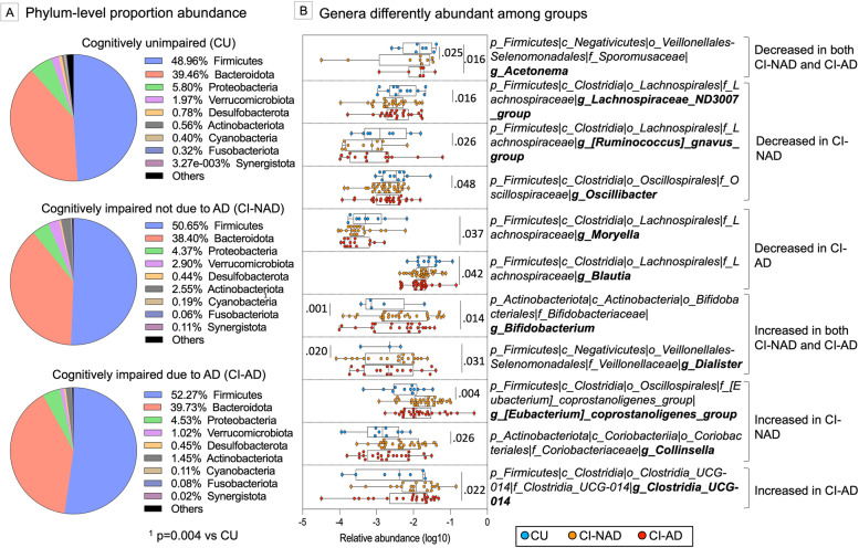
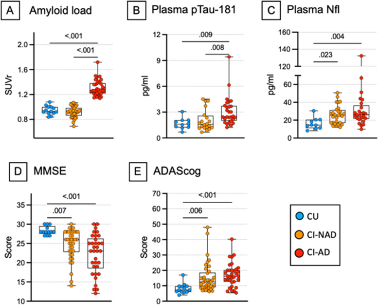
Results: CI-AD were characterized by (1) higher abundance of Clostridia_UCG-014 and decreased abundance of Moryella and Blautia (p < .04); (2) elevated levels of LPS (p < .03), upregulation of CAMs, Il1β, IL6, and TNFα, and downregulation of IL10 (p < .05); (3) increased brain amyloid, plasma pTau-181, and NfL (p < 0.004) compared with the other groups. CI-NAD showed (1) higher abundance of [Eubacterium] coprostanoligenes group and Collinsella and decreased abundance of Lachnospiraceae_ND3007_group, [Ruminococcus]_gnavus_group and Oscillibacter (p < .03); (2) upregulation of PECAM-1 and TNFα (p < .03); (4) increased plasma levels of NfL (p < .02) compared with CU. Different GM genera were associated with immune and endothelial markers in both CI-NAD and CI-AD but these mediators were widely related to amyloid cascade markers only in CI-AD.
Conclusions: Specific bacterial genera are associated with immune and endothelial MGBA mediators, and these are associated with amyloid cascade markers in sporadic AD. The physiological mechanisms linking the GM to the amyloid cascade should be further investigated to elucidate their potential therapeutic implications.
Keywords: Alzheimer’s disease; Cognitive impairment; Endothelial dysfunction; Gut microbiota; Lipopolysaccharide; Microbiota-gut-brain axis.
© 2023. The Author(s).
Conflict of interest statement
The authors declare no competing interests.
Figures


References
-
- Spadoni I, Zagato E, Bertocchi A, Paolinelli R, Hot E, Di Sabatino A, et al. A gut-vascular barrier controls the systemic dissemination of bacteria. Science. 2015;350:830–834. - PubMed
Publication types
MeSH terms
Substances
LinkOut - more resources
Full Text Sources
Medical
Research Materials
Miscellaneous
