Review
. 2023 May 31;23(1):53.
doi: 10.1186/s40644-023-00557-8.

Imaging in metastatic breast cancer, CT, PET/CT, MRI, WB-DWI, CCA: review and new perspectives

Affiliations
Review

Imaging in metastatic breast cancer, CT, PET/CT, MRI, WB-DWI, CCA: review and new perspectives

Dione Lother et al. Cancer Imaging. .

Abstract

Background: Breast cancer is the most frequent cancer in women and remains the second leading cause of death in Western countries. It represents a heterogeneous group of diseases with diverse tumoral behaviour, treatment responsiveness and prognosis. While major progress in diagnosis and treatment has resulted in a decline in breast cancer-related mortality, some patients will relapse and prognosis in this cohort of patients remains poor. Treatment is determined according to tumor subtype; primarily hormone receptor status and HER2 expression. Menopausal status and site of disease relapse are also important considerations in treatment protocols.

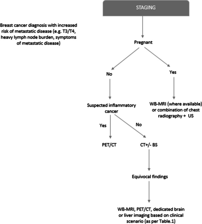
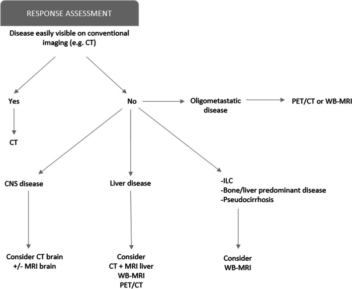
Main body: Staging and repeated evaluation of patients with metastatic breast cancer are central to the accurate assessment of disease extent at diagnosis and during treatment; guiding ongoing clinical management. Advances have been made in the diagnostic and therapeutic fields, particularly with new targeted therapies. In parallel, oncological imaging has evolved exponentially with the development of functional and anatomical imaging techniques. Consistent, reproducible and validated methods of assessing response to therapy is critical in effectively managing patients with metastatic breast cancer.

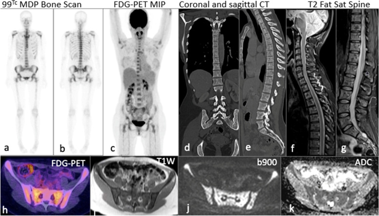
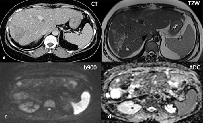
Conclusion: Major progress has been made in oncological imaging over the last few decades. Accurate disease assessment at diagnosis and during treatment is important in the management of metastatic breast cancer. CT (and BS if appropriate) is generally widely available, relatively cheap and sufficient in many cases. However, several additional imaging modalities are emerging and can be used as adjuncts, particularly in pregnancy or other diagnostically challenging cases. Nevertheless, no single imaging technique is without limitation. The authors have evaluated the vast array of imaging techniques - individual, combined parametric and multimodal - that are available or that are emerging in the management of metastatic breast cancer. This includes WB DW-MRI, CCA, novel PET breast cancer-epitope specific radiotracers and radiogenomics.

Keywords: Anatomo-functional imaging; Metastatic breast cancer; Multimodal; Multiparametric; Response assessment; Staging.


Conflict of interest statement

The authors declare that they have no competing interests.

Figures

Fig. 1
Fig. 1
Suggested decision-tree/diagnostic schema for initial staging (if appropriate)
Fig. 2
Fig. 2
Suggested decision-tree /diagnostic schema for assessing treatment response (restaging)
Fig. 3
Fig. 3
45-year-old female with multifocal grade 2 invasive ER positive HER2 negative metastatic lobular breast cancer: 99m Tc MDP planar bone scan (ab) and coronal maximum intensity projection PET/CT (c) show no uptake in the axial or appendicular skeleton. Coronal and sagittal CT reformats (de) performed 1 week later demonstrating subtle sclerotic changes. Sagittal fat saturated T2W sequence of the spine (fg) shows heterogenous marrow signal. Fused axial PET/CT (h) shows no FDG avidity. Widespread bone metastases in the same patient on WB DW-MRI (i) axial T1W (j) b900 and (k) ADC
Fig. 4
Fig. 4
Pseudocirrhosis of the liver in 59-year-old female with relapsed grade 3 ER positive HER2 negative metastatic invasive ductal carcinoma of the left breast. Axial CT performed in May 2020 demonstrates a nodular hepatic contour with capsular retraction (a). Increasing hypodensity within the right lobe was felt to represent disease progression. WB DWI-MRI performed 3 weeks later in June shows high b900 signal and corresponding increased ADC values (cd) at sites of previously identified disease in the right lobe of liver in keeping with maintained treatment response
Fig. 5
Fig. 5
Peritoneal and serosal disease in 47-year-old female with BRCA 2 mutation and bilateral metastatic lobular breast cancer. No measurable peritoneal or serosal disease on axial CT (a). WB DWI-MRI performed within 2 weeks demonstrates thickening of the right peritoneal reflection on axial T2W sequence (b), restricted diffusion along the caecum and appendix on the b900 sequence (c) and corresponding low ADC values (d) in keeping with peritoneal and serosal metastatic disease
Fig. 6
Fig. 6
56-year-old female with grade 2 ER positive HER 2 negative invasive ductal carcinoma of the right breast: comparison of pre and post treatment CT (a-f) showing ‘new sclerotic ‘bone metastases in T10 and L1. Pre and post treatment T1W sagittal MRI sequences (g, h) show apparently new sclerotic foci (occult on CT). Post treatment WB DWI-MRI unequivocally proves treatment response, with low signal on axial fat fraction imaging (i, l), high b900 signal (j, m) and high ADC values (k, n) at the sites of previously identified disease
Fig. 7
Fig. 7
65-year-old patient with HER2 positive invasive ductal carcinoma of the right breast with metastases to the brain, lung and bone. Resected right frontal lobe metastasis and left parietal lobe deposit recently treated with stereotactic radiotherapy. Axial post-contrast FLAIR sequence (a) demonstrates confluent signal abnormality within the left parietal lobe. Axial post-contrast T1W sequence (b) shows heterogenous enhancement at the site of treated metastasis in the left partial lobe. CCA (c) shows corresponding contrast accumulation (red arrow) at the site of contrast enhancement, unequivocal for radiation necrosis rather than persistent tumoral activity

References

    1. Siegel RL, Miller KD, Jemal A. Cancer statistics, 2016. CA Cancer J Clin. 2016;66(1):7–30. doi: 10.3322/caac.21332. - DOI - PubMed
    1. Kennecke H, Yerushalmi R, Woods R, et al. Metastatic behavior of breast cancer subtypes. J Clin Oncol. 2010;28(20):3271–7. doi: 10.1200/JCO.2009.25.9820. - DOI - PubMed
    1. Pan H, Gray R, Braybrooke J, et al. EBCTCG. 20-Year Risks of Breast-Cancer Recurrence after Stopping Endocrine Therapy at 5 Years. N Engl J Med. 2017;377 (19):1836–46. doi: 10.1056/NEJMoa1701830. - DOI - PMC - PubMed
    1. Pesapane F, Downey K, Rotili A, Cassano E, Koh DM. Imaging diagnosis of metastatic breast cancer. Insights Imaging. 2020;11(1):79. doi: 10.1186/s13244-020-00885-4. - DOI - PMC - PubMed
    1. Amin MB, Greene FL, Edge SB, et al. The Eighth Edition AJCC Cancer staging Manual: continuing to build a bridge from a population-based to a more “personalized” approach to cancer staging. CA Cancer J Clin. 2017;67(2):93–9. doi: 10.3322/caac.21388. - DOI - PubMed

MeSH terms