. 2023 Aug;80(8):1683-1696.
doi: 10.1161/HYPERTENSIONAHA.122.20449. Epub 2023 May 31.

Notch3/Hes5 Induces Vascular Dysfunction in Hypoxia-Induced Pulmonary Hypertension Through ER Stress and Redox-Sensitive Pathways

Affiliations

Notch3/Hes5 Induces Vascular Dysfunction in Hypoxia-Induced Pulmonary Hypertension Through ER Stress and Redox-Sensitive Pathways

Hannah E Morris et al. Hypertension. 2023 Aug.

Abstract

Background: Notch3 (neurogenic locus notch homolog protein 3) is implicated in vascular diseases, including pulmonary hypertension (PH)/pulmonary arterial hypertension. However, molecular mechanisms remain elusive. We hypothesized increased Notch3 activation induces oxidative and endoplasmic reticulum (ER) stress and downstream redox signaling, associated with procontractile pulmonary artery state, pulmonary vascular dysfunction, and PH development.

Methods: Studies were performed in TgNotch3R169C mice (harboring gain-of-function [GOF] Notch3 mutation) exposed to chronic hypoxia to induce PH, and examined by hemodynamics. Molecular and cellular studies were performed in pulmonary artery smooth muscle cells from pulmonary arterial hypertension patients and in mouse lung. Notch3-regulated genes/proteins, ER stress, ROCK (Rho-associated kinase) expression/activity, Ca2+ transients and generation of reactive oxygen species, and nitric oxide were measured. Pulmonary vascular reactivity was assessed in the presence of fasudil (ROCK inhibitor) and 4-phenylbutyric acid (ER stress inhibitor).

Results: Hypoxia induced a more severe PH phenotype in TgNotch3R169C mice versus controls. TgNotch3R169C mice exhibited enhanced Notch3 activation and expression of Notch3 targets Hes Family BHLH Transcription Factor 5 (Hes5), with increased vascular contraction and impaired vasorelaxation that improved with fasudil/4-phenylbutyric acid. Notch3 mutation was associated with increased pulmonary vessel Ca2+ transients, ROCK activation, ER stress, and increased reactive oxygen species generation, with reduced NO generation and blunted sGC (soluble guanylyl cyclase)/cGMP signaling. These effects were ameliorated by N-acetylcysteine. pulmonary artery smooth muscle cells from patients with pulmonary arterial hypertension recapitulated Notch3/Hes5 signaling, ER stress and redox changes observed in PH mice.

Conclusions: Notch3 GOF amplifies vascular dysfunction in hypoxic PH. This involves oxidative and ER stress, and ROCK. We highlight a novel role for Notch3/Hes5-redox signaling and important interplay between ER and oxidative stress in PH.

Keywords: calcium signaling; endoplasmic reticulum; hypoxia; pulmonary hypertension; reactive oxygen species.


Conflict of interest statement

Disclosures None.

Figures

Figure 1.
Figure 1.
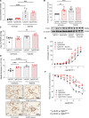
Hypoxia recapitulates increased gain-of-function (GOF) Notch3 (neurogenic locus notch homolog protein 3)-Hes Family BHLH Transcription Factor 5 (Hes5) axis signaling and impaired vascular function in TgNotch3WT mice and induces a more pronounced hypoxic pulmonary hypertension (PH) phenotype in TgNotch3R169C mice. TgNotch3 mice were exposed to 14 days of chronic hypobaric hypoxia (10%). Notch3 signaling was assessed in the lung. A, Notch3 mRNA expression by real-time quantitative polymerase chain reaction normalized to GAPDH (n=5–7; 1-way ANOVA with Bonferroni post-test). B, Notch3 target Hes5 protein expression by immunoblot normalized to α-tubulin. Upper, Quantification of Hes5. Lower, Representative Hes5 immunoblot. n=8; 1-way ANOVA with Bonferroni post-test. Hemodynamics were measured in vivo via right-heart catheterization. C, Right ventricular systolic pressure (RVSP) (n=5; 1-way ANOVA with Bonferroni post-test). D, Small pulmonary artery medial layer thickness by α-SMA (α-smooth muscle actin) immunohistochemistry. Upper, Semiquantitative medial layer thickness analysis (% total vessel diameter [TVD])±hypoxia. Lower, Representative images of increasing medial layer thickness (n=4–5 per group, average of 6 vessels in triplicate per animal). E and F, Pulmonary artery wire myography in normoxic and hypoxic TgNotch3 mice. Cumulative concentration-response curves of contraction to (E) endothelin 1 (ET-1), and relaxation of U46619 preconstriction to (F) acetylcholine (Ach; n=3–9; nonlinear regression). Results are mean±SEM. *P<0.05 vs TgNotch3WT, #P<0.05 vs TgNotch3R169C. WT indicates wild type.
Figure 2.
Figure 2.
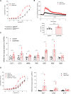
Baseline pulmonary vascular contraction is enhanced in TgNotch3R169C mice alongside altered calcium-dependent and -independent contractile mechanisms. Vascular reactivity was assessed in TgNotch3 pulmonary arteries by wire myography±fasudil (1 μmol/L), inhibitor of ROCK (Rho-associated kinase). A, Cumulative concentration-response curve to endothelin-1 (ET-1; n=7–9; nonlinear regression). B, Intracellular Ca2+ transients to ET-1 in TgNotch3 pulmonary artery smooth muscle cells (PASMCs) by live-cell fluorescence. Upper, Pulmonary artery smooth muscle cell (PASMC) intracellular calcium levels ([Ca2+]i) responses (ET-1, 100 nmol/L). Lower, [Ca2+]i expressed as area under the curve (AUC; n=6; 1-way ANOVA with Bonferroni post-test). C, L-type calcium channel, subunit α1S (Cav1.1), T-type calcium channel, subunit α1G (Cav3.1), IP3R (inositol 1,4,5-trisphosphate receptor), TRPM2 (voltage-dependent Transient receptor potential cation channel, subfamily M, member 2), RyR1 (ryanodine receptor 1), RyR2, and RyR3 Ca2+ channel gene expression by real-time quantitative polymerase chain reaction, normalized to GAPDH, in TgNotch3 lung (n=7–8; unpaired t test). D, Cumulative concentration-response curves to endothelin-1 (ET-1) following fasudil pretreatment (n=7–9; nonlinear regression). E, Gene expression for PDZ, LARG, and p115 Rho-GEFs, normalised to GAPDH (n=8–9; unpaired t test). Results are mean±SEM. *P<0.05 vs TgNotch3WT, #P<0.05 vs TgNotch3R169C. Larg indicates leukemia-associated RhoGEF; Pdz, Pdz RhoGEF; and wt, wild type.
Figure 3.
Figure 3.
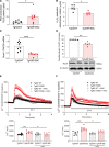
Endoplasmic reticulum (ER) stress in TgNotch3R169C mice contributes to altered pulmonary vascular reactivity. Vascular contraction to endothelin 1 (ET-1; A) was assessed by TgNotch3 pulmonary artery wire myography±ER stress inhibitor 4-phenylbutyric acid (4-PBA; 1 mmol/L). n=7–9; nonlinear regression. B, Intracellular Ca2+ transients to ET-1 in TgNotch3 pulmonary artery smooth muscle cells (PASMCs) by live-cell fluorescence±24-hour 4-PBA (1 mmol/L). Upper, Representative PASMC intracellular calcium levels ([Ca2+]i) responses to ET-1 (100 nmol/L) +24-hour 4-PBA. Lower, [Ca2+]i expressed as area under the curve (AUC; n=6; 1-way ANOVA with Bonferroni post-test). ER stress markers were assessed by real-time quantitative polymerase chain reaction and immunoblotting in TgNotch3 lung. C, Gene expression for BiP (binding immunoglobulin protein), XBP1 (X-box binding protein 1), and activating transcription factor 6 (ATF6) normalized to GAPDH (n=8; unpaired t test). D, upper, Quantification of ER chaperone BiP protein normalised to α-tubulin (n=11; unpaired t test). Lower, representative BiP immunoblot. E, upper, quantification of ER sensor IRE1 (inositol-requiring enzyme 1) phosphorylation (p-IRE1) normalised to total IRE1 (n=6; unpaired t test). Lower, Representative p-IRE1 immunoblot. Results are mean±SEM. *P<0.05 vs TgNotch3WT, #P<0.05 vs TgNotch3R169C. WT indicates wild type.
Figure 4.
Figure 4.
TgNotch3R169C gain-of-function mutation is associated with altered redox signaling and reactive oxygen species (ROS) levels. ROS production was measured in TgNotch3 lung by lucigenin-enhanced chemiluminescence and Amplex red assay, lipid peroxidation was assessed TBARS assay normalized by total protein. A, ROS production by lucigenin (n=7–8; unpaired t test). B, H2O2 levels by Amplex red (n=5; unpaired t test). Nox4 (NAPDH oxidase 4) expression in TgNotch3 lung was assessed by (C) real-time quantitative PCR normalized to GAPDH (n=5–8; unpaired t test) and (D) immunoblot normalized to α-tubulin. D, Upper, Quantification of Nox4 protein expression. Lower, Representative Nox4 immunoblot (n=5; unpaired t test). TgNotch3 pulmonary artery smooth muscle cell (PASMC) intracellular Ca2+ transients to (E) endothelin 1 (ET-1) and (F) 5-hydroxytryptamine (5-HT) were measured by live-cell fluorescence ±24-hour antioxidant N-acetylcysteine (NAC; 10 µmol/L). Upper, PASMC intracellular calcium levels ([Ca2+]i) responses to ET-1 (100 nmol/L) or 5-HT (1 μmol/L) +24-hour NAC. Lower, [Ca2+]i calculated as area under the curve (n=6; 1-way ANOVA with Bonferroni post-test). Results are mean±SEM. *P<0.05. AUC indicates area under the curve; RLU, relative light units; and WT, wild type.
Figure 5.
Figure 5.
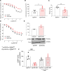
Impaired endothelial-dependent and -independent vasorelaxation in TgNotch3R169C arteries involves altered NO/cGMP signaling. Vascular reactivity was assessed in TgNotch3 pulmonary arteries by wire myography, responses expressed as percentage relaxation of preconstriction. A, Endothelium-dependent acetylcholine (ACh) vasorelaxation (n=7–9; nonlinear regression). B, Levels of 3-nitrotyrosine modified proteins in TgNotch3 lung, normalized to total protein (n=5; unpaired t test). C, NO levels by total nitrite/nitrate in TgNotch3 lung. D, Endothelium-independent sodium nitroprusside vasorelaxation in TgNotch3 pulmonary arteries (n=9–10; nonlinear regression). E, Reversible sGCβ1 (soluble guanylyl cyclase β1) oxidation by affinity capture of sulfenylated proteins in whole lung from TgNotch3WT and TgNotch3R169C mice (pool of 3 animals per sample). F, cGMP ELISA in TgNotch3 pulmonary artery smooth muscle cells±antioxidant N-acetylcysteine (NAC; 10 µM). Results are mean±SEM. *P<0.05 vs TgNotch3WT, **P<0.01 vs TgNotch3WT.
Figure 6.
Figure 6.
Upregulation of signaling associated with Notch3 (neurogenic locus notch homolog protein 3)-Hes Family BHLH Transcription Factor 5 (Hes5), endoplasmic reticulum (ER) stress, and reactive oxygen species (ROS) in pulmonary artery smooth muscle cells (PASMCs) from patients with pulmonary arterial hypertension (PAH). Protein expression in PAH vs non-PAH PASMCs by immunoblot normalized to α-tubulin, data expressed as % of non-PAH, comparisons by unpaired t test (n=5). A, upper, Quantification of Notch3 protein in PAH vs non-PAH PASMCs. Lower, Representative immunoblot of total Notch3. B, upper, Quantification of Notch3 transmembrane intracellular (TMIC) domain. Lower, Representative immunoblot of Notch3 TMIC. C, Upper, Quantification of Notch3 target Hes5. Lower, Representative Hes5 immunoblot. D, Upper, Quantification of ER stress chaperone BiP (binding immunoglobulin protein). Lower, Representative BiP immunoblot. E, NADPH-dependent ROS and (F) H2O2 by lucigenin and Amplex Red assay respectively ±24-hour Notch inhibitor GSI (n=5 per group, 1-way ANOVA with Bonferroni postcorrection). GSI indicates γ-secretase; and RLU, relative light units. Results are mean±SEM. *P<0.05.

References

    1. Artavanis-Tsakonas S, Rand MD, Lake RJ. Notch signaling: cell fate control and signal integration in development. Science. 1999;284:770–776. doi: 10.1126/science.284.5415.770 - PubMed
    1. Baeten J, Lilly B. Notch signaling in vascular smooth muscle cells. Adv Pharmacol. 2017;78:351–382. doi: 10.1016/bs.apha.2016.07.002 - PMC - PubMed
    1. Coupland K, Lendahl U, Karlström H. Role of NOTCH3 mutations in the cerebral small vessel disease cerebral autosomal dominant arteriopathy with subcortical infarcts and leukoencephalopathy. Stroke. 2018;49:2793–2800. doi: 10.1161/STROKEAHA.118.021560 - PubMed
    1. Morris H, Neves K, Montezano A, MacLean M, Touyz R. Notch3 signalling and vascular remodelling in pulmonary arterial hypertension. Clin Sci (Lond). 2019;133:2481–2498. doi: 10.1042/cs20190835 - PMC - PubMed
    1. Joutel A, Corpechot C, Ducros A, Vahedi K, Chabriat H, Mouton P, Alamowitch S, Domenga V, Cecillion M, Marechal E, et al. . Notch3 mutations in CADASIL, a hereditary adult-onset condition causing stroke and dementia. Nature. 1996;383:707–710. doi: 10.1038/383707a0 - PubMed

Publication types

MeSH terms