Review
. 2024 Apr 2;12(2):2219605.
doi: 10.1080/21688370.2023.2219605. Epub 2023 May 31.

(Zebra)fishing for nephrogenesis genes

Affiliations
Review

(Zebra)fishing for nephrogenesis genes

Brooke E Chambers et al. Tissue Barriers. .

Abstract

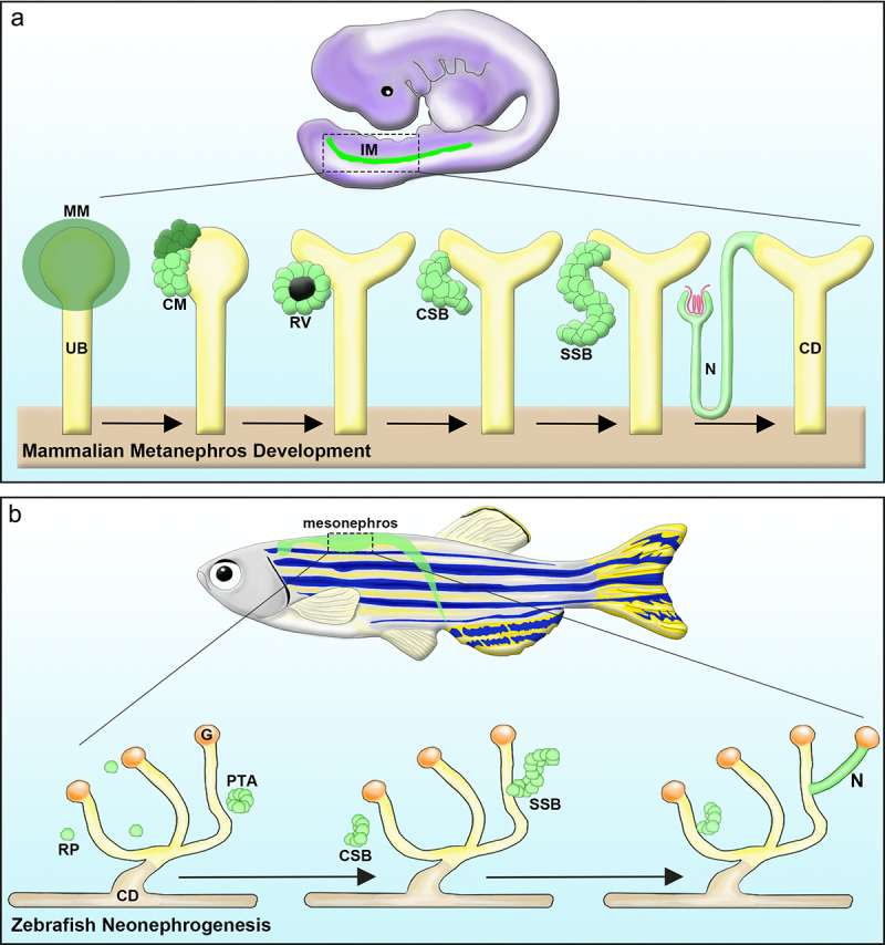
Kidney disease is a devastating condition affecting millions of people worldwide, where over 100,000 patients in the United States alone remain waiting for a lifesaving organ transplant. Concomitant with a surge in personalized medicine, single-gene mutations, and polygenic risk alleles have been brought to the forefront as core causes of a spectrum of renal disorders. With the increasing prevalence of kidney disease, it is imperative to make substantial strides in the field of kidney genetics. Nephrons, the core functional units of the kidney, are epithelial tubules that act as gatekeepers of body homeostasis by absorbing and secreting ions, water, and small molecules to filter the blood. Each nephron contains a series of proximal and distal segments with explicit metabolic functions. The embryonic zebrafish provides an ideal platform to systematically dissect the genetic cues governing kidney development. Here, we review the use of zebrafish to discover nephrogenesis genes.

Keywords: kidney; nephrogenesis; nephron; renal progenitor; segmentation; zebrafish.


Conflict of interest statement

No potential conflict of interest was reported by the author(s).

Figures

Figure 1.
Figure 1.
Nephron segmentation is conserved across vertebrate species. (a) Schematic of adult human kidney (left) and segmented nephron (right). Circle insets depicts a proximal tubule cell (above, gray) and thick ascending limb cell (below, teal) with specialized solute transport proteins (orange, purple) on the apical surfaces. (b) Developing mouse at embryonic day 17.5 and color-coded nephron segment compartments. (c) 24 hpf zebrafish embryo and color-coded pronephros segment compartments. Matching segment colors indicate the current working model of analogous structures between mammals and zebrafish. Abbreviations: PCT = proximal convoluted tubule, PST = proximal straight tubule, TAL = thick ascending limb, DE = distal early segment, MD = macula densa, CS = Corpuscle of Stannius, DCT = distal convoluted tubule, DL = distal late segment, CD = collecting duct, PD = pronephric duct, C = cloaca. Schematics adapted in part from..
Figure 2.
Figure 2.
Mammalian nephrogenesis (top) and adult zebrafish neonephrogenesis (bottom) involve similar morphogenetic events. (a) During mammalian kidney development, the MM is derived from the IM. Upon UB (yellow) invasion, the MM (dark green) condenses to form the CM, a renal progenitor population. These progenitors receive signals to self-renew (dark green) or differentiate (light green). Cells receiving differentiation signals are organized into an epithelialized RV. Upon further maturation, these cells form the CSB, then an SSB, and finally the N. Concurrently, the UB undergoes progressive branching to form the CD system (yellow). (b) Adult zebrafish possess the unique ability to generate new nephrons during adulthood. Neonephrogenesis in the zebrafish kidney mimics the cellular dynamics of mammalian nephrogenesis. RPs (green) cluster to create a PTA, which polarizes and undergoes epithelialization. This structure changes morphology and forms a CSB, SSB, and eventually a mature N structure. IM: Intermediate mesoderm; MM: Metanephric mesenchyme; UB: Ureteric bud; CM: Cap mesenchyme; RV: Renal vesicle; CSB: Comma-shaped body; SSB: S-shaped body; N: Nephron; CD: Collecting duct; Renal Progenitor: RP; PTA: Pre-tubular aggregate; G: Glomerulus. Schematics adapted from..

References

    1. Preuss HG. Basics of renal anatomy and physiology. Clin Lab Med. 1993;13(1):1–29. doi:10.1016/S0272-2712(18)30456-6. - DOI - PubMed
    1. van der Wijst J, Belge H, Bindels RJM, Devuyst O. Learning physiology from inherited kidney disorders. Physiol Rev. 2019;99(3):1575–1653. doi:10.1152/physrev.00008.2018. - DOI - PubMed
    1. Bertram JF, Douglas-Denton RN, Diouf B, Hughson MD, Hoy WE. Human nephron number: implications for health and disease. Pediatr Nephrol. 2011;26(9):1529–1533. doi:10.1007/s00467-011-1843-8. - DOI - PubMed
    1. Balzer MS, Rohacs T, Susztak K. How many cell types are in the kidney and what do they do?Annu. Annu Rev Physiol. 2022;84(1):507–531. doi:10.1146/annurev-physiol-052521-121841. - DOI - PMC - PubMed
    1. Little MH, Howden SE, Lawlor KT, Vanslambrouck JM. Determining lineage relationships in kidney development and disease. Nat Rev Nephrol. 2022;18(1):8–21. doi:10.1038/s41581-021-00485-5. - DOI - PubMed

Publication types

LinkOut - more resources