Review
. 2023 May 15;150(10):dev201507.
doi: 10.1242/dev.201507. Epub 2023 May 31.

Human embryo implantation

Affiliations
Review

Human embryo implantation

Joanne Muter et al. Development. .

Abstract

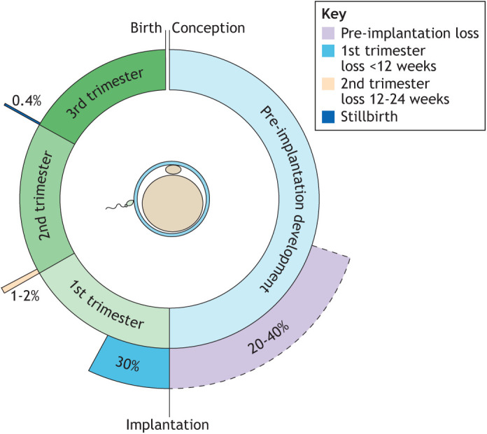
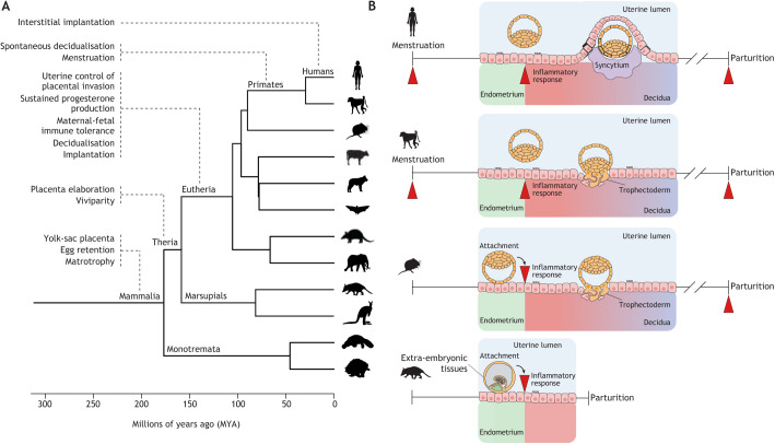
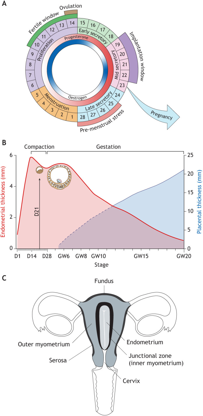
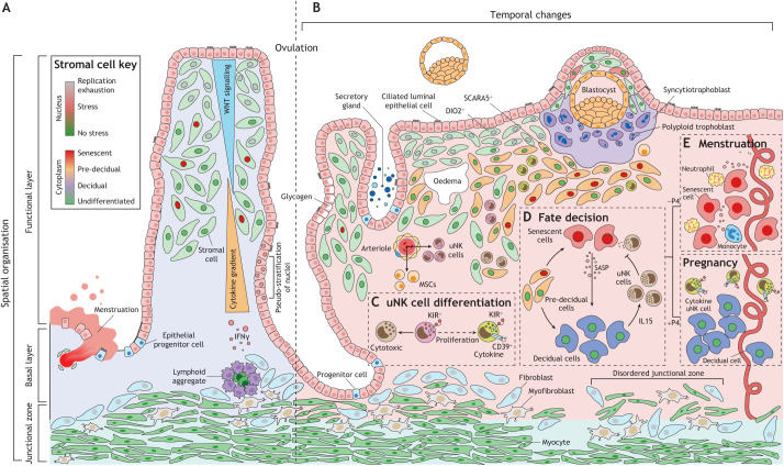
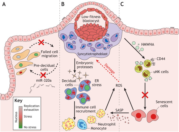
Embryo implantation in humans is interstitial, meaning the entire conceptus embeds in the endometrium before the placental trophoblast invades beyond the uterine mucosa into the underlying inner myometrium. Once implanted, embryo survival pivots on the transformation of the endometrium into an anti-inflammatory placental bed, termed decidua, under homeostatic control of uterine natural killer cells. Here, we examine the evolutionary context of embryo implantation and elaborate on uterine remodelling before and after conception in humans. We also discuss the interactions between the embryo and the decidualising endometrium that regulate interstitial implantation and determine embryo fitness. Together, this Review highlights the precarious but adaptable nature of the implantation process.

Keywords: Aneuploidy; Decidualisation; Implantation; Pregnancy; Uterine remodelling.


Conflict of interest statement

Competing interests The authors declare no competing or financial interests.

Figures

Fig. 1.
Fig. 1.
Reproductive failure and success. Estimates of the rates of pre-implantation embryo loss, miscarriage before and after 12 weeks of gestation, and stillbirth.
Fig. 2.
Fig. 2.
Evolution of embryo implantation. (A) Phylogenetic tree showing major reproductive innovations in the evolution of pregnancy in Mammalia, Theria, Eutheria and higher primates, including humans (adapted from Mika et al., 2022). (B) Attachment of extra-embryonic tissues to the endometrial luminal epithelium triggers uterine inflammation (red arrowhead) and parturition in marsupials, such as the grey short-tailed opossum (bottom). In eutherians with haemochorial placenta, such as mice, inflammatory reprogramming of endometrial stromal cells upon embryo attachment gives rise to decidual cells that accommodate invasive trophectoderm (placental precursors) throughout gestation. Consequently, pregnancy in eutherians is demarcated by two inflammatory uterine events, coinciding with implantation and parturition (note the two arrowheads). The emergence of an endogenous (endometrial) deciduogenic signal leads to spontaneous decidualisation and accounts for menstruation in higher primates. Menstruation imposes an additional inflammatory event on the uterus that marks the start of each reproductive (menstrual) cycle (note the three arrowheads). In humans and great apes (top), endometrial invasion is not limited to placental cells, but the entire embryo embeds in the stroma at implantation (interstitial implantation).
Fig. 3.
Fig. 3.
Uterine remodelling. (A) The menstrual cycle. The cycle is depicted as being 28 days long, although variations in cycle length are common between women and across the reproductive years (Bull et al., 2019). The rise and fall in oestradiol and progesterone levels are indicated in the inner circles. The fertile window indicates the days preceding ovulation when intercourse is most likely to result in pregnancy. The window of implantation coincides with the mid-secretory phase of the cycle. (B) Dynamic changes in endometrial thickness from menstruation until 20 weeks of pregnancy. Superimposed are the diameters of the conceptus at implantation and 6 weeks of pregnancy, as well as placental thickness. This composite figure is based on data extracted from several studies (Raine-Fenning et al., 2004a; Tongsong and Boonyanurak, 2004). D, day of menstrual cycle; GW, gestational week. (C) Uterine zonal anatomy based on T2-weighted magnetic resonance imaging.
Fig. 4.
Fig. 4.
Cellular dynamics in the endometrium before and during embryo implantation. (A) Endometrial regeneration leads to cell specification and tissue patterning. While oestradiol drives rapid endometrial growth prior to ovulation, its actions are modified by morphogen and cytokine gradients emanating from the luminal epithelium and lymphoid aggregates in the basal layer, respectively. Consequently, proliferative activity is positional, resulting in the emergence of exhausted, stressed and non-stressed subpopulations as indicated by the colour of nuclei. Morphogen gradients and cell–cell signalling also lead to specification of epithelial cells, exemplified by the emergence of ciliated cells. Rapid growth accounts for the pseudostratified appearance of glands. (B) Following the post-ovulatory rise in progesterone, the endometrium undergoes sequential morphological changes, starting with the appearance of subnuclear ‘vacuoles’ in glandular epithelium in the early secretory phase. The mid-secretory window of implantation coincides with a decidual reaction, characterised by apocrine glandular secretion, stromal oedema and accumulation of uterine natural killer (uNK) cells and bone marrow-derived mesenchymal stem cells (MSCs). Subluminal DIO2+ stromal cells are progesterone resistant whereas underlying progesterone responsive (SCARA5+) stromal cells decidualise (pre-decidual cells). (C) Differentiation of uNK cells into functionally distinct subpopulations. For detailed explanation, see text. KIR, killer cell immunoglobulin-like receptors. (D) Pre-decidual cells emerge as decidual cells upon closure of the implantation window, although some cells damaged by replication stress give rise to decidual-like senescent cells producing a complex inflammatory secretome (senescence associated secretory phenotype, SASP). The SASP induces secondary senescence in decidual cells, a process countered by decidual cells through activation of uNK cells that target and eliminate stressed/senescent cells. (E) In the late-secretory phase, falling progesterone (−P4) levels lead to expansion of decidual-like senescent cells, influx of neutrophils and monocytes, and menstruation, whereas sustained progesterone signalling (+P4) upon implantation transforms the endometrium into the decidua of pregnancy.
Fig. 5.
Fig. 5.
Natural selection of human embryos at implantation. Multiple mechanisms in decidualising endometrium ensure rapid elimination of low-fitness embryos. (A) Pre-decidual cells migrate and actively encapsulate embryos that breached the luminal epithelium. This migratory response is disrupted upon implantation of a poor-quality embryo, which has been linked to the lack of embryonic secretion of hsa-miR-320a. (B) Proteases secreted by low-fitness embryos induce prolonged and disordered Ca2+ signalling in pre-decidual cells, causing endoplasmic reticulum (ER) stress, attenuated secretion of implantation factors and induction of chemokines involved in recruitment of neutrophils and monocytes. (C) Low-fitness embryos secrete high molecular weight hyaluronic acid (HMWHA), which, upon binding to CD44 on uNK cells, blocks targeting and elimination of stressed/senescent cells, thereby causing sterile tissue inflammation through secondary senescence and menstruation-like breakdown of the endometrium, irrespective of circulating progesterone levels. Further, reactive oxygen species (ROS) produced by decidual-like senescent cells and immune cells may damage the conceptus and impair or preclude further development.

Similar articles

Cited by

References

    1. Abbas, Y., Carnicer-Lombarte, A., Gardner, L., Thomas, J., Brosens, J. J., Moffett, A., Sharkey, A. M., Franze, K., Burton, G. J. and Oyen, M. L. (2019). Tissue stiffness at the human maternal-fetal interface. Hum. Reprod. 34, 1999-2008. 10.1093/humrep/dez139 - DOI - PMC - PubMed
    1. Aberkane, A., Essahib, W., Spits, C., De Paepe, C., Sermon, K., Adriaenssens, T., Mackens, S., Tournaye, H., Brosens, J. J. and Van De Velde, H. (2018). Expression of adhesion and extracellular matrix genes in human blastocysts upon attachment in a 2D co-culture system. Mol. Hum. Reprod. 24, 375-387. 10.1093/molehr/gay024 - DOI - PubMed
    1. Al-Sabbagh, M., Fusi, L., Higham, J., Lee, Y., Lei, K., Hanyaloglu, A. C., Lam, E. W., Christian, M. and Brosens, J. J. (2011). NADPH oxidase-derived reactive oxygen species mediate decidualization of human endometrial stromal cells in response to cyclic AMP signaling. Endocrinology 152, 730-740. 10.1210/en.2010-0899 - DOI - PMC - PubMed
    1. Amorim, S., Reis, C. A., Reis, R. L. and Pires, R. A. (2021). Extracellular matrix mimics using hyaluronan-based biomaterials. Trends Biotechnol. 39, 90-104. 10.1016/j.tibtech.2020.06.003 - DOI - PubMed
    1. Aplin, J. D. and Ruane, P. T. (2017). Embryo-epithelium interactions during implantation at a glance. J. Cell Sci. 130, 15-22. 10.1242/jcs.175943 - DOI - PubMed

Publication types