

Microbial Pathogenesis in the Era of Spatial Omics
- PMID: 37255461
- PMCID: PMC10353406
- DOI: 10.1128/iai.00442-22
Microbial Pathogenesis in the Era of Spatial Omics
Abstract
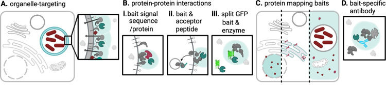
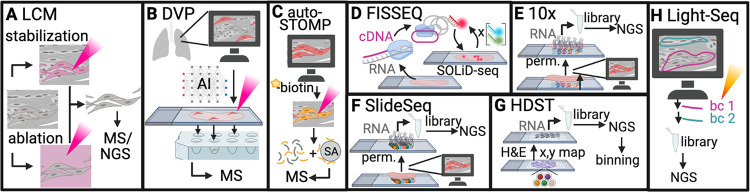
The biology of a cell, whether it is a unicellular organism or part of a multicellular network, is influenced by cell type, temporal changes in cell state, and the cell's environment. Spatial cues play a critical role in the regulation of microbial pathogenesis strategies. Information about where the pathogen is-in a tissue or in proximity to a host cell-regulates gene expression and the compartmentalization of gene products in the microbe and the host. Our understanding of host and pathogen identity has bloomed with the accessibility of transcriptomics and proteomics techniques. A missing piece of the puzzle has been our ability to evaluate global transcript and protein expression in the context of the subcellular niche, primary cell, or native tissue environment during infection. This barrier is now lower with the advent of new spatial omics techniques to understand how location regulates cellular functions. This review will discuss how recent advances in spatial proteomics and transcriptomics approaches can address outstanding questions in microbial pathogenesis.
Keywords: host-pathogen interactions; microbial pathogenesis; spatial proteomics; spatial transcriptomics.
Conflict of interest statement
The authors declare no conflict of interest.
Figures

Similar articles
-
Proteomics and integrative omic approaches for understanding host-pathogen interactions and infectious diseases.Mol Syst Biol. 2017 Mar 27;13(3):922. doi: 10.15252/msb.20167062. Mol Syst Biol. 2017. PMID: 28348067 Free PMC article. Review.
-
Adenovirus in the omics era - a multipronged strategy.FEBS Lett. 2020 Jun;594(12):1879-1890. doi: 10.1002/1873-3468.13710. Epub 2020 Jan 26. FEBS Lett. 2020. PMID: 31811727 Review.
-
Perusal of parasitic nematode 'omics in the post-genomic era.Mol Biochem Parasitol. 2017 Jul;215:11-22. doi: 10.1016/j.molbiopara.2016.11.003. Epub 2016 Nov 22. Mol Biochem Parasitol. 2017. PMID: 27887974 Free PMC article. Review.
-
Foodomics as part of the host-microbiota-exposome interplay.J Proteomics. 2016 Sep 16;147:3-20. doi: 10.1016/j.jprot.2016.04.033. Epub 2016 Apr 26. J Proteomics. 2016. PMID: 27130534 Review.
-
Integration of 'omics' data: does it lead to new insights into host-microbe interactions?Future Microbiol. 2010 Feb;5(2):313-28. doi: 10.2217/fmb.10.1. Future Microbiol. 2010. PMID: 20143952 Review.
Cited by
-
Molecular Advances in Microbial Metabolism 2.0.Int J Mol Sci. 2024 Jan 22;25(2):1361. doi: 10.3390/ijms25021361. Int J Mol Sci. 2024. PMID: 38279361 Free PMC article.
-
Trypanosoma cruzi-derived exovesicles contribute to parasite infection, tissue damage, and apoptotic cell death during ex vivo infection of human placental explants.Front Cell Infect Microbiol. 2024 Oct 14;14:1437339. doi: 10.3389/fcimb.2024.1437339. eCollection 2024. Front Cell Infect Microbiol. 2024. PMID: 39469456 Free PMC article.
-
Contribution of the infection ecosystem and biogeography to antibiotic failure in vivo.NPJ Antimicrob Resist. 2024;2(1):45. doi: 10.1038/s44259-024-00063-2. Epub 2024 Dec 4. NPJ Antimicrob Resist. 2024. PMID: 39649078 Free PMC article. Review.
-
Enablers and challenges of spatial omics, a melting pot of technologies.Mol Syst Biol. 2023 Nov 9;19(11):e10571. doi: 10.15252/msb.202110571. Epub 2023 Oct 16. Mol Syst Biol. 2023. PMID: 37842805 Free PMC article. Review.
References
-
- Gouin E, Gantelet H, Egile C, Lasa I, Ohayon H, Villiers V, Gounon P, Sansonetti PJ, Cossart P. 1999. A comparative study of the actin-based motilities of the pathogenic bacteria Listeria monocytogenes, Shigella flexneri and Rickettsia conorii. J Cell Sci 112:1697–1708. doi: 10.1242/jcs.112.11.1697. - DOI - PubMed
Publication types
MeSH terms
Grants and funding
LinkOut - more resources
Full Text Sources
