. 2023 May 15;1(5):1043-1051.
doi: 10.1021/acsaom.3c00100. eCollection 2023 May 26.

Transparent Wood Biocomposite of Well-Dispersed Dye Content for Fluorescence and Lasing Applications

Affiliations

Transparent Wood Biocomposite of Well-Dispersed Dye Content for Fluorescence and Lasing Applications

Martin Höglund et al. ACS Appl Opt Mater. .

Abstract

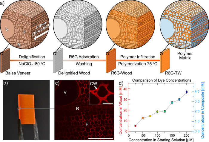
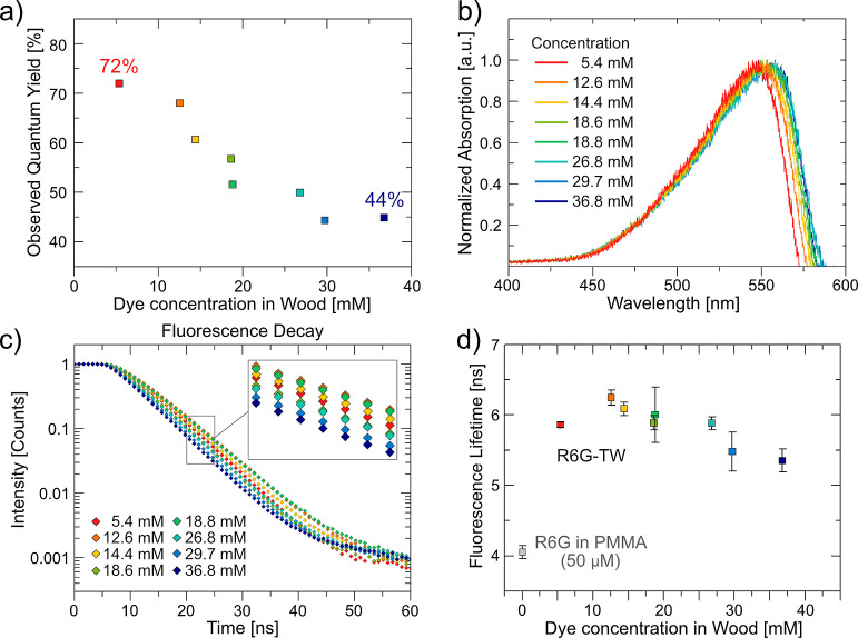
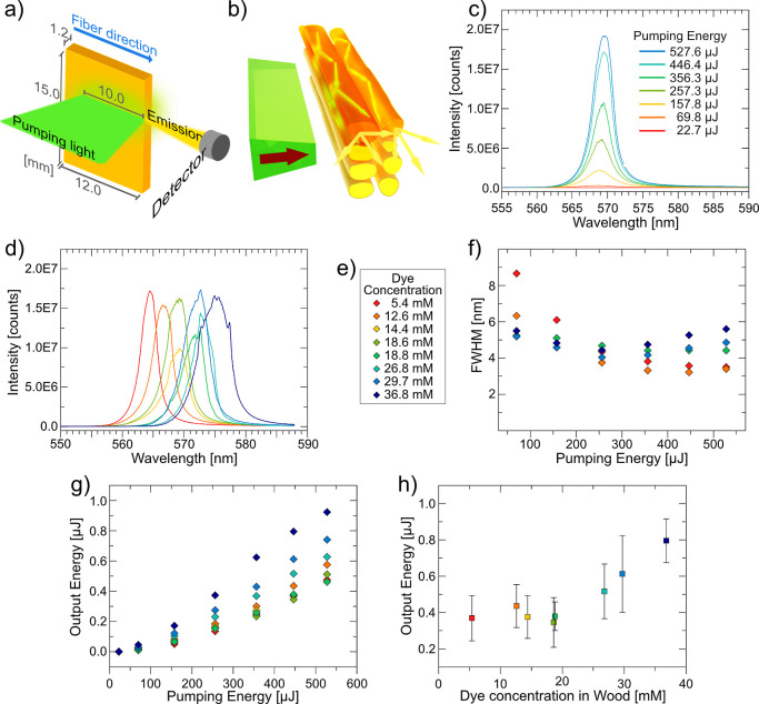
Aggregation-induced quenching often restricts emissive performance of optically active solid materials with embedded fluorescent dyes. Delignified and nanoporous wood readily adsorbs organic dyes and is investigated as a host material for rhodamine 6G (R6G). High concentration of R6G (>35 mM) is achieved in delignified wood without any ground-state dye aggregation. To evaluate emissive performance, a solid-state random dye laser is prepared using the dye-doped wood substrates. The performance in terms of lasing threshold and efficiency was improved with increased dye content due to the ability of delignified wood to disperse R6G.


Conflict of interest statement

The authors declare no competing financial interest.

Figures

Figure 1
Figure 1
(a) Preparation of dye-doped transparent wood biocomposites, R6G-TW. Illustrations of the anisotropic wood structure. Fibers, rays, and vessels are marked as F, R, and V, respectively. In the R6G-TW structure at the far right, the polymer matrix (blue) and dye (orange) distributions are illustrated. (b) Photograph of R6G-TW of 26.8 mM dye concentration in wood. The gray square indicates the cross section of the R6G-TW structure. (c) Cross-sectional confocal fluorescence micrographs of R6G-TW of 36.8 mM dye concentration in wood. V, R, F, and CW refer to vessel, rays, fibers, and cell wall, respectively. The scale bar represents 200 μm. The inset shows a higher magnification micrograph, with a scale bar of 20 μm. (d) Comparative graph of R6G dye concentration in wood and in the composite, versus starting concentration in solution (average of three samples).
Figure 2
Figure 2
(a) Observed fluorescence QY of R6G-TW samples versus dye concentration in wood. (b) Normalized absorption spectra of R6G-TW samples. (c) Characteristic fluorescence decay curves of R6G-TW. The inset shows magnification of the marked area. (d) Fluorescence lifetimes of R6G-TW (average of six measurements).
Figure 3
Figure 3
(a) Schematic layout of pumping setup for investigation of lasing properties of R6G-TW. (b) Illustration of the wave-guiding effect of wood fibers in TW. (c) Emission spectra of a 5.4 mM sample pumped with increasing pumping energies from a second harmonic generation Nd/YAG laser. (d) Characteristic lasing spectra of R6G-TW samples. (e) Color legend for graphs (d,f–h). (f) FWHM of selected R6G-TW samples as a function of pumping energy. (g) Output energy vs pumping energy of R6G-TW samples. (h) Comparison of output energy from R6G-TW samples with different dye concentrations pumped with 446 μJ (average of three samples, six positions).
Figure 4
Figure 4
Energy diagrams showing the main excitation and radiative relaxation events for (a) fluorescence where reabsorption occurs due to dye molecules primarily populating the electronic ground state and (b) lasing where stimulated emission occurs because of population inversion (excited electronic states are primarily populated).

Similar articles

References

    1. Chen S.; Lin C.; Cheng T.; Tseng W. 6-Mercaptopurine-Induced Fluorescence Quenching of Monolayer MoS2 Nanodots: Applications to Glutathione Sensing, Cellular Imaging, and Glutathione-Stimulated Drug Delivery. Adv. Funct. Mater. 2017, 27, 1702452.10.1002/adfm.201702452. - DOI
    1. Baldo M. A.; O’Brien D. F.; You Y.; Shoustikov A.; Sibley S.; Thompson M. E.; Forrest S. R. Highly efficient phosphorescent emission from organic electroluminescent devices. Nature 1998, 395, 151–154. 10.1038/25954. - DOI
    1. Gerega A.; Milej D.; Weigl W.; Botwicz M.; Zolek N.; Kacprzak M.; Wierzejski W.; Toczylowska B.; Mayzner-Zawadzka E.; Maniewski R.; Liebert A. Multiwavelength time-resolved detection of fluorescence during the inflow of indocyanine green into the adult’s brain. J. Biomed. Opt. 2012, 17, 087001.10.1117/1.jbo.17.8.087001. - DOI - PubMed
    1. Duarte F. J. Tunable organic dye lasers: Physics and technology of high-performance liquid and solid-state narrow-linewidth oscillators. Prog. Quant. Electron. 2012, 36, 29–50. 10.1016/j.pquantelec.2012.03.002. - DOI
    1. Kuehne A. J. C.; Gather M. C. Organic Lasers: Recent Developments on Materials, Device Geometries, and Fabrication Techniques. Chem. Rev. 2016, 116, 12823–12864. 10.1021/acs.chemrev.6b00172. - DOI - PubMed