Case Reports
. 2023 Sep:31:101857.
doi: 10.1016/j.ajoc.2023.101857. Epub 2023 May 18.

The spectrum of COVID-19-associated chorioretinal vasculopathy

Affiliations
Case Reports

The spectrum of COVID-19-associated chorioretinal vasculopathy

Piero Carletti et al. Am J Ophthalmol Case Rep. 2023 Sep.

Abstract

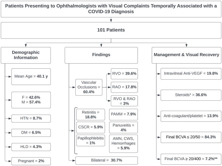
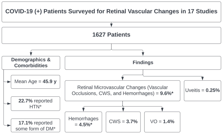
Purpose: Although conjunctivitis represents the most common ocular manifestation of COVID-19 infection, sight-threatening retinal involvement has been reported. Herein, we report and characterize with multimodal retinal imaging 5 cases of acute vision loss secondary to presumed chorioretinal vasculopathy temporally associated with COVID-19 infection with varying severity, visual morbidity, and treatment response, and review the available literature on the association between COVID-19 infection and retinal microvascular changes.

Design: Observational case series and literature review.

Methods: Multicenter case series of 5 patients who presented to academic centers and private offices with acute vision loss temporally associated with COVID-19 infection. A review of the literature was conducted using online databases.

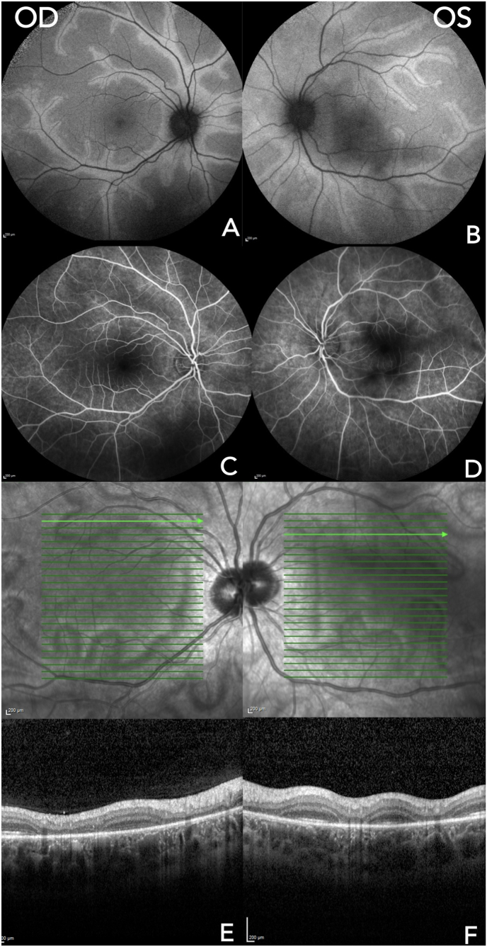
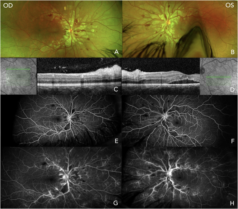
Results: 10 eyes of 5 patients, 3 men and 2 women, with a mean age of 30.8 years (median 33, range 16-44) were described. All patients had a recently preceding episode of COVID-19, with symptomatology ranging from mild infection to life-threatening encephalopathy. Treatment for their retinal disease included topical, oral, intravitreal, and intravenous steroids, steroid-sparing immunosuppression, retinal photocoagulation, antivirals, and antiplatelet and anticoagulant agents. Treatment response and visual recovery ranged from complete recovery of baseline acuity to permanent vision loss and need for chronic immunosuppression.

Conclusions and importance: Clinicians should be mindful of the potential for vision-threatening retinal involvement after COVID-19 infection. If found, treatment with both anti-inflammatory therapy and anticoagulation should be considered, in addition to close monitoring, as some patients with this spectrum of disease may require chronic immune suppression and/or anti-VEGF therapy.

Keywords: COVID-19; Chorioretinal vasculopathy; Retinal microvascular changes.


Conflict of interest statement

The authors declare that they have no known competing financial interests or personal relationships that could have appeared to influence the work reported in this paper.

Figures

Fig. 1
Fig. 1
Multimodal Imaging in Case #1. A and B: Optos ultra-widefield (UWF) fundus photos of the right eye (OD) and left eye (OS) showed the presence of macular edema and perivascular sheathing with areas of focal whitening along the vessels in both eyes and an area of macular ischemia OS. C and D: initial late phase Fluorescein Angiography (FA) showed focal venous leakage, small vessel/capillary leakage, and widespread arteriole obliteration in both eyes. E and F: one-month follow-up late phase FA with improved vascular leakage but progressive occlusion in both eyes. G and H: initial indocyanine green angiography (ICG) showed retinal and choroidal vessel staining and hypercyanescence in both eyes. I and F: Optical Coherence Tomography (OCT) images showed inner retinal edema, disruption of the retinal layers and ellipsoid zone, and cystoid macular edema in both eyes. (For interpretation of the references to colour in this figure legend, the reader is referred to the Web version of this article.)
Fig. 2
Fig. 2
Multimodal Imaging in Case #2. A and B: Fundus Autofluorescence (FAF) images showed hyperautofluorescence along retinal venules throughout the posterior pole in both eyes. C and D: Fluorescein Angiography (FA) images showed subtle retinal pigment epithelium (RPE) transmission defects adjacent to retinal venules with no vascular staining, optic disc staining, or macular leakage in either eye. E and F: en face and cross-sectional Optical Coherence Tomography (OCT) images showed a perivenular pattern of retinal thinning and ellipsoid zone disruption with preservation of normal macular architecture in both eyes.
Fig. 3
Fig. 3
Multimodal Imaging in Case #3. A and B: Optos ultra-widefield (UWF) fundus photos of the right eye (A) showed vitreous haze, disc and macular edema, cotton-wool spots, diffuse perivenular sheathing, and pre-retinal hemorrhages; the left eye (B) showed mild patchy periphlebitis. C and D: Optical Coherence Tomography (OCT) of the right eye (C) showed a subfoveal collection of hypo- and hyperreflective material, diffuse non-cystoid edema, and multiple hyperreflective dots in the inner retinal layers. The left eye (D) showed a normal contour hyperreflective dots along the posterior hyaloid, pre-foveal vitreous, and the inner retinal layers. E: initial Fluorescein Angiography (FA) of the right eye showed capillary dilation, early leakage from neovascularization along the inferonasal arcade, focal areas of capillary nonperfusion, and late diffuse vascular staining. F: initial FA of the left eye, which showed focal venular vasculitis, and late staining of the nerve. G and H: two-month follow-up FA showed increased neovascularization in the right eye (G); and increased peripheral microaneurysms and capillary nonperfusion in the left eye (H).
Fig. 4
Fig. 4
Multimodal Imaging in Case #4. A and B: Optos ultra-widefield (UWF) fundus photos showed bilateral numerous cotton wool spots with perivascular clearing, scattered retinal hemorrhages, and optic disc edema. Polygonal areas of retinal whitening consistent with Purtscher flecken were noted. C and D: Optical Coherence Tomography (OCT) images showed inner retinal hyperreflectivity and thickening, cysts, and subretinal fluid worse in the left eye compared to the right. E and F: early phase Fluorescein Angiography (FA) images showed bilateral scattered hypofluorescent patches along the vascular arcades likely due to retinal hemorrhages and inner retinal edema. G and H: late phase FA images showed bilateral perivascular hyperfluorescence suggestive of periphlebitis.
Fig. 5
Fig. 5
Multimodal Imaging in Case #5. A and B: Optos ultra-widefield (UWF) fundus photos showed multifocal hypopigmented placoid lesions involving bilateral macula and periphery. C and D: Optical Coherence Tomography (OCT) scans showed borders of outer retinal hyporeflectivity (C) that surround active lesions seen as severe outer retinal and ellipsoid zone atrophy (D). E and F: Fundus Autofluorescent (FAF) scans showed hyper and hypo-autofluorescent patches corresponding with the placoid lesions. The lesions abutting bilateral foveas had indistinct borders while the ones inferonasal to optic nerves were more consolidated. G and H: Fluorescein Angiography (FA) early phase showed bilateral hyperfluorescent patches and mid-peripheral hypofluorescent spots. I and J: FA late phase showed bilateral hyperfluorescent patches without leakage and subsequent hyperfluorescence of the previously hypofluorescent mid-peripheral spots.
Fig. 6
Fig. 6
Breakdown of demographic information, retinal findings, therapeutic interventions, and visual recovery from 54 reports of 83 patients who presented to ophthalmologists with visual complaints surrounding a COVID-19 diagnosis. Abbreviations: HTN = hypertension, DM = diabetes mellitus, HDL = hyperlipidemia, RVO = retinal vein occlusion, RAO = retinal artery occlusion, PAMM = paracentral acute middle maculopathy, CSCR = central serous chorioretinopathy, AMN = acute macular neuroretinopathy, CWS = cotton-wool spots, Anti-VEGF = anti-vascular endothelial growth factor, BCVA = best-corrected visual acuity. *Topical, intravitreal, sub-tenon's, and oral. **All patients with BCVA ≥20/400 had some form of retinal artery occlusions and had a visual acuity better than 20/200 before the onset of symptoms.
Fig. 7
Fig. 7
Diagram breaking down the incidence of retinal microvascular changes in 862 COVID-19 (+) patients whose retinas were surveyed in 14 studies. Abbreviations: HTN = hypertension, DM = diabetes mellitus, VO = vascular occlusions, CWS = cotton-wool spots.

References

    1. Sen S., Kannan N.B., Kumar J., et al. Retinal manifestations in patients with SARS-CoV-2 infection and pathogenetic implications: a systematic review. Int Ophthalmol. Aug. 2021;11:1–14. doi: 10.1007/s10792-021-01996-7. - DOI - PMC - PubMed
    1. Becker R.C. COVID-19-associated vasculitis and vasculopathy. J Thromb Thrombolysis. Oct 2020;50(3):499–511. doi: 10.1007/s11239-020-02230-4. - DOI - PMC - PubMed
    1. McGonagle D., Bridgewood C., Ramanan A.V., Meaney J.F.M., Watad A. COVID-19 vasculitis and novel vasculitis mimics. Lancet Rheumatol. 2021;3(3):e224–e233. doi: 10.1016/s2665-9913(20)30420-3. - DOI - PMC - PubMed
    1. Cheruiyot I., Kipkorir V., Ngure B., Misiani M., Munguti J., Ogeng'o J. Arterial thrombosis in coronavirus disease 2019 patients: a rapid systematic review. Ann Vasc Surg. 2021;70:273–281. doi: 10.1016/j.avsg.2020.08.087. - DOI - PMC - PubMed
    1. Malas M.B., Naazie I.N., Elsayed N., Mathlouthi A., Marmor R., Clary B. Thromboembolism risk of COVID-19 is high and associated with a higher risk of mortality: a systematic review and meta-analysis. EClinicalMedicine. 2020;29doi doi: 10.1016/j.eclinm.2020.100639. - DOI - PMC - PubMed

Publication types