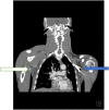
Ischemic stroke at first presentation of Takayasu arteritis in a young African male from Kenya, East Africa: Case report and brief literature review
- PMID: 37255613
- PMCID: PMC10225614
- DOI: 10.1002/ccr3.7412
Ischemic stroke at first presentation of Takayasu arteritis in a young African male from Kenya, East Africa: Case report and brief literature review
Abstract
Key clinical message: This case highlights the need for thorough clinical examination to rule out Takayasu arteritis (TA) as a cause of stroke in a young asymptomatic East-African male. Available clinical management guidelines should guide management of TA patients.
Abstract: We present a case of a young, previously asymptomatic East-African Black male presenting with large territory ischemic infarct at first diagnosis of TA. To our knowledge, this is the first published report of a male patient in East Africa with a stroke as the first presentation of TA.
Keywords: Takayasu arteritis; cerebrovascular accident; stroke; vasculitis.
© 2023 The Authors. Clinical Case Reports published by John Wiley & Sons Ltd.
Conflict of interest statement
The authors declare that they have no conflicts of interest.
Figures


References
-
- Couture P, Chazal T, Rosso C, et al. Cerebrovascular events in Takayasu arteritis: a multicenter case‐controlled study. J Neurol. 2018;265:757‐763. - PubMed
-
- Arnaud L, Haroche J, Limal N, et al. Takayasu arteritis in France: a single‐center retrospective study of 82 cases comparing white, North African, and black patients. Medicine (Baltimore). 2010;89(1):1‐17. https://pubmed.ncbi.nlm.nih.gov/20075700/ - PubMed
-
- Sugiyama K, Ijiri S, Tagawa S, Shimizu K. Takayasu disease on the centenary of its discovery. Jpn J Ophthalmol. 2009;53(2):81‐91. - PubMed
-
- Arnaud L, Haroche J, Mathian A, Gorochov G, Amoura Z. Pathogenesis of Takayasu's arteritis: a 2011 update. Autoimmun Rev. 2011;11(1):61‐67. https://pubmed.ncbi.nlm.nih.gov/21855656/ - PubMed
Publication types
LinkOut - more resources
Full Text Sources

