Review
. 2023 May 28;4(3):e288.
doi: 10.1002/mco2.288. eCollection 2023 Jun.

Targeting the p53 signaling pathway in cancers: Molecular mechanisms and clinical studies

Affiliations
Review

Targeting the p53 signaling pathway in cancers: Molecular mechanisms and clinical studies

Jinze Shen et al. MedComm (2020). .

Abstract

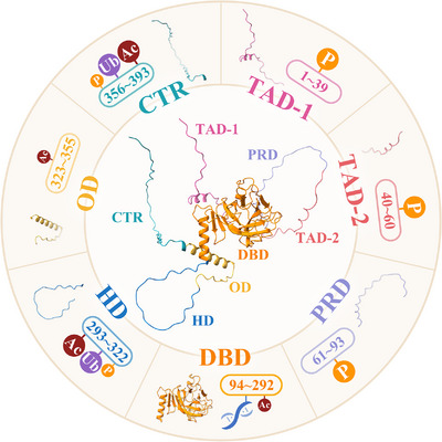
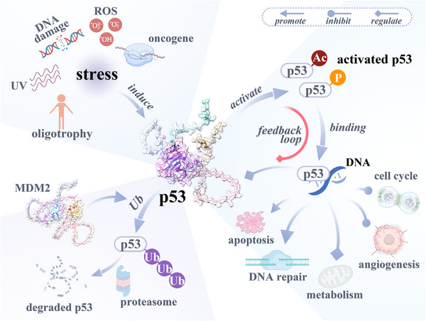
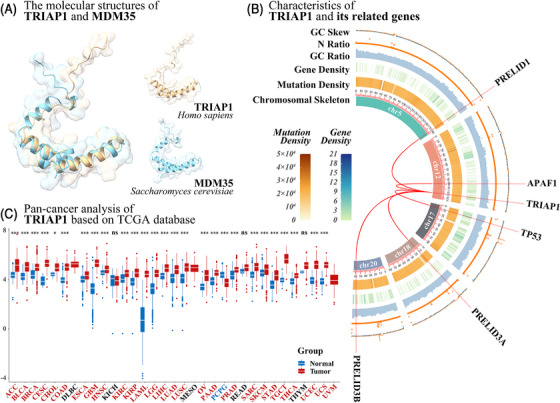
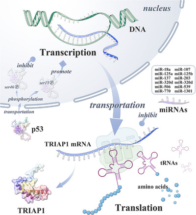
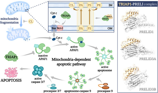
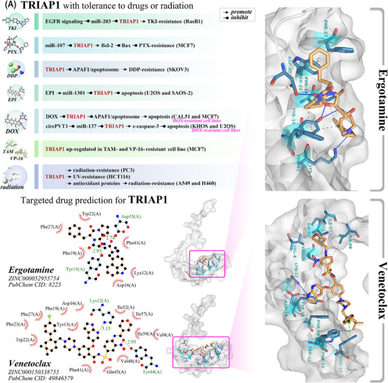
Tumor suppressor p53 can transcriptionally activate downstream genes in response to stress, and then regulate the cell cycle, DNA repair, metabolism, angiogenesis, apoptosis, and other biological responses. p53 has seven functional domains and 12 splice isoforms, and different domains and subtypes play different roles. The activation and inactivation of p53 are finely regulated and are associated with phosphorylation/acetylation modification and ubiquitination modification, respectively. Abnormal activation of p53 is closely related to the occurrence and development of cancer. While targeted therapy of the p53 signaling pathway is still in its early stages and only a few drugs or treatments have entered clinical trials, the development of new drugs and ongoing clinical trials are expected to lead to the widespread use of p53 signaling-targeted therapy in cancer treatment in the future. TRIAP1 is a novel p53 downstream inhibitor of apoptosis. TRIAP1 is the homolog of yeast mitochondrial intermembrane protein MDM35, which can play a tumor-promoting role by blocking the mitochondria-dependent apoptosis pathway. This work provides a systematic overview of recent basic research and clinical progress in the p53 signaling pathway and proposes that TRIAP1 is an important therapeutic target downstream of p53 signaling.

Keywords: TRIAP1; apoptosis; cancer biomarker; molecular mechanism; p53; targeted therapy.


Conflict of interest statement

The authors declare that there are no competing interests.

Figures

FIGURE 1
FIGURE 1
Functional domains of p53. p53 consists of 393 amino acids, which are usually divided into seven functional domains from N‐terminal to C‐terminal, including TAD‐1 (amino acid 1–39), TAD‐2 (amino acid 40–60), PRD (amino acid 61–93), DBD (amino acid 94–292), HD (amino acid 293–322), OD (amino acid 323–355), and CTR (amino acid 356–393). Phosphorylation (P) occurs in TAD‐1, TAD‐2, and PRD, and acetylation (Ac) and ubiquitination (Ub) occur in HD and CTR. DBD can bind DNA and is the domain of p53 to exert transcriptional activity.
FIGURE 2
FIGURE 2
The function of p53 and its regulation mechanism. p53 can be activated under various stresses, including oligotrophy, UV radiation, DNA damage, ROS, activation of an oncogene, and so on. Activated p53 can bind and induce target gene transcription on DNA, thereby promoting apoptosis and DNA repair, inhibiting cell cycle and angiogenesis, and regulating various metabolisms. Some downstream target genes of p53 are also involved in the regulation of p53 levels, thereby forming a feedback loop. MDM2 is a partner protein of p53, which ubiquitinates p53 and subsequently causes p53 to be degraded by the proteasome. UV, ultraviolet; ROS, reactive oxygen species; Ac, acetylation; P, phosphorylation; Ub, ubiquitination.
FIGURE 3
FIGURE 3
The structure, related genes, and a pan‐cancer analysis of TRIAP1. (A) Molecular structure comparison of human protein TRIAP1 (gold) and yeast protein MDM35 (cyan). TRIAP1 and MDM35 have similar protein structures, but the N‐terminus of MDM35 has one more short α‐helix structure than TRIAP1, and the C‐terminus is more flexible than TRIAP1. (B) The sequence characteristics of TRIAP1 and its related genes in chromosomes. (C) A pan‐cancer analysis of TRIAP1 based on TCGA database. *** means adjusted p < 0.001; ** means adjusted p < 0.01; * means adjusted p < 0.05; ns means no significant difference. TRIAP1 was significantly upregulated (adjusted p < 0.05) in 26 tumors, including ACC, BLCA, BRCA, CESC, CHOL, COAD, ESCA, GBM, HNSC, KIRC, KIRP, LAML, LGG, LIHC, LUAD, LUSC, OV, PAAD, PRAD, SARC, SKCM, STAD, TGCT, THCA, THYM, UCEC, and UCS. TRIAP1 was significantly downregulated in PCPG (adjusted p < 0.05). TRIAP1 was not significantly changed in three tumors (KICH, READ, and THYM). There were no controls in three tumors (DLBC, MESO, and UVM). Please check GDC (https://gdc.cancer.gov/resources‐tcga‐users/tcga‐code‐tables/tcga‐study‐abbreviations) for the full name of the TCGA abbreviations.
FIGURE 4
FIGURE 4
The regulatory factors of TRIAP1 expression. TRIAP1 is regulated in different stages during the expression process, including p53 protein regulating TRIAP1 at the transcriptional stage, and 12 miRNAs targeting TRIAP1 at the translational stage.
FIGURE 5
FIGURE 5
TRIAP1 inhibits mitochondria‐dependent apoptosis. TRIAP1 exerts antiapoptotic effects by inhibiting mitochondria‐dependent apoptosis pathways through various regulatory effects. TRIAP1 can ensure CL accumulation by promoting phospholipid transport in the mitochondrial intermembrane space and inhibiting mitochondrial lysis; TRIAP1 can also directly interact with APAF1 and inhibit the activation of apoptosomes. Cyt c, cytochrome c; PA, phosphatidic acid; PS, phosphatidylserine; PE, phosphatidylethanolamine; CL, cardiolipin; OM, outer membrane; IM, inner membrane.
FIGURE 6
FIGURE 6
The potential role of TRIAP1 in chemotherapy and radiotherapy. (A) TRIAP1 can promote the resistance of cancer cells to radiation and various anticancer drugs, including DOX, DDP, TKI, TAM, and PTX. PTX, taxol; DDP, cisplatin; EPI, epirubicin; DOX, doxorubicin; TAM, tamoxifen; VP‐16, etoposide. (B) Interaction of Ergotamine and Venetoclax with protein production. The protein backbone is shown in silver gray, the ligands are shown in orange, and the active site amino acids are shown in cyan. In the 2D diagram, the green dotted line shows the hydrogen bonds and their bond lengths, and the red arcs and black atoms represent the residues and atoms involved in hydrophobic interactions, respectively. In the 3D diagram, the gray dashed lines represent hydrophobic interactions, the blue solid lines represent hydrogen bonds, and the green dashed lines represent π–π stacking bonds. Ergotamine has a hydrogen bond interaction with Tyr15, Phe19, Trp22, Phe27, Asp35, and Phe41 of TRIAP1, a hydrophobic interaction with Lys12, Tyr15, Asp35, Phe19, Phe27, Trp22, Phe41, and Asp16 of TRIAP1, and forms a stacking bond with Trp22 of TRIAP1. Venetoclax has hydrogen bond interactions with Lys12, Asp16, and Tyr44 of TRIAP1. Venetoclax forms hydrophobic interactions with Val4, Tyr15, Asp16, Phe19, Trp22, Phe23, Phe27, Phe41, Gln45, Val48, Ile52, Ile57, and Ile59 of TRIAP1. Venetoclax forms π–π stacking bonds with Tyr15 and Phe27 of TRIAP1.

Similar articles

Cited by

References

    1. Amendolare A, Marzano F, Petruzzella V, et al. The underestimated role of the p53 pathway in renal cancer. Cancers (Basel). 2022;14(23) - PMC - PubMed
    1. Kern SE. p53: tumor suppression through control of the cell cycle. Gastroenterology. 1994;106(6):1708‐11. - PubMed
    1. Sengupta S, Harris CC. p53: traffic cop at the crossroads of DNA repair and recombination. Nat Rev Mol Cell Biol. 2005;6(1):44‐55. - PubMed
    1. Speidel D. Transcription‐independent p53 apoptosis: an alternative route to death. Trends Cell Biol. 2010;20(1):14‐24. - PubMed
    1. Hafner A, Bulyk ML, Jambhekar A, Lahav G. The multiple mechanisms that regulate p53 activity and cell fate. Nat Rev Mol Cell Biol. 2019;20(4):199‐210. - PubMed