. 2023 May 31;19(5):e1011421.
doi: 10.1371/journal.ppat.1011421. eCollection 2023 May.

Airway proteolytic control of pneumococcal competence

Affiliations

Airway proteolytic control of pneumococcal competence

Haley Echlin et al. PLoS Pathog. .

Abstract

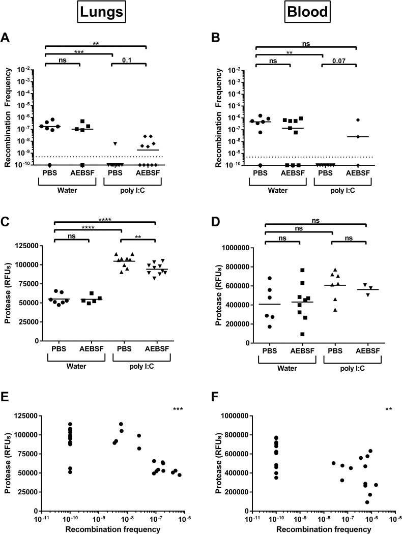
Streptococcus pneumoniae is an opportunistic pathogen that colonizes the upper respiratory tract asymptomatically and, upon invasion, can lead to severe diseases including otitis media, sinusitis, meningitis, bacteremia, and pneumonia. One of the first lines of defense against pneumococcal invasive disease is inflammation, including the recruitment of neutrophils to the site of infection. The invasive pneumococcus can be cleared through the action of serine proteases generated by neutrophils. It is less clear how serine proteases impact non-invasive pneumococcal colonization, which is the key first step to invasion and transmission. One significant aspect of pneumococcal biology and adaptation in the respiratory tract is its natural competence, which is triggered by a small peptide CSP. In this study, we investigate if serine proteases are capable of degrading CSP and the impact this has on pneumococcal competence. We found that CSP has several potential sites for trypsin-like serine protease degradation and that there were preferential cleavage sites recognized by the proteases. Digestion of CSP with two different trypsin-like serine proteases dramatically reduced competence in a dose-dependent manner. Incubation of CSP with mouse lung homogenate also reduced recombination frequency of the pneumococcus. These ex vivo experiments suggested that serine proteases in the lower respiratory tract reduce pneumococcal competence. This was subsequently confirmed measuring in vivo recombination frequencies after induction of protease production via poly (I:C) stimulation and via co-infection with influenza A virus, which dramatically lowered recombination events. These data shed light on a new mechanism by which the host can modulate pneumococcal behavior and genetic exchange via direct degradation of the competence signaling peptide.


Conflict of interest statement

The authors have declared that no competing interests exist.

Figures

Fig 1
Fig 1. Serine proteases cleave competence-stimulating peptide of S. pneumoniae.
(A) Sequence of competence-stimulating peptide 1 (CSP1) and peptide 2 (CSP2). Red arrows indicate potential cleavage sites by trypsin-like serine proteases. (B) Mass spectrometry analysis of cleaved residues of CSP1 upon incubation with no trypsin (grey) or with porcine trypsin (black) for 30 minutes. (C) Mass spectrometry analysis of cleaved residues of CSP1 upon incubation with no trypsin (grey) or with human airway trypsin (HAT) (black) for 30 minutes. (D) Mass spectrometry analysis of cleaved residues of CSP2 upon incubation with no trypsin (grey) or with porcine trypsin (black) for 30 minutes. (E) Mass spectrometry analysis of cleaved residues of CSP2 upon incubation with no trypsin (grey) or with HAT (black) for 30 minutes. (B-E) The fraction of each peptide detected by mass spectrometry was calculated by dividing the area of the cleaved peptide by the total area of all peptides. In the case of two cleavage sites on the same peptide, the fraction was equally attributed to both cleavage sites. Each bar represents the fraction of identified peptides that contained a cleavage event after the amino acid depicted under the bar. Bars above the final amino acid represent uncleaved CSP1 or CSP2.
Fig 2
Fig 2. Serine proteases reduce recombination frequency of S. pneumoniae in a dose dependent manner.
Recombination frequency upon incubation of CSP1 or CSP2 with increasing concentrations of serine proteases with and without 0.1mM of the serine protease inhibitor, AEBSF. D39x transformed with CSP1 incubated with (A) porcine pancreatic trypsin (PPT) or (B) human airway trypsin (HAT). D39x comC- transformed with CSP1 incubated with (C) PPT or (D) HAT. TIGR4 transformed with CSP2 incubated with (E) PPT or (F) HAT. TIGR4 comC- transformed with CSP2 incubated with (G) PPT or (H) HAT. Negative controls included no addition of CSP1 and no addition of gDNA. Lines represent median value. Dotted line represents lowest point of detection. Recombination frequencies of increasing concentrations of protease within each group (0 mM AEBSF or 0.1 mM AEBSF) were compared using Kruskal-Wallis one-way ANOVA; *p = 0.05–0.01, **p = 0.01–0.001, ***p = 0.001–0.0001.
Fig 3
Fig 3. Serine proteases reduce expression of CSP induced luciferase in S. pneumoniae.
Luminescence (RLU) of DLA3 grown in the presence of CSP1 incubated with increasing concentrations of serine proteases with and without inhibitor AEBSF. Incubation of CSP1 with PPT (A) without AEBSF or (B) with AEBSF. Incubation of CSP1 with HAT (C) without AEBSF or (D) with AEBSF. Experiment was repeated in triplicate. The mean value of RLU of each 30-minute timepoint is reported; error bars are SEM. Luminescence of increasing concentrations of protease within each group (0 mM AEBSF or 0.1 mM AEBSF) were compared using two-way ANOVA; ****p<0.0001.
Fig 4
Fig 4. Modified CSP alters impact of protease on recombination frequency.
Recombination frequency with modified CSP1. (A) Transformation of D39x comC- with CSP1 with modifications R3H, K6H, R9H, and R15H. Transformation of D39x comC- with modified CSP1 incubated with increasing concentrations of (B) PPT or (C) HAT. Recombination frequency reported as % of 0 trypsin. Negative controls included no addition of CSP1 and no addition of gDNA. Lines represent mean value. Recombination frequencies of increasing concentrations of protease within each modified CSP were compared using one-way ANOVA; ***p = 0.001–0.0001, ****p<0.0001. Changes in the concentrations of protease between modified CSP was compared using two-way ANOVA; ##p = 0.01–0.001, ###p = 0.001–0.0001.
Fig 5
Fig 5. Modification of R9 of CSP1 alters protease digestion profile.
Mass spectrometry analysis of cleaved residues of CSP1 (black) or CSP1 R9A (striped) upon incubation with (A) porcine trypsin or with (B) HAT. The fraction of each peptide detected by mass spectrometry was calculated by dividing the area of the cleaved peptide by the total area of all peptides. In the case of two cleavage sites on the same peptide, the fraction was equally attributed to both cleavage sites. Each bar represents the fraction of identified peptides that contained a cleavage event after the amino acid depicted under the bar. Bars above the final amino acid represent uncleaved CSP1.
Fig 6
Fig 6. Inhibition of proteases from mouse lungs increase recombination frequency of S. pneumoniae.
Recombination frequency and protease levels upon incubation of CSP1 with homogenized mouse lungs with increasing concentrations of inhibitor AEBSF. (A) Recombination frequency of D39x transformed with CSP1 incubated with homogenized mouse lungs. (B) Protease levels in homogenized mouse lungs upon incubation with AEBSF used in D39x transformation; determined by fluorescence of substrate t-Butyloxycarbonyl Phe-Ser-Arg 7-amino-4methyl coumarin (BOC). (C) Correlation of recombination frequency of D39x with the protease levels in the same homogenized mouse lung. (D) Recombination frequency of D39x comC- transformed with CSP1 incubated with homogenized mouse lung. (E) Protease levels in homogenized mouse lungs upon incubation with AEBSF used in D39x comC- transformations; determined by fluorescence of substrate BOC. (F) Correlation of recombination frequency of D39x comC- with the protease levels in the same homogenized mouse lung. (A,D) Line represents median; dotted line represents lowest point of detection; recombination frequency of 1 mM and 2 mM AEBSF were compared to 0 mM AEBSF using Kruskal-Wallis one-way ANOVA; ****p<0.0001. (B,E) Lines represent mean; protease levels of 1 mM and 2 mM AEBSF were compared to 0 mM AEBSF using one-way ANOVA. (C,F) Correlation was compared using two-tailed spearman; *** p = 0.0004, **** p<0.0001.
Fig 7
Fig 7. Stimulation of proteases in vivo reduces ex vivo recombination frequency of S. pneumoniae.
Recombination frequency upon incubation of CSP1 with homogenized lungs from mice that were administered poly (I:C) in vivo, treated with inhibitor AEBSF in vivo, and then treated ex vivo with increasing concentrations of inhibitor AEBSF. (A) Recombination frequency of D39x transformed with CSP1 incubated with homogenized lungs from mice that received no stimulant (water) or poly (I:C), and either received no inhibitor (PBS) or inhibitor AEBSF. (B) The same lungs were then treated with either 0, 1, or 2 mM AEBSF ex vivo prior to incubation with CSP1 and recombination frequency was determined. The 0 mM AEBSF are the same data used in Fig 7A and were included here for comparison. (C) Recombination frequency of D39x comC- transformed with CSP1 incubated with homogenized lungs from mice that received no stimulant (water) or poly (I:C), and either received no inhibitor (PBS) or inhibitor AEBSF. (D) The same lungs were then treated with either 0, 1, or 2 mM AEBSF ex vivo prior to incubation with CSP1 and recombination frequency was determined. The 0 mM AEBSF are the same data used in Fig 7C and were included here for comparison. Line represents median; dotted line represents lowest point of detection. (A,C) Recombination frequencies were compared pairwise using nonparametric Mann-Whitney t test; *p = 0.05–0.01, **p = 0.01–0.001. (B,D) Recombination frequency of 1 mM and 2 mM AEBSF were compared to 0 mM AEBSF of each group using Kruskal-Wallis one-way ANOVA; *p = 0.05–0.01, **p = 0.01–0.001, ***p = 0.001–0.0001, ****p<0.0001.
Fig 8
Fig 8. Stimulation of protease production in vivo reduces recombination frequency of S. pneumoniae.
Recombination frequency of S. pneumoniae from (A) the lungs and (B) the blood of mice that received no stimulant (water) or poly (I:C), and either received no inhibitor (PBS) or inhibitor AEBSF. Protease levels in the same mouse lungs (C) and blood (sera) (D); determined by fluorescence of substrate BOC. Correlation of recombination frequency in lungs (E) and blood (F) with the protease levels in the same tissue. (A,B) Line represents median; dotted line represents lowest point of detection; recombination frequencies of all groups were compared pairwise for each tissue using nonparametric Mann-Whitney t test; **p = 0.01–0.001, ***p = 0.001–0.0001. (C,D) Lines represent mean; protease levels of all groups were compared pairwise for each tissue using unpaired t test; **p = 0.01–0.001, ****p<0.0001. (E,F) Correlation was compared using two-tailed spearman; **p = 0.004, ***p = 0.0001.
Fig 9
Fig 9. Co-infection with influenza in vivo reduces recombination frequency of S. pneumoniae.
Recombination frequency of S. pneumoniae from (A) the lungs and (B) the blood of mice infected with and without influenza (Flu). The total number of recombinant colonies per mL used to calculate recombination frequency enumerated from (C) the lungs and (D) the blood of mice infected with and without influenza (Flu). Lines represent median. Dotted line represents lowest point of detection. Recombination frequency and total number of recombinants from mice infected with influenza was compared to that without influenza for each tissue using nonparametric Mann-Whitney t test; **** p<0.0001.
Fig 10
Fig 10. Proposed model of impact of host serine proteases on S. pneumoniae adaptation to host epithelium and invasion.
Created with BioRender.com.

References

    1. Henriques-Normark B, Tuomanen EI. The pneumococcus: epidemiology, microbiology, and pathogenesis. Cold Spring Harb Perspect Med. 2013;3(7). doi: 10.1101/cshperspect.a010215 ; PubMed Central PMCID: PMC3685878. - DOI - PMC - PubMed
    1. Weiser JN, Ferreira DM, Paton JC. Streptococcus pneumoniae: transmission, colonization and invasion. Nat Rev Microbiol. 2018;16(6):355–67. Epub 2018/03/31. doi: 10.1038/s41579-018-0001-8 ; PubMed Central PMCID: PMC5949087. - DOI - PMC - PubMed
    1. Bogaert D, De Groot R, Hermans PW. Streptococcus pneumoniae colonisation: the key to pneumococcal disease. Lancet Infect Dis. 2004;4(3):144–54. doi: 10.1016/S1473-3099(04)00938-7 . - DOI - PubMed
    1. Henriques-Normark B, Normark S. Commensal pathogens, with a focus on Streptococcus pneumoniae, and interactions with the human host. Exp Cell Res. 2010;316(8):1408–14. Epub 2010/03/17. doi: 10.1016/j.yexcr.2010.03.003 . - DOI - PubMed
    1. Loughran AJ, Orihuela CJ, Tuomanen EI. Streptococcus pneumoniae: Invasion and Inflammation. Microbiol Spectr. 2019;7(2). Epub 2019/03/16. doi: 10.1128/microbiolspec.GPP3-0004-2018 ; PubMed Central PMCID: PMC6422050. - DOI - PMC - PubMed

Publication types