. 2023 Jun 2;9(22):eadg2834.
doi: 10.1126/sciadv.adg2834. Epub 2023 May 31.

PAXX binding to the NHEJ machinery explains functional redundancy with XLF

Affiliations

PAXX binding to the NHEJ machinery explains functional redundancy with XLF

Murielle Seif-El-Dahan et al. Sci Adv. .

Erratum in

Abstract

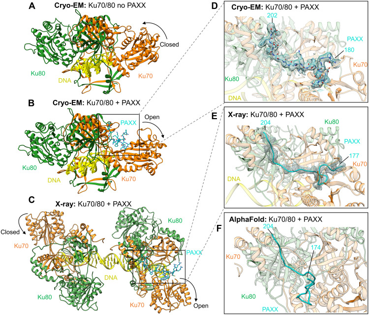
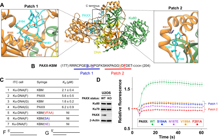
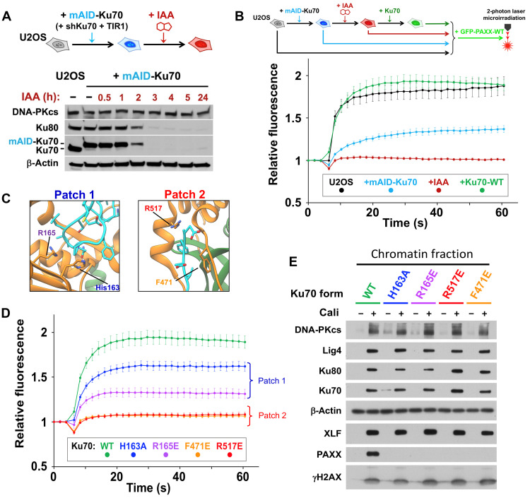
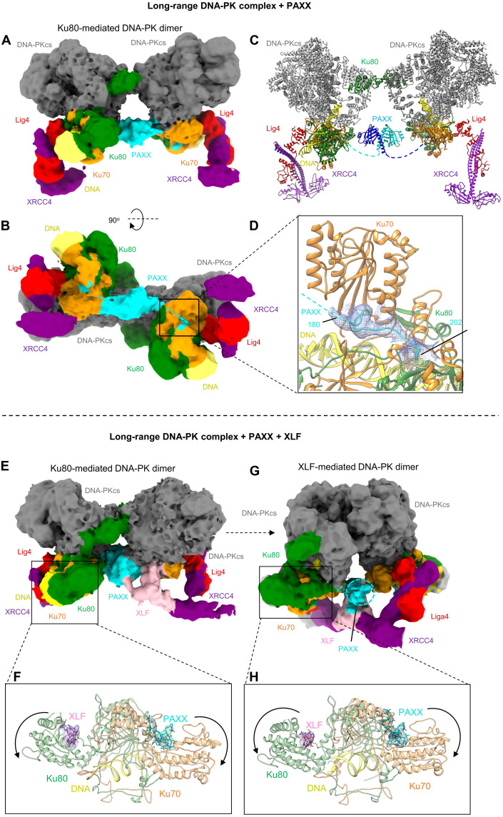
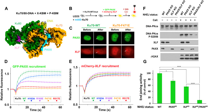
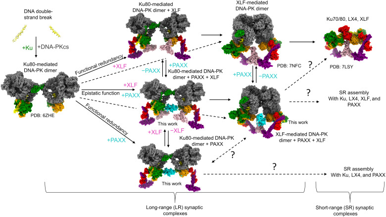
Nonhomologous end joining is a critical mechanism that repairs DNA double-strand breaks in human cells. In this work, we address the structural and functional role of the accessory protein PAXX [paralog of x-ray repair cross-complementing protein 4 (XRCC4) and XRCC4-like factor (XLF)] in this mechanism. Here, we report high-resolution cryo-electron microscopy (cryo-EM) and x-ray crystallography structures of the PAXX C-terminal Ku-binding motif bound to Ku70/80 and cryo-EM structures of PAXX bound to two alternate DNA-dependent protein kinase (DNA-PK) end-bridging dimers, mediated by either Ku80 or XLF. We identify residues critical for the Ku70/PAXX interaction in vitro and in cells. We demonstrate that PAXX and XLF can bind simultaneously to the Ku heterodimer and act as structural bridges in alternate forms of DNA-PK dimers. Last, we show that engagement of both proteins provides a complementary advantage for DNA end synapsis and end joining in cells.


Figures

Fig. 1.
Fig. 1.. Structures of the PAXX KBM bound to Ku70/80.
(A) Cryo-EM structure of Ku70/80-DNA with no PAXX bound to 2.7-Å resolution (PDB: 7ZVT). (B) Cryo-EM structure of Ku70/80-DNA with the P-KBM bound to 2.8-Å resolution. (C) X-ray crystallography structure of Ku70/80, with the P-KBM bound to one of the heterodimers in the crystallographic unit to 2.97-Å resolution. (D) Enlarged view of the P-KBM bound to Ku70/80 in the cryo-EM structure with the density for the peptide shown as mesh. (E) Enlarged view of the P-KBM bound to Ku70/80 in the x-ray structure with the density shown as a mesh. (F) AF model of the P-KBM bound to Ku70/80. Ku70 is shown in orange, Ku80 in green, DNA in yellow, and PAXX in cyan.
Fig. 2.
Fig. 2.. Mutagenesis of residues within the P-KBM of PAXX affects Ku binding and recruitment to DNA damage sites.
(A) The cryo-EM structure of Ku70/80-DNA bound to the P-KBM of PAXX, with interaction of Patches 1 and 2 illustrated, and insets show them enlarged with residues labeled. Ku70 is in orange, Ku80 is in green, DNA is in yellow, and PAXX is in cyan. (B) The sequence of the C terminus of PAXX, with interaction of Patches 1 and 2 identified and residues mutated colored, in bold and underlined. (C) Isothermal titration calorimetry (ITC) data table for Ku70/80-DNA (either F or G DNA shown below the table) with PAXX (full-length, KBM, or mutants), Kd values are indicated in millimolars. NI: no interaction. (D) Laser microirradiation experiment monitoring the recruitment of WT PAXX or mutants to sites of DNA damage in U2OS PAXX knockout (KO) cells. Results were plotted as mean values ± SEM. Ku70 mutants of the P-KBM of PAXX are colored accordingly. Left: Western blot confirming PAXX KO in U2OS cells.
Fig. 3.
Fig. 3.. Mutagenesis of PAXX-binding sites in Ku70 impairs PAXX recruitment at DSBs using a degron approach.
(A) Depiction of the system used for Ku degradation upon auxin (IAA) addition in U2OS cells. (B) Principle of the microirradiation experiment (top) and quantifications (bottom) of GFP-PAXX accumulation at laser-induced DNA damage in control U2OS cells or U2OS cells expressing mAID-Ku70 without or with WT-Ku70 complementation, under conditions without or with IAA. Results were plotted as mean values ± SEM. (C) Structural maps of mutated residues in the Ku70 interface with PAXX. (D) Quantification of GFP-PAXX accumulation at laser-induced DNA damage in U2OS cells expressing WT or mutated Ku70 as indicated. Results were plotted as mean values ± SEM. Ku70 mutants are colored accordingly. (E) Western blot of chromatin fractions from U2OS cells expressing WT or mutated Ku70 as indicated, treated or not with 3 nM calicheamicin (Cali) for 1 hour.
Fig. 4.
Fig. 4.. Cryo-EM structures of DNA-PK LR synaptic complexes with PAXX.
(A and B) Two views of the DNA-PK Ku80-mediated LR cryo-EM map with PAXX. DNA-PKcs is shown in gray, Ku70 in orange, Ku80 in green, DNA in yellow, XRCC4 in purple, Lig4 in red, and PAXX in cyan. (C) Structure of the DNA-PK LR Ku80-mediated dimer with PAXX. Each half of the PAXX homodimer is colored different shades of blue, and the dashed lines indicate where the C-terminal tail links to the P-KBM within Ku70. (D) Close up view of the P-KBM of PAXX bound to Ku70 in the LR DNA-PK Ku80-mediated dimer, density is shown as a mesh. (E) Cryo-EM map of the Ku80-mediated DNA-PK LR synaptic dimer with PAXX (cyan) and XLF (pink). (F) Enlarged view of Ku70/80 from the LR Ku80-mediated DNA-PK complex, with the peptide of XLF shown as pink sticks and the density as pink mesh and the peptide of PAXX in cyan sticks and cyan mesh. (G) Cryo-EM map of the XLF-mediated DNA-PK LR synaptic dimer with PAXX and XLF bound. (H) Enlarged view of Ku70/80 from the LR Ku80-mediated DNA-PK complex, with the same color codes for the XLF and PAXX peptides than in (F).
Fig. 5.
Fig. 5.. Simultaneous binding of PAXX and XLF to Ku70/80.
(A) Cryo-EM map (2.68-Å overall resolution) of Ku70/80-DNA with the X-KBM peptide from XLF shown in pink and the P-KBM peptide from PAXX shown in cyan. Ku70 is in orange, Ku80 in green, and DNA in yellow. (B) Principle of the laser microirradiation experiment and (C) representative images before and after irradiation of nuclei from U2OS cells expressing WT or mutated Ku70, cotransfected with GFP-PAXX and mCherry-XLF. (D and E) Quantification of fluorescence accumulation at laser-induced DNA damage sites in cells expressing GFP-PAXX and mCherry-XLF. Results were plotted as mean values ± SEM. Ku70 mutants are colored accordingly. (F) Western blotting on whole cell extracts from HEK-293T cells as stated, treated or not with 150 pM calicheamicin (Cali) for 1 hour. (G) Histogram with mean values ± SEM of end joining efficiency in HEK-293T cells as indicated, quantified by fluorescence expression analyzed by flow cytometry 48 hours after cell cotransfection with Cas9-targeted reporter and control circular plasmids. Values were set at 100% for the WT condition and statistical analysis was performed using the unpaired Student’s t-test. The P values for PAXXKO vs WT and XLFKO vs WT were 0.0188 (*) and 0.0011 (**), respectively. The P values for XLFKO/PAXXKO vs WT and XLFKO/PAXXKO vs XLFKO were <0.0001 (****).
Fig. 6.
Fig. 6.. Snapshots of c-NHEJ assembly.
A model of the branched pathways and structures of the NHEJ assemblies dependent on the presence of either PAXX or XLF. DNA is in yellow, DNA-PKcs in gray, Ku70 in orange, Ku80 in green, XRCC4 in purple, XLF in pink, Lig4 in red, and PAXX in cyan.

References

    1. Zhao B., Rothenberg E., Ramsden D. A., Lieber M. R., The molecular basis and disease relevance of non-homologous DNA end joining. Nat. Rev. Mol. Cell Biol. 21, 765–781 (2020). - PMC - PubMed
    1. Stinson B. M., Loparo J. J., Repair of DNA double-strand breaks by the nonhomologous end joining pathway. Annu. Rev. Biochem. 90, 137–164 (2021). - PMC - PubMed
    1. Ghosh D., Raghavan S. C., Nonhomologous end joining: New accessory factors fine tune the machinery. Trends Genet. 37, 582–599 (2021). - PubMed
    1. Meek K., Dang V., Lees-Miller S. P., DNA-PK: The means to justify the ends? Adv. Immunol. 99, 33–58 (2008). - PubMed
    1. Jette N., Lees-Miller S. P., The DNA-dependent protein kinase: A multifunctional protein kinase with roles in DNA double strand break repair and mitosis. Prog. Biophys. Mol. Biol. 117, 194–205 (2015). - PMC - PubMed

Substances