. 2023 Jun 15;388(24):2241-2252.
doi: 10.1056/NEJMoa2202318. Epub 2023 May 31.

Variant STAT4 and Response to Ruxolitinib in an Autoinflammatory Syndrome

Affiliations

Variant STAT4 and Response to Ruxolitinib in an Autoinflammatory Syndrome

Hratch Baghdassarian et al. N Engl J Med. .

Abstract

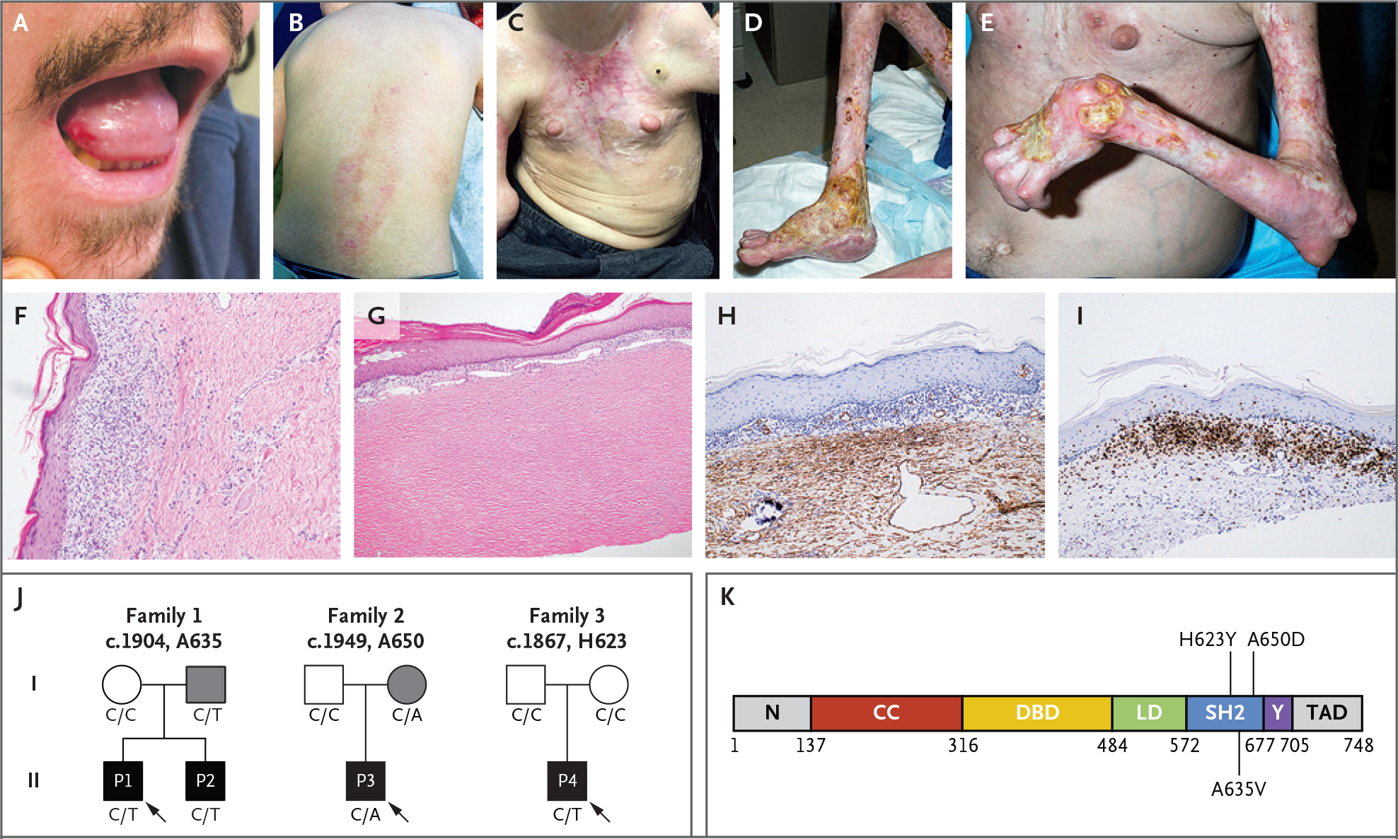
Background: Disabling pansclerotic morphea (DPM) is a rare systemic inflammatory disorder, characterized by poor wound healing, fibrosis, cytopenias, hypogammaglobulinemia, and squamous-cell carcinoma. The cause is unknown, and mortality is high.

Methods: We evaluated four patients from three unrelated families with an autosomal dominant pattern of inheritance of DPM. Genomic sequencing independently identified three heterozygous variants in a specific region of the gene that encodes signal transducer and activator of transcription 4 (STAT4). Primary skin fibroblast and cell-line assays were used to define the functional nature of the genetic defect. We also assayed gene expression using single-cell RNA sequencing of peripheral-blood mononuclear cells to identify inflammatory pathways that may be affected in DPM and that may respond to therapy.

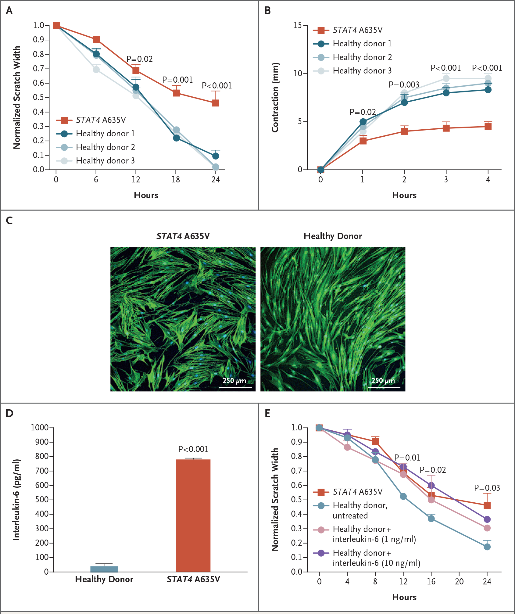
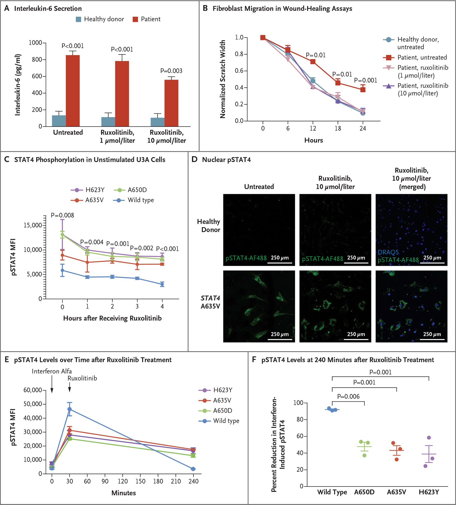
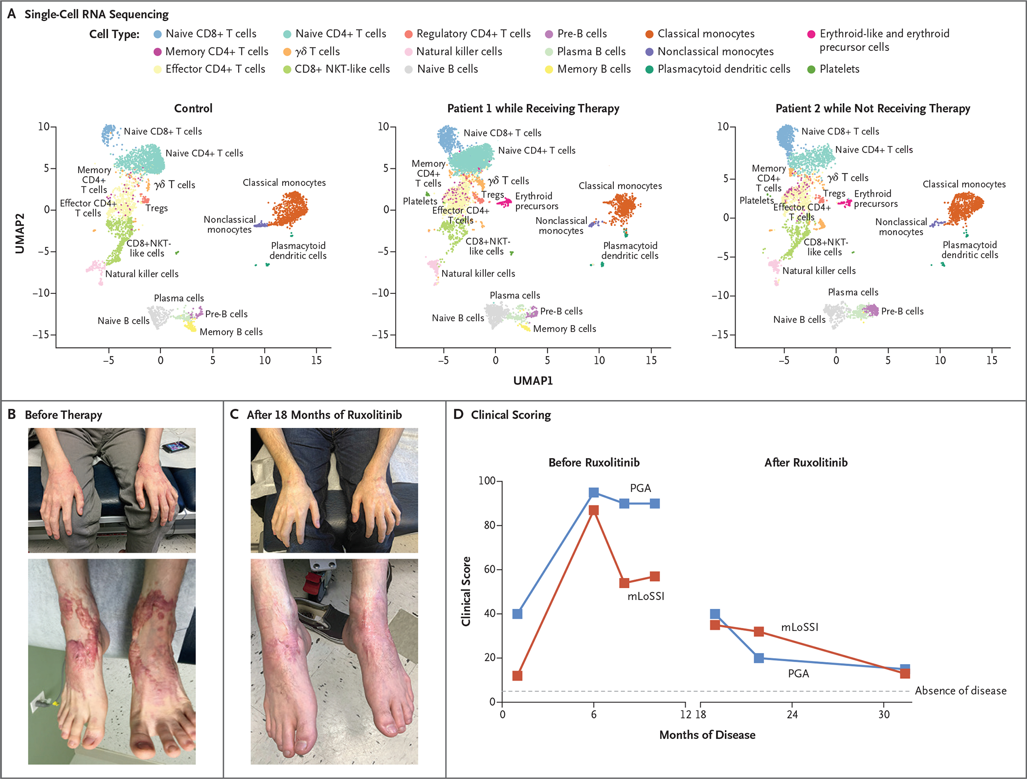
Results: Genome sequencing revealed three novel heterozygous missense gain-of-function variants in STAT4. In vitro, primary skin fibroblasts showed enhanced interleukin-6 secretion, with impaired wound healing, contraction of the collagen matrix, and matrix secretion. Inhibition of Janus kinase (JAK)-STAT signaling with ruxolitinib led to improvement in the hyperinflammatory fibroblast phenotype in vitro and resolution of inflammatory markers and clinical symptoms in treated patients, without adverse effects. Single-cell RNA sequencing revealed expression patterns consistent with an immunodysregulatory phenotype that were appropriately modified through JAK inhibition.

Conclusions: Gain-of-function variants in STAT4 caused DPM in the families that we studied. The JAK inhibitor ruxolitinib attenuated the dermatologic and inflammatory phenotype in vitro and in the affected family members. (Funded by the American Academy of Allergy, Asthma, and Immunology Foundation and others.).


Figures

Figure 1.
Figure 1.. Clinical Manifestations of Disabling Pansclerotic Morphea (DPM).
Clinical images show oral ulceration and limitation in tongue protrusion (Panel A); spreading waxy, hypopigmented lesions on the back and waxy hypopigmented “tank top” sign on the chest (Panels B and C); and ulcerations with articular ankylosis of the legs and arms (Panels D and E). Histologic sections of skin-biopsy samples show prominent inflammation (Panel F) and dermal thickening and hyalinization of morphea (Panel G). Images are shown of immunohistochemical staining for smooth-muscle action (Panel H) and CD3 (Panel I) in skin-biopsy samples before use of ruxolitinib. Family pedigrees (Panel J) are shown, with probands indicated by arrows. Circles represent female family members, squares male family members, and solid symbols persons who have received a diagnosis of DPM. Grey shading indicates persons with the STAT4 variants but with milder symptoms. The genotype at the specified locus is indicated under each person. A linear protein model (Panel K) shows the approximate locations of the identified variants in the SRC homology 2 (SH2) domain. CC denotes coiled-coil domain, DBD DNA-binding domain, LD linker domain, N the N-terminal domain, TAD transactivation domain, and Y the phosphotyrosyl-tail segment.
Figure 2
Figure 2. Effects of STAT4 Variants on Phosphorylation.
Panel A shows absolute luciferase emission from the interleukin-6 Leeporter cell line transfected with vector carrying wild-type or variant STAT4. Transcriptional activity was enhanced in the presence of STAT4 A635V, H623Y, and A650D, as compared with wild-type STAT4 and the nontransfected cell line, with or without stimulation with lipopolysaccharide (LPS) or interleukin-6. Panels B and C show STAT4 phosphorylation in U3A cells stably transfected with wild-type or variant STAT4. Flow cytometry that was used to measure the mean fluorescence intensity (MFI) of phosphorylated STAT4 (pSTAT4) showed increased pSTAT4 in unstimulated cells (Panel B) that were transfected with A635V (red), A650D (green), and H623Y (purple) variants, as compared with wild-type STAT4 (blue). In response to interferon alfa (Panel C), STAT4 phosphorylation persisted in variant cells at 240 minutes as compared with wild-type cells. In Panels A and C, error bars indicate standard errors. Panel D shows HEK293T cells transiently transfected with plasmids containing wild-type, H623Y, A635V, or phospho-dead Y693A STAT4 tagged with green fluorescent protein. Unstimulated cells that were transfected with H623Y or A635V variants had a greater accumulation of STAT4 in the nucleus than those transfected with wild-type or Y693A STAT4. Panel E shows that primary skin fibroblasts from a patient with the A635V variant had prominent pSTAT4 (green), as compared with fibroblasts from a healthy donor. Phosphorylated STAT4 staining of patient fibroblasts persisted in a perinuclear location with interleukin-6 stimulation. Nuclear staining was performed with DRAQ5 (blue).
Figure 3.
Figure 3.. Evaluation of the Function of Primary Skin Fibroblasts In Vitro.
Panel A shows that wound healing as measured by scratch assay was reduced in fibroblasts from patients with STAT4 A635V (red), as compared with fibroblasts from healthy donors (blue). For the scratch assays, three experiments were performed with six scratches each. Panel B shows that transforming growth factor β–induced contraction of collagen matrix by patient-derived fibroblasts (red) was reduced relative to fibroblasts from healthy donors. Panel C shows that in F-actin immunocytochemical analysis, cell size was increased in primary skin fibroblasts from a patient with STAT4 A635V, as compared with fibroblasts from healthy donors. Panel D shows that patient fibroblasts had enhanced interleukin-6 secretion in the absence of stimulation. Panel E shows that wound healing was reduced in primary skin fibroblasts from healthy donors when treated with varying concentrations of interleukin-6, with rates approaching those of cells from patients with STAT4 A635V (three experiments with six scratches each). Throughout the figure, P values were calculated by means of two-way analysis of variance. Error bars indicate standard errors.
Figure 4
Figure 4. Janus Kinase Inhibition, Reduction of STAT4 Phosphorylation, and Wound Healing.
Panel A shows that patient fibroblasts had enhanced interleukin-6 secretion that was responsive to ruxolitinib. Data for healthy donors are a summary of three such donors. The P value for patient cells treated with ruxolitinib as compared with untreated patient cells is 0.02. Panel B shows that pretreatment with ruxolitinib led to enhanced fibroblast migration in wound-healing assays, with closure at 24 hours that was similar to that in unaffected fibroblasts (three experiments with six scratches each). Panel C shows that STAT4 phosphorylation in unstimulated U3A cells was reduced after treatment with ruxolitinib. Panel D shows that nuclear pSTAT4 was reduced in response to ruxolitinib treatment of patient fibroblasts. Panels E and F show that interferon alfa–stimulated U3A cells expressing variant STAT4 had higher levels of pSTAT4 than cells expressing wild-type STAT4 at 240 minutes. Ruxolitinib treatment decreased phosphorylation in cell lines expressing variant STAT4 and in those expressing wild-type STAT4. In Panel F, mean decreases and standard errors are shown. Throughout the figure, P values were calculated by means of two-way analysis of variance. Error bars indicate standard errors.
Figure 5
Figure 5. Ruxolitinib Treatment and Resolution of the Inflammatory Phenotype.
Shown are peripheral-blood mononuclear cells analyzed by means of single-cell RNA sequencing (Panel A), plotted in uniform manifold approximation and projection (UMAP) space and clustered for each of the control and patient samples. NKT cells denote natural killer T cells, and Tregs regulatory T cells. Photographs show waxy, erythematous nodular lesions on bilateral hands and feet before (Panel B) and after (Panel C) initiation of ruxolitinib therapy. Shown are clinical scores (Panel D) on the modified Localized Scleroderma Skin Severity Index (mLoSSI, red) and the Physician Global Assessment (PGA, blue) of disease activity. Scores on both scales range from 0 to 100, with higher scores indicating greater disease activity.

Comment in

References

    1. Wollina U, Looks A, Uhlemann C, Wollina K. Pansclerotic morphea of childhood-follow-up over 6 years. Pediatr Dermatol 1999;16:2 45–7. - PubMed
    1. Moll M, Holzer U, Zimmer C, Rieber N, Kuemmerle-Deschner JB. Autologous stem cell transplantation in two children with disabling pansclerotic morphea. Pediatr Rheumatol Online J 2011; 9: Suppl 1:77. abstract.
    1. Gruss C, Stücker M, Kobyletzki G, Schreiber D, Altmeyer P, Kerscher M. Low dose UVA1 phototherapy in disabling pansclerotic morphoea of childhood. Br J Dermatol 1997;136:293–4. - PubMed
    1. Kowal-Bielecka O, Distler O. Use of methotrexate in patients with scleroderma and mixed connective tissue disease. Clin Exp Rheumatol 2010;2 8: Suppl 61:S160–S163. - PubMed
    1. Forsea A-M, Cretu A-N, Ionescu R, Giurcaneanu C. Disabling pansclerotic morphea of childhood — unusual case and management challenges. J Med Life 2008;1:348–54. - PMC - PubMed

Publication types

MeSH terms