. 2023 Jun 13;56(6):1410-1428.e8.
doi: 10.1016/j.immuni.2023.05.007. Epub 2023 May 16.

Multi-omics blood atlas reveals unique features of immune and platelet responses to SARS-CoV-2 Omicron breakthrough infection

Affiliations

Multi-omics blood atlas reveals unique features of immune and platelet responses to SARS-CoV-2 Omicron breakthrough infection

Hong Wang et al. Immunity. .

Abstract

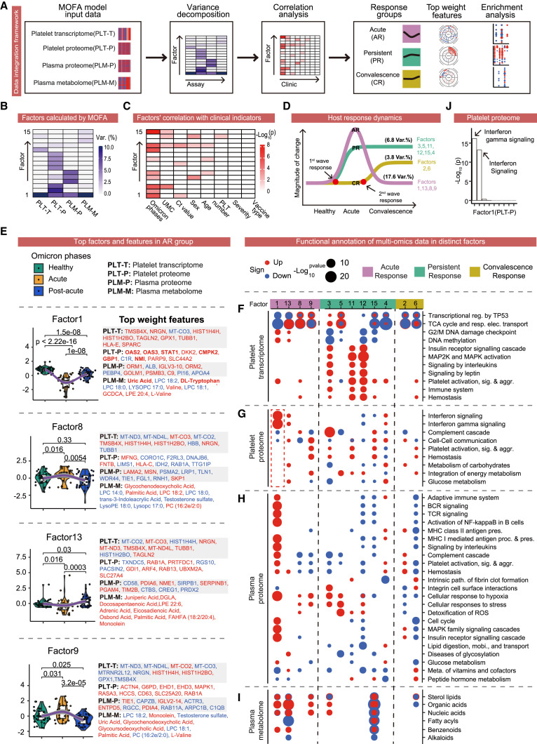
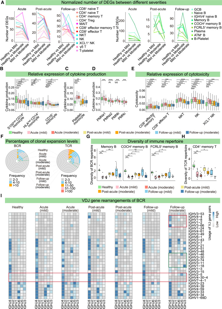
Although host responses to the ancestral SARS-CoV-2 strain are well described, those to the new Omicron variants are less resolved. We profiled the clinical phenomes, transcriptomes, proteomes, metabolomes, and immune repertoires of >1,000 blood cell or plasma specimens from SARS-CoV-2 Omicron patients. Using in-depth integrated multi-omics, we dissected the host response dynamics during multiple disease phases to reveal the molecular and cellular landscapes in the blood. Specifically, we detected enhanced interferon-mediated antiviral signatures of platelets in Omicron-infected patients, and platelets preferentially formed widespread aggregates with leukocytes to modulate immune cell functions. In addition, patients who were re-tested positive for viral RNA showed marked reductions in B cell receptor clones, antibody generation, and neutralizing capacity against Omicron. Finally, we developed a machine learning model that accurately predicted the probability of re-positivity in Omicron patients. Our study may inspire a paradigm shift in studying systemic diseases and emerging public health concerns.

Keywords: COVID-19; Omicron, platelet; blood; metabolome; plasma; platelet-leukocyte aggregate; proteome; re-positive; single-cell RNA-seq.


Conflict of interest statement

Declaration of interests A patent related to this work has been filed.

Figures

Figure 1
Figure 1
Study design for multi-dimensional dissection of the blood ecosystem in SARS-CoV-2 Omicron patients (A) Overview of assay modalities and Omicron disease phases analyzed. (B) Summary of omics measurements, and clinical parameters for patients enrolled in the omics study. The y axis displays patient IDs from XGO_001 to XGO_110, while the x axis shows days since disease onset. See also Table S1.
Figure 2
Figure 2
Plasma proteome and metabolome analyses unveil evident platelet-associated dysregulation in Omicron patients (A) Heatmap of 447 differentially expressed metabolites (DEMs) (ANOVA B-H-adjusted p < 0.05) clustered using mFuzz into five discrete significant clusters. (B) Super classes enrichment based on the metabolites in (A) using MetaboAnalyst. (C) Heatmap of 449 differentially expressed proteins (DEPs) (ANOVA B-H adjusted p < 0.05) clustered using mFuzz into four discrete significant clusters. (D and E) Networks generated by String database analysis using dysregulated proteins in clusters 2 and 3 for (D) and (E), respectively. See also Figures S2 and S3 and Table S2.
Figure 3
Figure 3
Deep platelet proteome and transcriptome analyses reveal enhanced immune signatures and reduced thrombosis in Omicron breakthrough compared with ancestral strain patients (A) Heatmap showing the four dynamic expression patterns of 415 DEPs (B-H-adjusted p < 0.05), clustered using mFuzz, during different phases of Omicron breakthrough infection. (B) Dot plot showing representative gene ontology (GO) terms enriched in the four clusters. (C) Boxplot displaying expression levels of proteins related to “response to virus” (GO:0009615) in distinct phases of infection. The y axis shows log2 normalized protein expression. Wilcoxon test: p ≤ 0.05; ∗∗ p ≤ 0.01; ∗∗∗ p ≤ 0.001. (D) Boxplots showing expression of IFIT1, IFIT5, and PARP14 in different phases of Omicron breakthrough infection. (E) Heatmap of differentially expressed genes (DEGs) in platelets from healthy participants and COVID-19 patients with Omicron breakthrough and ancestral SARS-CoV-2 infections (Wilcoxon test B-H-adjusted p < 0.05 and fold change > 2). (F) Bar plots showing GO terms for platelet DEGs enriched in patients with Omicron breakthrough or ancestral SARS-CoV-2 infections. (G) Boxplots showing expression of representative DEGs in (E). (H) Schematic diagram showing the differences in platelets between patients with ancestral COVID-19 and Omicron breakthrough infection. See also Figure S3 and Tables S3 and S4.
Figure 4
Figure 4
Multi-omics integration reveals the landscape of time-resolved host responses (A) Workflow of MOFA integrating the four omics datasets. (B) MOFA deconstructed vast variances into 15 factors in PLT-T, PLT-P, PLM-T, and PLM-M. (C) Correlation analysis of the top 15 MOFA factors with clinical parameters. (D) Top 15 MOFA factors were classified into three groups and two waves of host responses. AR, acute response; PR, persistent response; CR, convalescence response; and Var.%, percentage of variances explained. (E) Differences of factors among distinct Omicron phases (left) along with top-weighted features from the four omics datasets (right) in the AR group. Red font indicates positive weighting, whereas blue font shows negative weighting. (F–I) Pathway enrichment analyses utilizing the four omics datasets for each factor from the three response groups. Red dots indicate enrichment in positive changes, whereas blue dots show enrichment in negative changes in Omicron patients compared with healthy controls. (J) Pathway enrichment of the platelet proteome in factor 1. The x axis shows enriched pathways, whereas the y axis shows the −log (10) p value. See also Figure S4 and Table S5.
Figure 5
Figure 5
Single-cell transcriptome landscape of peripheral blood cells in Omicron patients (A) Schematic diagram showing sample information for single-cell RNA-seq coupled with scBCR-seq and scTCR-seq analyses of frozen PBMCs and fresh neutrophil extraction. (B) UMAP plots displaying the clusters of the total of 462,937 frozen PBMCs (left) and PBMCs from healthy donors and mild and moderate patients (right). Colors indicate cell determined by unsupervised Leiden clustering. (C) Variation in cellular composition measured by log2 (fold-change) for each cluster of cells comparing Omicron patients to healthy donors across the acute, post-acute, and follow-up phases. (D) Variation in cellular composition for each cluster of neutrophils from mild- and moderate-severity infectious patients. (E) Normalized number of DEGs between the two phases for NK and T cells, B cells, monocytes, and platelets in mild (top) and moderate (bottom) patients. For each cell cluster, the number of DEGs was scaled by (X-min, max-min) + 1. See also Figure S5 and Table S6.
Figure 6
Figure 6
Immune responses to Omicron variant at single-cell resolution (A) Number of DEGs in NK and T cells (left) and B cells (right) from mild- and moderate-severity infection patients in the acute, post-acute, and follow-up phases. (B–E) Relative expression gene of cytokines in CD14+ and CD16+ monocytes (B), platelet-monocytes (C), and neutrophils (D), and cytotoxicity-related genes in CD8+ Teff and NK cells (E) from healthy donors and mild and moderate patients during the acute, post-acute, and follow-up phases. Wilcoxon test: p ≤ 0.05; ∗∗ p ≤ 0.01; ∗∗∗ p ≤ 0.001; ∗∗∗∗ p ≤ 0.0001. (F) Percentages of different clonal expansion levels for BCR (left) and TCR (right) across seven severity and stage conditions. (G and H) Diversities of BCR repertoires in memory B cells (G) and TCR repertoires in memory T cells (H) across the seven conditions. Diversity was evaluated as Shannon entropy = ΣPlog2(1/P), in which P represents the frequency of a given BCR and TCR clone among that of all BCR and TCR clones. Wilcoxon test: p ≤ 0.05; ∗∗ p ≤ 0.01; ∗∗∗ p ≤ 0.001; ∗∗∗∗ p ≤ 0.0001. (I) Heatmap showing the VDJ gene rearrangements of BCRs across the seven conditions. Red and green rectangles indicate biased and declined usage, respectively. See also Figure S6 and Table S7.
Figure 7
Figure 7
Machine-learning models accurately predict re-positive patients with decreased immunity (A) Variation in cellular composition in each cluster of cells from re-positive patients (RPs) and non-re-positive patients (NRPs). (B–G) Relative expression of cytokine production in CD14+ and CD16+ monocytes (B), platelet-monocytes (C), and neutrophils (D), cytotoxicity-related genes in CD8+ memory Teff, CD8+ Teff, and NK cells (E), exhaustion-related genes in CD8+ Teff and NK cells (F), and apoptosis signaling pathway-related genes in CD8+ memory Teff and NK cells (G) from healthy donors, RP and NRP. Wilcoxon test: p ≤ 0.05; ∗∗ p ≤ 0.01; ∗∗∗ p ≤ 0.001; ∗∗∗∗ p ≤ 0.0001. (H) Percentages of different clonal expansion levels for BCR (top) and TCR (bottom) in healthy donors, RP and NRP. (I and J) Boxplot showing levels of antibodies against Omicron spike (S) protein (I) and the inhibition effects of neutralization antibodies (J) in RP and NRP, as detected by ELISA assay. (K) Workflow of the machine-learning models, and performance of the model in a test cohort of 26 Omicron patients. (L–N) Top four clinical indicators (L), top four metabolites (M), and top 10 proteins (N) prioritized by random forest analysis and ranked by the Gini index. (O) Importance of three types of features, as determined by combined coefficient of the model. (P and Q) Boxplot displaying the abundance of four metabolites (P) and four representative proteins (Q) in the RP and NRP groups in the training cohort. See also Figure S7.

References

    1. Tirosh I., Izar B., Prakadan S.M., Wadsworth M.H., Treacy D., Trombetta J.J., Rotem A., Rodman C., Lian C., Murphy G., et al. Dissecting the multicellular ecosystem of metastatic melanoma by single-cell RNA-seq. Science. 2016;352:189–196. doi: 10.1126/science.aad0501. - DOI - PMC - PubMed
    1. Lloyd-Price J., Arze C., Ananthakrishnan A.N., Schirmer M., Avila-Pacheco J., Poon T.W., Andrews E., Ajami N.J., Bonham K.S., Brislawn C.J., et al. Multi-omics of the gut microbial ecosystem in inflammatory bowel diseases. Nature. 2019;569:655–662. doi: 10.1038/s41586-019-1237-9. - DOI - PMC - PubMed
    1. Wagner J., Rapsomaniki M.A., Chevrier S., Anzeneder T., Langwieder C., Dykgers A., Rees M., Ramaswamy A., Muenst S., Soysal S.D., et al. A single-cell atlas of the tumor and immune ecosystem of human breast cancer. Cell. 2019;177:1330–1345.e18. doi: 10.1016/j.cell.2019.03.005. - DOI - PMC - PubMed
    1. Sun Y., Wu L., Zhong Y., Zhou K., Hou Y., Wang Z., Zhang Z., Xie J., Wang C., Chen D., et al. Single-cell landscape of the ecosystem in early-relapse hepatocellular carcinoma. Cell. 2021;184:404–421.e16. doi: 10.1016/j.cell.2020.11.041. - DOI - PubMed
    1. López-Otín C., Kroemer G. Hallmarks of health. Cell. 2021;184:1929–1939. doi: 10.1016/j.cell.2021.03.033. - DOI - PubMed

Publication types

Supplementary concepts