. 2023 May 1;37(9-10):418-431.
doi: 10.1101/gad.350400.123. Epub 2023 May 31.

Germ cell-specific eIF4E1b regulates maternal mRNA translation to ensure zygotic genome activation

Affiliations

Germ cell-specific eIF4E1b regulates maternal mRNA translation to ensure zygotic genome activation

Guanghui Yang et al. Genes Dev. .

Abstract

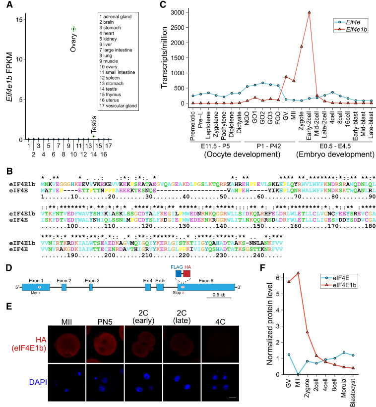
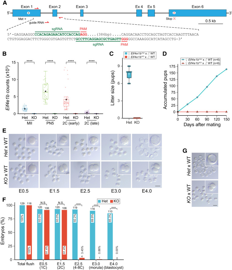
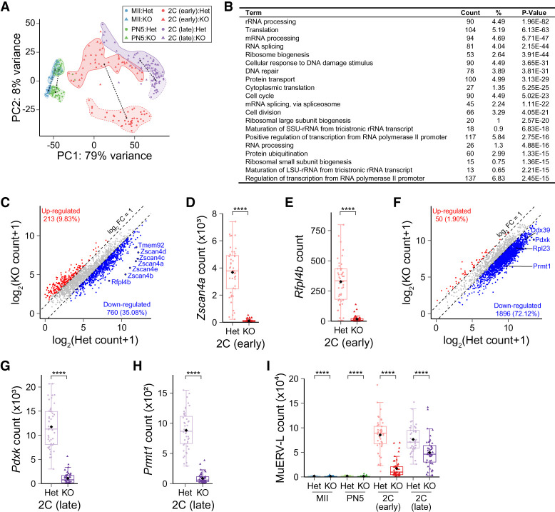
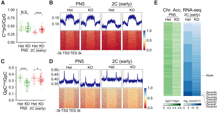
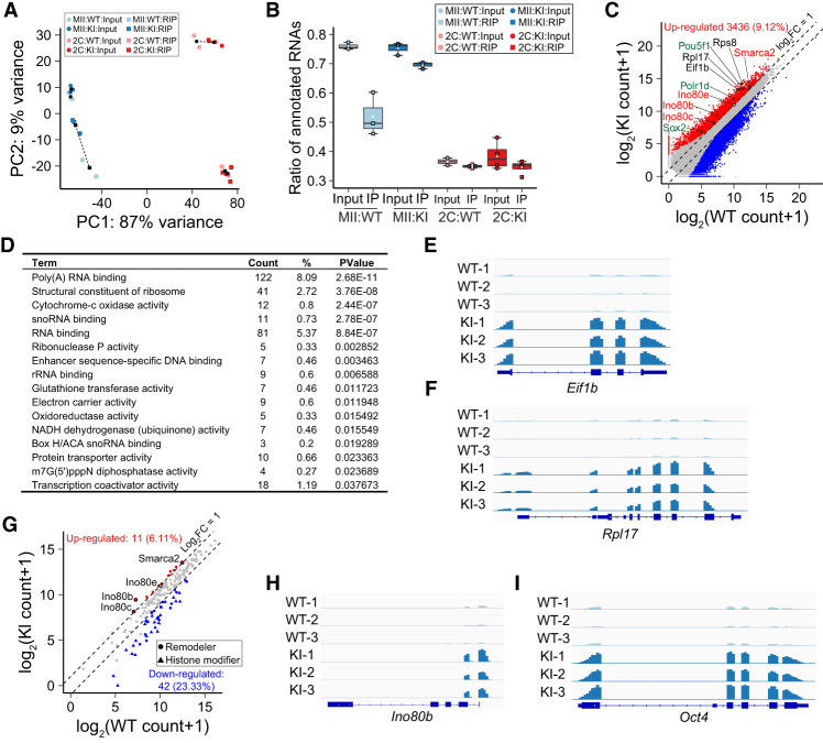
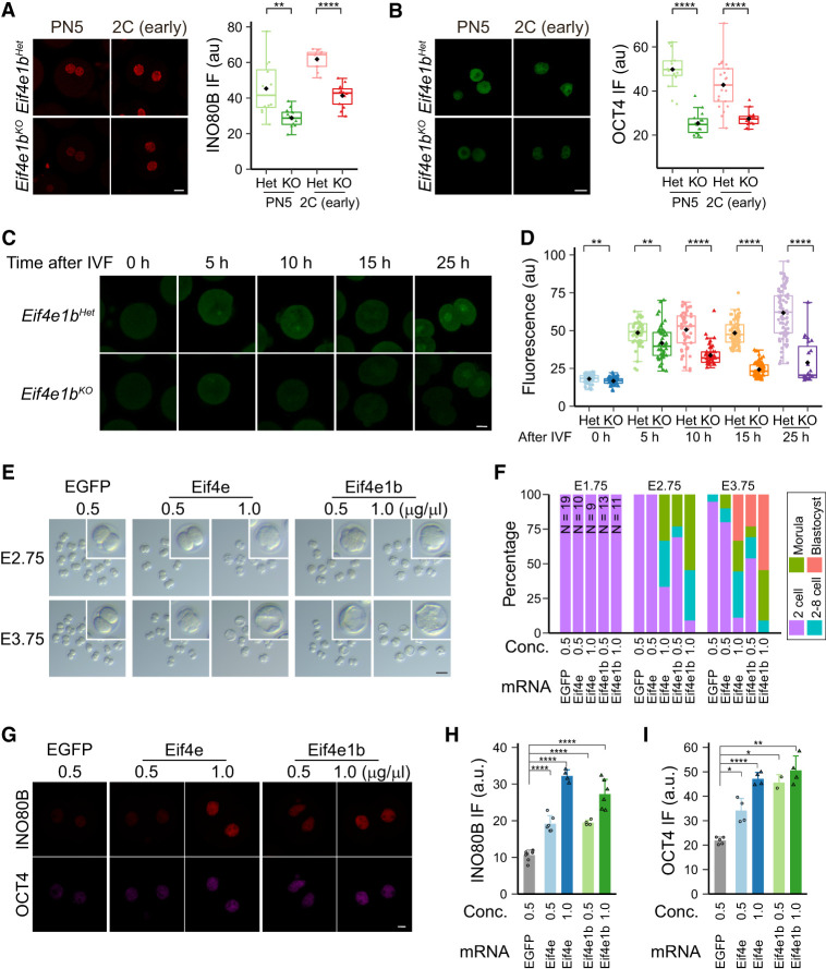
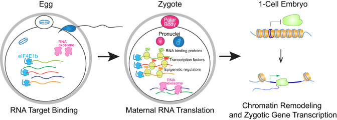
Translation of maternal mRNAs is detected before transcription of zygotic genes and is essential for mammalian embryo development. How certain maternal mRNAs are selected for translation instead of degradation and how this burst of translation affects zygotic genome activation remain unknown. Using gene-edited mice, we document that the oocyte-specific eukaryotic translation initiation factor 4E family member 1b (eIF4E1b) is the regulator of maternal mRNA expression that ensures subsequent reprogramming of the zygotic genome. In oocytes, eIF4E1b binds to transcripts encoding translation machinery proteins, chromatin remodelers, and reprogramming factors to promote their translation in zygotes and protect them from degradation. The protein products are thought to establish an open chromatin landscape in one-cell zygotes to enable transcription of genes required for cleavage stage development. Our results define a program for rapid resetting of the zygotic epigenome that is regulated by maternal mRNA expression and provide new insights into the mammalian maternal-to-zygotic transition.

Keywords: Eif4e1b; maternal RNA translation; mouse embryo; zygotic genome activation.


Figures

Figure 1.
Figure 1.
Eif4e1b expresses late in oogenesis and persists until two-cell embryos. (A) Abundance (FPKM [fragments per kilobase of transcript per million mapped reads]) of Eif4e1b transcripts in different tissues from published data. (B) Alignment of mouse eIF4E1b (NP_001028441.1) protein sequence with that of eIF4E (NP_031943.3). (C) Abundance (TPM [transcripts per million mapped reads]) of Eif4e1a (Eif4e) and Eif4e1b transcripts during mouse oogenesis and early embryo development. Eif4e1b transcripts are more abundant from GV to the early two-cell stage, while Eif4e is more abundant at other stages. (D) Schematic of the Eif4e1b gene locus in the Eif4e1bKI mouse line with FLAG and HA tags at the C terminus. (*) Initiator methionine, (x) stop codon. (E) Immunofluorescence of eggs and embryos derived from Eif4e1bKI female mice. Anti-HA antibody and DAPI were used to visualize the eIF4E1b fusion protein and nuclear DNA, respectively. Scale bar, 20 µm. (F) Protein levels of eIF4E and eIF4E1b from published proteomics results from GV oocyte to blastocyst stage.
Figure 2.
Figure 2.
Maternal deletion of Eif4e1b leads to developmental arrest at two cells. (A) Schematic of the Eif4e1b gene (top) and sequences of sgRNAs (bottom) for generation of Eif4e1bKO mouse lines. (*) Initiator methionine, (x) stop codon. (B) Abundance of read overlaps with Eif4e1b exon 1 or 2. Residual Eif4e1b transcripts in Eif4e1bKO female mice lack exons 1 and 2 and produce no functional eIF4E1b protein. (****) P < 0.0001; two-tailed t-test. (C) Six-week-old female Eif4e1bHet (control) and homozygous Eif4e1bKO mice were mated with WT males to determine litter sizes. (D). Accumulated pups from Eif4e1bHet and Eif4e1bKO females during 5 mo of continuous mating. (E) Representative images of in vitro cultured embryos from Eif4e1bHet and Eif4e1bKO females after mating with WT males at embryonic day 0.5 (E0.5), E1.5, E2.5, E3.0, and E4.0. Inset magnification, 2.5×. Scale bar, 100 µm. (F) Quantification of embryos in E. Ratio of embryos at different stages is plotted. Total number of embryos is above each bar. (N.S.) Not significant, (****) P < 0.0001; two-sided proportion test. (G) Images of embryos flushed from Eif4e1bHet and Eif4e1bKO female reproductive tracts at E3.5 after successful in vivo mating. Inset magnification, 2.5×. Scale bar, 100 µm.
Figure 3.
Figure 3.
Maternal deletion of Eif4e1b impairs ZGA. (A) Principal component analysis (PCA) plot of RNA-seq results of single embryos from Eif4e1bHet (control) or Eif4e1bKO female mice at different developmental stages. The lengths of dashed lines between cluster centers represent differences between samples. (B) Top gene ontology (GO) terms of down-regulated genes in late two-cell embryos from Eif4e1bKO females. Top 20 molecular function terms as well as the number and ratio of genes and P-values are shown. (C) Scatter plot documenting differentially expressed RNAs expected to be transcribed during minor ZGA in early two-cell embryos. mRNAs from multiple well-known minor ZGA genes are labeled in the plots. (D,E) Abundance of Zscan4a and Rfpl4b, two minor ZGA genes, at the early two-cell stage. (F) Scatter plot documenting differentially expressed RNAs expected to be transcribed during major ZGA in late two-cell embryos. mRNAs from multiple well-known major ZGA genes are labeled. (G,H) Abundance of Pdxk and Prmt1, two major ZGA genes, at the late two-cell stage. (I) Abundance of transcripts from the MuERV-L transposon in embryos from control or Eif4e1bKO female mice at different developmental stages. RNAs that have log2 fold change >2 or <−2 with adjusted P value of <0.01 are considered significantly up-regulated or down-regulated in C and F and are shown as red and blue dots, respectively. The total number of up-regulated or down-regulated RNAs is labeled in each plot. (****) P < 0.0001; two-tailed t-test.
Figure 4.
Figure 4.
Maternal eIF4E1b reprograms zygotic chromatin accessibility. (A) Ratio of endogenous methylated CpG among all CpG motifs to show global DNA methylation patterns in PN5 zygotes and early two-cell embryos from Eif4e1bHet (control) and Eif4e1bKO females. (B) DNA methylation profile around minor ZGA gene loci in PN5 zygotes and early two-cell embryos from control and Eif4e1bKO females. (N.S.) Not significant, (*) P < 0.05, (****) P < 0.0001; two-tailed t-test. (C) Same as A, but for global chromatin accessibility using an exogenous GpC methyltransferase (M.CviPI) to methylate accessible GpC. (D) Same as B, but for global chromatin accessibility. (E) Heat map showing chromatin accessibility at promoter regions (left) and expression of corresponding minor ZGA genes (right) in early two-cell embryos. Individual minor ZGA genes are annotated.
Figure 5.
Figure 5.
eIF4E1b binds to a subset of mRNAs in MII eggs. (A) PCA of input and immunoprecipitated transcripts after eIF4E1b RIP. MII eggs or early two-cell embryos from Eif4e1bKI female mice were used, and WT eggs/embryos served as controls. (B) Ratio of input and immunoprecipitated (RIP) reads that can be mapped to annotated RNAs in RIP-seq. (C) Scatter plot documenting all differentially expressed RNAs as determined by the RIP-seq using WT and Eif4e1bKI MII eggs. Transcripts that have a log2 fold change of >1 or <−1 with adjusted P-value of <0.1 are considered significantly up-regulated and down-regulated and are shown as red and blue dots, respectively. Up-regulated RNAs represent potential Eif4e1b mRNA targets. Three groups of them are labeled in different colors (red, black, and green). (D) Top gene ontology (GO) terms of potential eIF4E1b RNA targets. Top 16 molecular function terms with P-values of <0.05 are shown. The number and ratio of genes that fall into each GO term are shown as well. (E,F) Integrated Genomics Viewer (IGV) view of eIF4E1b RIP-seq results at Eif1b and Rpl17 loci in WT and Eif4e1bKI samples. (G) Scatter plot as in C documenting differentially expressed RNAs coding all known chromatin remodeling complex subunits and histone modification enzymes as determined by RIP-seq experiments using WT and Eif4e1bKI MII eggs. Several potential eIF4E1b mRNA targets are labeled. (H,I) Same as E, but for Ino80b and Oct4 (Pou5f1) loci.
Figure 6.
Figure 6.
eIF4E1b controls translation of maternal mRNA in mouse zygotes. (A,B) INO80B and OCT4 protein expression in embryos from Eif4e1bHet and Eif4e1bKO females at different developmental stages. Scale bar, 20 µm. The nuclear fluorescent signals are quantified at the right. (C) Imaging of nascent proteins in embryos derived from Eif4e1bHet and Eif4e1bKO females at different time points after IVF. Scale bar, 20 µm. (D) Fluorescent signal from OPP (O-propargyl-puromycin) incorporation indicating that protein translation from C was quantified. (E) Morphology of in vitro cultured embryos after microinjection of mRNAs. The number of embryos is quantified in F. Inset magnification, 2.8×. Scale bar, 100 µm. (G) Immunostaining showing expression of two eIF4E1b targets in early two-cell embryos. Scale bar, 10 µm. (H,I) Quantification of nuclear INO80B and OCT4 signals in G. (*) P < 0.05, (**) P < 0.01, (****) P < 0.0001; two-tailed t-test.
Figure 7.
Figure 7.
Working model. eIF4E1b binds a subset of essential maternal RNAs in MII eggs to protect them from degradation. After fertilization, eIF4E1b-bound mRNAs are translated and remodel chromatin into an open state to enable transcription of the zygotic genes that establish early developmental programs.

Similar articles

Cited by

References

    1. Abe K, Yamamoto R, Franke V, Cao M, Suzuki Y, Suzuki MG, Vlahovicek K, Svoboda P, Schultz RM, Aoki F. 2015. The first murine zygotic transcription is promiscuous and uncoupled from splicing and 3′ processing. EMBO J 34: 1523–1537. 10.15252/embj.201490648 - DOI - PMC - PubMed
    1. Abe KI, Funaya S, Tsukioka D, Kawamura M, Suzuki Y, Suzuki MG, Schultz RM, Aoki F. 2018. Minor zygotic gene activation is essential for mouse preimplantation development. Proc Natl Acad Sci 115: E6780–E6788. 10.1073/pnas.1804309115 - DOI - PMC - PubMed
    1. Alda-Catalinas C, Bredikhin D, Hernando-Herraez I, Santos F, Kubinyecz O, Eckersley-Maslin MA, Stegle O, Reik W. 2020. A single-cell transcriptomics CRISPR-activation screen identifies epigenetic regulators of the zygotic genome activation program. Cell Syst 11: 25–41.e9. 10.1016/j.cels.2020.06.004 - DOI - PMC - PubMed
    1. Alizadeh Z, Kageyama S, Aoki F. 2005. Degradation of maternal mRNA in mouse embryos: selective degradation of specific mRNAs after fertilization. Mol Reprod Dev 72: 281–290. 10.1002/mrd.20340 - DOI - PubMed
    1. Aoki F, Hara KT, Schultz RM. 2003. Acquisition of transcriptional competence in the 1-cell mouse embryo: requirement for recruitment of maternal mRNAs. Mol Reprod Dev 64: 270–274. 10.1002/mrd.10227 - DOI - PubMed

Publication types

Substances