. 2023 May;11(5):e006264.
doi: 10.1136/jitc-2022-006264.

Cell surface marker-based capture of neoantigen-reactive CD8+ T-cell receptors from metastatic tumor digests

Affiliations

Cell surface marker-based capture of neoantigen-reactive CD8+ T-cell receptors from metastatic tumor digests

Praveen D Chatani et al. J Immunother Cancer. 2023 May.

Abstract

Background: Cellular immunotherapies using autologous tumor-infiltrating lymphocytes (TIL) can induce durable regression of epithelial cancers in selected patients with treatment-refractory metastatic disease. As the genetic engineering of T cells with tumor-reactive T-cell receptors (TCRs) comes to the forefront of clinical investigation, the rapid, scalable, and cost-effective detection of patient-specific neoantigen-reactive TIL remains a top priority.

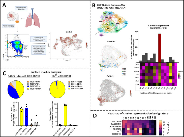
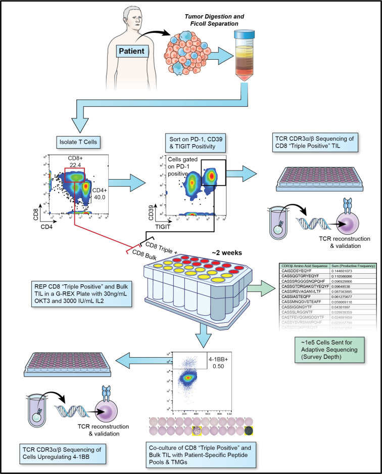
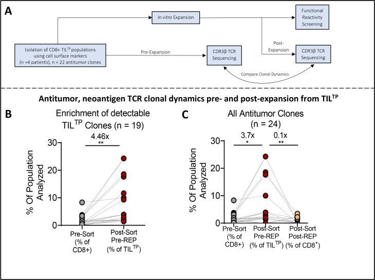
Methods: We analyzed the single-cell transcriptomic states of 31 neoantigen-specific T-cell clonotypes to identify cell surface dysfunction markers that best identified the metastatic transcriptional states enriched with antitumor TIL. We developed an efficient method to capture neoantigen-reactive TCRs directly from resected human tumors based on cell surface co-expression of CD39, programmed cell death protein-1, and TIGIT dysfunction markers (CD8+ TILTP).

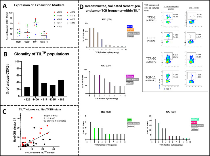
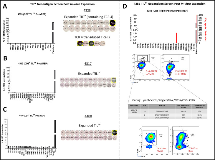
Results: TILTP TCR isolation achieved a high degree of correlation with single-cell transcriptomic signatures that identify neoantigen-reactive TCRs, making it a cost-effective strategy using widely available resources. Reconstruction of additional TILTP TCRs from tumors identified known and novel antitumor TCRs, showing that at least 39.5% of TILTP TCRs are neoantigen-reactive or tumor-reactive. Despite their substantial enrichment for neoantigen-reactive TCR clonotypes, clonal dynamics of 24 unique antitumor TILTP clonotypes from four patients indicated that most in vitro expanded TILTP populations failed to demonstrate neoantigen reactivity, either by loss of neoantigen-reactive clones during TIL expansion, or through functional impairment during cognate neoantigen recognition.

Conclusions: While direct usage of in vitro-expanded CD8+ TILTP as a source for cellular therapy might be precluded by profound TIL dysfunction, isolating TILTP represents a streamlined effective approach to rapidly identify neoantigen-reactive TCRs to design engineered cellular immunotherapies against cancer.

Keywords: CD8-Positive T-Lymphocytes; Cell Engineering; Immunity, Cellular; Immunotherapy; Lymphocytes, Tumor-Infiltrating.


Conflict of interest statement

Competing interests: FJL, SK, PR, and SAR are listed on an international patent application filed based on the NeoTCR8 signature described in this study. All other authors declare no competing interests.

Figures

Figure 1
Figure 1
(A) CD8+ TIL populations were isolated from fresh tumor digest. Single-cell transcriptomic analysis was performed on the patient’s TIL from tumor digest and known neoantigen-reactive clonotypes and signature-enriched cells were projected onto a Uniform Manifold Approximation and Projection (UMAP). (B) Integrated single-cell transcriptomic analysis showing UMAP-based projection of CD8+ TIL states from five patient samples (4400, 4385, 4382, 4323, 4317; left, top); cluster 7 (C7) indicated by arrow. Back-projection of 31 known neoantigen-reactive TCRs (NeoTCR) clones onto UMAP plot (left, middle). UMAP transcriptomic map of CD8+ TIL shows NeoTCR8 transcriptomic state showing co-localization of CXCL13-expressing cells (left, bottom). Bar graph indicating the percentage of known neoantigen-reactive TCRs (NeoTCRs) within each cluster, with the bar for NeoTCR8 state C7 denoted in red (right, top). Heatmap indicating average transcriptomic expression of inhibitory genes within each cluster (right, bottom). (C) Surface marker analysis of CD8+ CD39+CD103+ (double positive) and ‘triple positive’ (CD8+ PD-1+ TIGIT+ CD39+) TILTP cells of four patients (4400, 4385, 4323, 4393). Pie chart of proportion of cells expressing TIGIT and PD-1 within the CD39+CD103+ TIL subset; bar graph with the median of individual patient data (n=4) represented by black dots (left). Pie chart of proportion of cells expressing CD39 and CD103 within the TILTP subset; bar graph with the median of individual patient data represented by black dots (right). (D) Heatmap showing the relative frequency of cells within each cluster from single-cell RNA from figure 1B, that can be identified by the top decile within each signature. ‘All Cells’ includes all cells present in the transcriptomic UMAP. ‘Random50’ and ‘Random500’ represent signatures derived from random genes sets of 50 and 500 genes as negative control signatures, respectively. TCR, T-cell receptor; TIL, tumor-infiltrating lymphocytes.
Figure 2
Figure 2
Overview of experimental pipeline for isolating, sequencing, expanding, and testing TILTP cells from single-cell suspensions of epithelial tumors. Single-cell PCR plates of pre-REP TILTP populations were performed to capture candidate neoantigen-reactive clones for reconstruction and testing. Survey sequencing was performed on post-REP TILTP to analyze clonal dynamics of TILTP clones before and after in vitro REP expansion. In vitro expanded post-REP TILTP populations were analyzed for the presence of known neoantigen-reactive clones and to identify new neoantigen-specific TCRs. PD-1, programmed cell death protein-1; REP, rapid expansion protocol; TCR, T-cell receptor; TIL, tumor-infiltrating lymphocytes.
Figure 3
Figure 3
(A) TIGIT, CD39, and PD-1 expression by flow cytometry across nine T-cell samples isolated from tumor digest demonstrates variability in the surface expression of dysfunction markers between patients. Median PD-1 % across the samples is higher than any other single marker (61.7%). (B) Clonality of CD8+ TILTP populations in each patient. Y-axis reflects the percentage of unique CDR3β clonotypes with respect to the number of legible TCRs sequenced. (C) Correlation of 69 clones from five patient TIL samples within the NeoTCR8 cluster by single-cell sequencing compared with their frequencies within the TILTP compartment (R2=0.4165). (D) TILTP sequenced from each patient’s tumor digest, ranked by frequency based on percentage of 96-well plate occupied. Reconstructed and experimentally vetted neoantigen-reactive and tumor-reactive TCR clonotypes are indicated. For patient 4317, FACS plots of activated TCR-transduced T cells (4-1BB+cells of CD8+ mTCR+ cells) co-culture results for TCR-2, TCR-5, TCR-10, and TCR-11 are shown with corresponded listed antigens (AURKAIP and PIK3CA), alongside pertinent negative controls. Unevaluated clones were unable to be reconstructed due to technical difficulties in Sanger sequencing. PD-1, programmed cell death protein-1; TCR, T-cell receptor; TIL, tumor-infiltrating lymphocytes.
Figure 4
Figure 4
Co-culture of TCRs reconstructed from CD8+ TILTP with tumor-specific TMGs and peptide pools failed to demonstrate reactivity via upregulation of 4-1BB and IFN-γ secretion detected by ELISpot assays in samples (A) 4323, (B) 4317, and (C) 4400. For 4323 TIL, inset also shows TMG-recognition by TCR4-transduced PBL in the same experiment (present within pre-REP TILTP) indicating poor functional response of the same clonotype within TILTP (D) Co-culture of in vitro-expanded post-REP CD8+ TILTP with tumor-specific TMGs and peptide pools demonstrated reactivity to TMG6 via IFN-γ release and low-level 4-1BB upregulation. CDR3β sequencing of 4-1BB-upregulating clonotypes resulted in the reconstruction and identification of the reactive TCR, which also recognized the patient-specific PDX tumor. Allo, Allogeneic control PDX; DMSO, control for peptide; IFN, interferon; Irr TMG, irrelevant control TMG; PMA, phorbol myristate acetate; PP, peptide pools; REP, rapid expansion protocol; TCR, T-cell receptor; TIL, tumor-infiltrating lymphocytes; TMG, tandem minigene.
Figure 5
Figure 5
(A) Workflow for assessing clonal dynamics of antitumor or neoantigen-specific TCR clonotypes before and after in vitro expansion of CD8+ TILTP populations. (B) In vitro tracking of the percentage of neoantigen-reactive and antitumor clonotypes (n=19 clones, samples 4323, 4317, 4400, 4385, and 4382) among CD8+ TILTP T cells relative to bulk CD8+ TIL in tumor digest. Only clones that were detectable in a TILTP FACS-based plate sort (96 wells) are included in this analysis. (C) In vitro clonal tracking of all antitumor, neoantigen-reactive clones (n=24) among bulk CD8+ TIL (left), CD8+ TILTP (middle), and post-REP CD8+ TILTP (right) for samples 4323, 4317, 4400, and 4385. The majority of known neoantigen-reactive clonotypes are enriched by triple-positive sorting but are unable to expand in the REP. P values indicate paired t-test between bulk versus pre-expansion (pre-REP) populations, and pre-REP versus post-expansion (post-REP) TILTP populations. Numbers above indicate fold-expansion between the populations analyzed. * = p<0.05 ** = p<0.01. REP, rapid expansion protocol; TCR, T-cell receptor; TIL, tumor-infiltrating lymphocytes.

References

    1. Tran E, Robbins PF, Rosenberg SA. "final common pathway'' of human cancer immunotherapy: targeting random somatic mutations. Nat Immunol 2017;18:255–62. 10.1038/ni.3682 - DOI - PMC - PubMed
    1. Colli LM, Machiela MJ, Myers TA, et al. . Burden of nonsynonymous mutations among TCGA cancers and candidate immune checkpoint inhibitor responses. Cancer Res 2016;76:3767–72. 10.1158/0008-5472.CAN-16-0170 - DOI - PMC - PubMed
    1. Yarchoan M, Hopkins A, Jaffee EM. Tumor mutational burden and response rate to PD-1 inhibition. N Engl J Med 2017;377:2500–1. 10.1056/NEJMc1713444 - DOI - PMC - PubMed
    1. Parkhurst MR, Robbins PF, Tran E, et al. . Unique neoantigens arise from somatic mutations in patients with gastrointestinal cancers. Cancer Discov 2019;9:1022–35. 10.1158/2159-8290.CD-18-1494 - DOI - PMC - PubMed
    1. Zacharakis N, Chinnasamy H, Black M, et al. . Immune recognition of somatic mutations leading to complete durable regression in metastatic breast cancer. Nat Med 2018;24:724–30. 10.1038/s41591-018-0040-8 - DOI - PMC - PubMed

Publication types

Substances