. 2023 May;11(5):e006509.
doi: 10.1136/jitc-2022-006509.

Single CAR-T cell treatment controls disseminated ovarian cancer in a syngeneic mouse model

Affiliations

Single CAR-T cell treatment controls disseminated ovarian cancer in a syngeneic mouse model

Diana Rose E Ranoa et al. J Immunother Cancer. 2023 May.

Abstract

Background: Treatment of some blood cancers with T cells that express a chimeric antigen receptor (CAR) against CD19 have shown remarkable results. In contrast, CAR-T cell efficacy against solid tumors has been difficult to achieve.

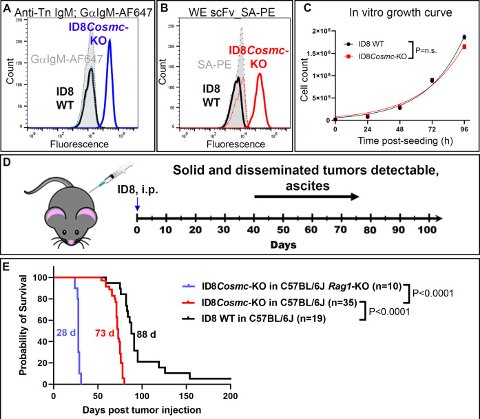
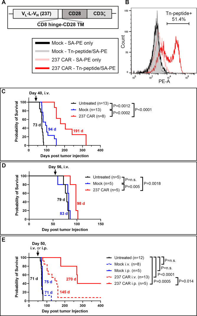
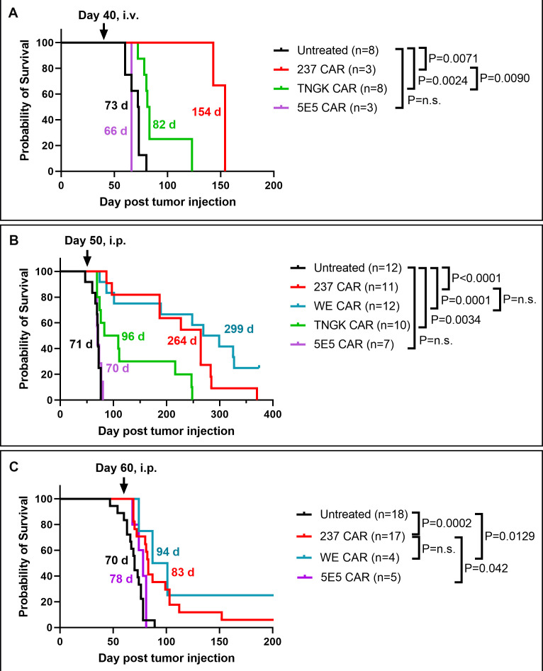
Methods: To examine the potential of CAR-T cell treatments against ovarian cancers, we used the mouse ovarian cancer cell line ID8 in an intraperitoneal model that exhibits disseminated solid tumors in female C57BL/6J mice. The CAR contained a single-chain Fv from antibody 237 which recognizes a Tn-glycopeptide-antigen expressed by ID8 due to aberrant O-linked glycosylation in the absence of the transferase-dependent chaperone Cosmc. The efficacy of four Tn-dependent CARs with varying affinity to Tn antigen, and each containing CD28/CD3ζ cytoplasmic domains, were compared in vitro and in vivo in this study.

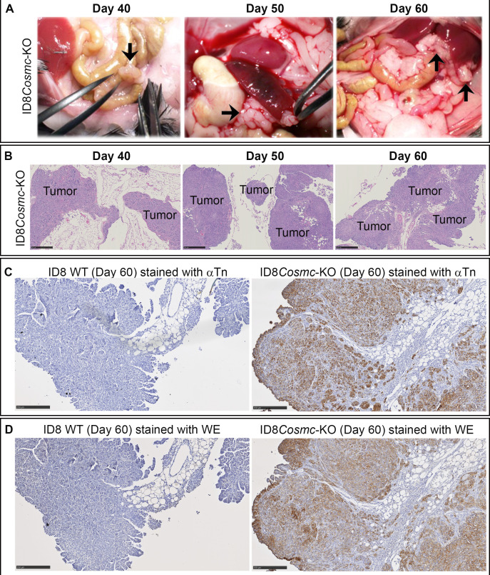
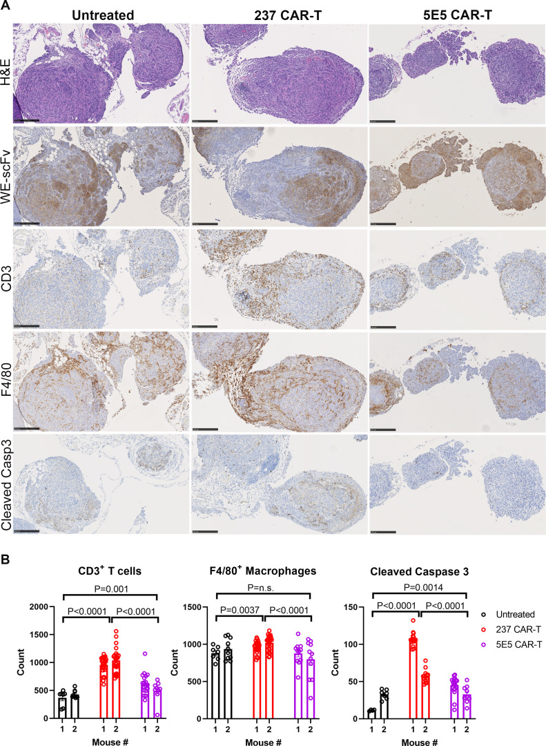
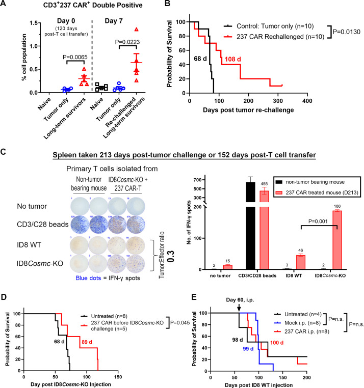
Results: In line with many observations about the impact of aberrant O-linked glycosylation, the ID8Cosmc knock-out (ID8Cosmc-KO) exhibited more rapid tumor progression compared with wild-type ID8. Despite the enhanced tumor growth in vivo, 237 CAR and a mutant with 30-fold higher affinity, but not CARs with lower affinity, controlled advanced ID8Cosmc-KO tumors. Tumor regression could be achieved with a single intravenous dose of the CARs, but intraperitoneal administration was even more effective. The CAR-T cells persisted over a period of months, allowing CAR-treated mice to delay tumor growth in a re-challenge setting. The most effective CARs exhibited the highest affinity for antigen. Antitumor effects observed in vivo were associated with increased numbers of T cells and macrophages, and higher levels of cleaved caspase-3, in the tumor microenvironment. Notably, the least therapeutically effective CAR mediated tonic signaling leading to antigen-independent cytokine expression and it had higher levels of the immunosuppressive cytokine interleukin10.

Conclusion: The findings support the development of affinity-optimized CAR-T cells as a potential treatment for established ovarian cancer, with the most effective CARs mediating a distinct pattern of inflammatory cytokine release in vitro. Importantly, the most potent Tn-dependent CAR-T cells showed no evidence of toxicity in tumor-bearing mice in a syngeneic, immunocompetent system.

Keywords: Antigens, Tumor-Associated, Carbohydrate; Cell Engineering; Immunotherapy; Receptors, Chimeric Antigen; T-Lymphocytes.


Conflict of interest statement

Competing interests: DMK is a member of the scientific advisory board of Affini-T Therapeutics and a consultant for Tempus. PS, HS, KS, and DMK are co-inventors on a patent application related to these chimeric antigen receptors. No potential conflicts of interest were disclosed by the other authors.

Figures

Figure 1
Figure 1
ID8Cosmc-KO as a model for ovarian cancer in C57BL/6J mice. Tn antigen expression on ID8Cosmc-KO was measured by flow cytometry using either (A) staining with monoclonal mouse IgM anti-Tn antibody 5F4 followed by goat anti-mouse IgM conjugated to Alexa Fluor 647 or (B) biotinylated anti-Tn-OTS8 WE scFv tetramer containing streptavidin-PE. (C) ID8 WT and ID8Cosmc-KO cell doubling times (19.2 hours and 21.8 hours, respectively) in vitro were measured over time by manual cell counting. Growth curves from two different experiments (total n=6) were combined and plotted using GraphPad Prism V.9.4.1 and cell doubling times were calculated using the exponential growth equation. (D) Timeline for ID8Cosmc-KO tumor growth in syngeneic C57BL/6J mice after intraperitoneal injection of 107 tumor cells. (E) Survival curves of C57BL/6J mice inoculated intraperitoneally with 107 ID8 WT or ID8Cosmc-KO. Median survival was 88 days and 73 days for ID8 WT (n=19) and ID8Cosmc-KO (n=35), respectively. For C57BL/6 Rag1-KO mice inoculated with 107 ID8Cosmc-KO (n=10), the median survival was 28 days. Survival curves were plotted using GraphPad Prism V.9.4.1 and effects were calculated using the log-rank (Mantel-Cox) test. i.p., intraperitoneal; KO, knock-out; scFv, single-chain Fv; WT, wild-type.
Figure 2
Figure 2
Pathological and histochemical analyses of tissue sections from ID8Cosmc-KO and ID8 WT mice at various times after tumor challenge. (A) At indicated days post-inoculation with 107 tumor cells, mice were euthanized, and organs in the peritoneal cavity were examined visually for tumor nodules (black arrows). (B) Formalin-fixed paraffin embedded tissue sections from mice inoculated with ID8Cosmc-KO cells at the indicated time points were stained with H&E. Tumor foci are labeled. Scale bar=500 µm. (C, D) ID8 WT or ID8Cosmc-KO tumors at day 60 post-tumor challenge were examined by immunohistochemistry staining using biotinylated anti-Tn IgM antibody 5F4 (C) or biotinylated WE scFv (D). Scale bar=250 µm. KO, knock-out; scFv, single-chain Fv; WT, wild-type.
Figure 3
Figure 3
CAR-T cell treatment of mice with established ID8Cosmc-KO tumors. (A) Schematic of 237 single-chain Fv linked by a CD8 hinge-CD28 transmembrane domain to the CD28 and CD3ζ cytoplasmic domains, cloned into the pMP71 retroviral vector. (B) Primary T cells isolated from splenocytes of naïve donor C57BL/6J mice were transduced with 237 CAR for 72 hours. Transduction efficiency was measured by flow cytometry using tetramers of biotinylated/Tn-glycosylated OTS8 peptide made with streptavidin-PE. Mock-transduced T cells were included as negative control. (C, D) Survival curves of ID8Cosmc-KO-bearing C57BL/6J mice either untreated (black), treated with mock-transduced T cells (blue) or 237 CAR-T cells (red) administered by retro-orbital intravenous injection of 5×10⁶ T cells at day 40 (C) or day 56 (D) post-tumor inoculation. Note that five of the mice in the 237 CAR group (C) were used at day 160 for a re-challenge experiment (figure 7B), such that their survival times may have been even longer without this re-challenge. (E) Comparison of intravenous (dashed red line) and intraperitoneal (solid red line) delivery of 237 CAR-T cells at day 50 post-tumor inoculation. All survival curves were plotted with GraphPad Prism V.9.4.1 using cumulative data across multiple experiments (two combined experiments for C and D, four combined experiments for E), with the indicated total n for each treatment arm. Median survival times are indicated; p values were calculated using the log-rank (Mantel-Cox) test. CAR, chimeric antigen receptor; i.p., intraperitoneal; i.v., intravenous; KO, knock-out.
Figure 4
Figure 4
Efficacy of engineered CAR T constructs with different affinities for the mouse Tn-OTS8 antigen in the ID8Cosmc-KO ovarian tumor model. (A) Survival curves of ID8Cosmc-KO-bearing C57BL/6J mice treated with 237 CAR T (red), TNGK CAR T (green), or 5E5 CAR T (purple) by intravenous injection of 5×10⁶ transduced T cells at day 40 post-tumor inoculation. (B, C) Survival curves of ID8Cosmc-KO-bearing C57BL/6J mice treated with different CAR-T cells by intraperitoneal injection of 5×10⁶ transduced T cells at day 50 (B) or day 60 (C) post tumor-inoculation. All survival curves were plotted with GraphPad Prism V.9.4.1 using cumulative data across multiple experiments (two combined experiments for A and C, four for B) with the indicated total n for each treatment arm. Median survival times are indicated; p values were calculated using the log-rank (Mantel-Cox) test. CAR, chimeric antigen receptor; i.p., intraperitoneal; i.v., intravenous; KO, knock-out.
Figure 5
Figure 5
In vitro cytokine and killing analyses of different CAR T cells. Mock, 237-CAR, WE-CAR, TNGK-CAR, or 5E5-CAR transduced primary T cells from C57BL/6J donor mice were co-cultured with ID8Cosmc-KO cells in a 96-well plate for 24 hours at various tumor:effector ratios. The number of effector T cells were kept constant at 12,000 cells/well across all conditions tested. After 24 hours, 25 uL culture supernatants were analyzed for multiple cytokines using a Luminex plate reader to measure IFN-γ (A), IL-2 (B), TNFα (C), IL-6 (D), IL-4 (E), and IL-10 (F). Plots shown are combined cytokine array data points from five experiments. Bar graphs were plotted using GraphPad Prism V.9.4.1. Error bars are SEM. To simplify the presentation, p values are shown only for 237 versus 5E5 CAR. A complete set of tumor:effector ratio titration plots for each of the five cytokine array experiments are shown in online supplemental figure S5. (G, H) The remaining ID8Cosmc-KO (G) or ID8 WT (H) adherent tumor cells on plates were fixed in ice-cold 10% trichloroacetic acid for the SRB viability assay. The percent growth inhibition was calculated using the formula outlined in the Methods section. SRB experiments were performed twice, each with a technical replicate of n=4. Bar graphs were plotted using GraphPad Prism V.9.4.1. Error bars are SEM. P values for 237, WE, TNGK were calculated against either mock or 5E5 at <0.0001 (or denoted by ***); p values for 5E5 against either mock T or TNGK are at <0.001 (**). CAR, chimeric antigen receptor; IFN, interferon; IL, interleukin; KO, knock-out; TNF, tumor necrosis factor; WT, wild-type.
Figure 6
Figure 6
Peritoneal organs were harvested from ID8Cosmc-KO inoculated mice, 3 days after intraperitoneal treatment with either 237 CAR-T or 5E5 CAR-T at day 54 post-tumor inoculation. Untreated mice were used as control. (A) Immunohistochemistry was performed to visualize Tn-OTS8-glycopeptide+ tumors, CD3+ T cells, F4/80+ macrophages, and apoptotic cells from adjacent FFPE tissue sections stained with biotinylated WE-scFv, anti-CD3, anti-F4/80, and anti-cleaved caspase 3 antibodies, respectively. H&E stained tissue sections were included as additional reference. Scale bar=250 µm. (B) ImageJ-quantified CD3+ T cells, F4/80+ macrophages, and cleaved caspase-3+ apoptotic cells from tissue sections of untreated, 237 CAR-T, 5E5 CAR-T treated mice. A total of 5–20 images at 20× magnification corresponding to tumors spotted lining the fatty tissues near stomach, small and large intestines of two mice per treatment group were analyzed and quantified using ImageJ. Dot plots and p values were generated using GraphPad Prism V.9.4.1. Error bars are SEM. Each dot represent one field of view at 20× magnification. CAR, chimeric antigen receptor; FFPE, formalin-fixed paraffin embedded; KO, knock-out; scFv, single-chain Fv.
Figure 7
Figure 7
Evidence for in vivo persistence of 237 CAR T cells. (A) 237 CAR T-treated ID8Cosmc-KO-bearing mice were re-challenged with 107 ID8Cosmc-KO cells at 120 days post-T cell transfer. Age-matched naïve mice were inoculated with 107 ID8Cosmc-KO cells and used as control. Blood samples were collected from some mice immediately before intraperitoneal injection of re-challenged tumors and 7 days after re-challenge; processed and stained with anti-CD3 antibody and Tn-glycosylated OTS8 peptide tetramer, and analyzed by flow cytometry. The percentage of CD3+ 237 CAR T cells were plotted using GraphPad Prism V.9.4.1. Each dot represents one mouse. Error bars are SEM. (B) Survival curve of 237 CAR T treated mice re-challenged with ID8Cosmc-KO at either 120 (n=5) or 160 days (n=5) after initial tumor injection, compared with age-matched C57BL/6 naïve mice that were inoculated with the same number of tumor cells. (C) Spleens were removed from 237 CAR T-treated ID8Cosmc-KO bearing mice up to 5 months after CAR treatment (152 days for the data shown). Spleen cells were co-cultured with either ID8 WT or ID8Cosmc-KO tumor cells in a 96-well plate at a tumor:effector ratio of 0.3. ELISpot assays were performed to measure IFN-gamma spots 24 hours after co-culture experiment. Splenocytes from age-matched mice were used as a control, while CD3/CD28 Dynabeads were added to T cells as a positive control for T cell activation. Plates were scanned with automated ImmunoSpot analyzer. Bar graph of quantified IFN-γ spots are plotted on the right panel. Results representative of five independent experiments. (D) Non-tumor bearing mice were injected intraperitoneally with 237 CAR T (see online supplemental figure S2B) or untreated. At day 60, mice were challenged with 107 ID8Cosmc-KO cells and the survival curve was compared with that of mice inoculated with tumor that did not receive prior 237 CAR T injections (untreated). (E) Survival curve of ID8 WT-bearing C57BL/6J mice either untreated (black), treated with mock-transduced T cells (blue), or treated with 237 CAR T (red), administered by intraperitoneal injection of 5×10⁶ T cells at day 60. Data for D and E are from two combined experiments. All survival curves were plotted using GraphPad Prism V.9.4.1 with the indicated total n for each treatment arm. Median survival times are indicated; p values were calculated using the log-rank (Mantel-Cox) test. CAR, chimeric antigen receptor; IFN, interferon; i.p., intraperitoneal; KO, knock-out; WT, wild-type.

References

    1. June CH, Riddell SR, Schumacher TN. Adoptive cellular therapy: a race to the finish line. Sci Transl Med 2015;7:280. 10.1126/scitranslmed.aaa3643 - DOI - PubMed
    1. Finck AV, Blanchard T, Roselle CP, et al. . Engineered cellular Immunotherapies in cancer and beyond. Nat Med 2022;28:678–89. 10.1038/s41591-022-01765-8 - DOI - PMC - PubMed
    1. Anderson KG, Stromnes IM, Greenberg PD. Obstacles posed by the tumor Microenvironment to T cell activity: A case for synergistic therapies. Cancer Cell 2017;31:311–25. 10.1016/j.ccell.2017.02.008 - DOI - PMC - PubMed
    1. Philip M, Schietinger A. Cd8+ T cell differentiation and dysfunction in cancer. Nat Rev Immunol 2022;22:209–23. 10.1038/s41577-021-00574-3 - DOI - PMC - PubMed
    1. Rodriguez-Garcia A, Minutolo NG, Robinson JM, et al. . T-cell target antigens across major gynecologic cancers. Gynecol Oncol 2017;145:426–35. 10.1016/j.ygyno.2017.03.510 - DOI - PubMed

Publication types