

Mitochondrial alterations in the cochlea of Cdk5rap1-knockout mice with age-related hearing loss
- PMID: 37258461
- PMCID: PMC10315731
- DOI: 10.1002/2211-5463.13655
Mitochondrial alterations in the cochlea of Cdk5rap1-knockout mice with age-related hearing loss
Abstract
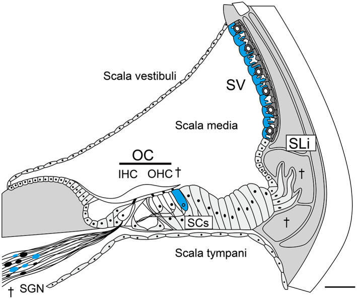
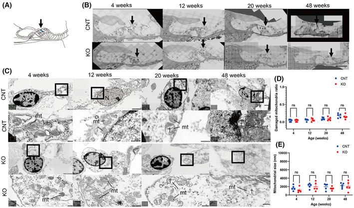
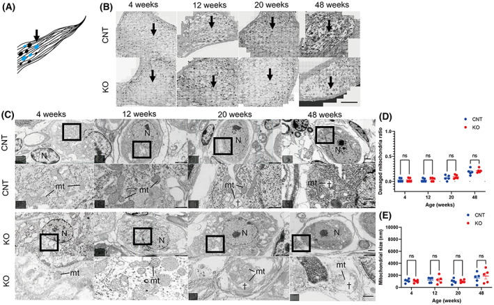
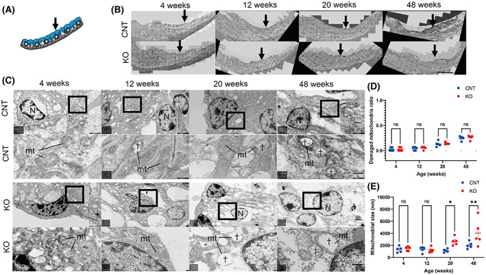
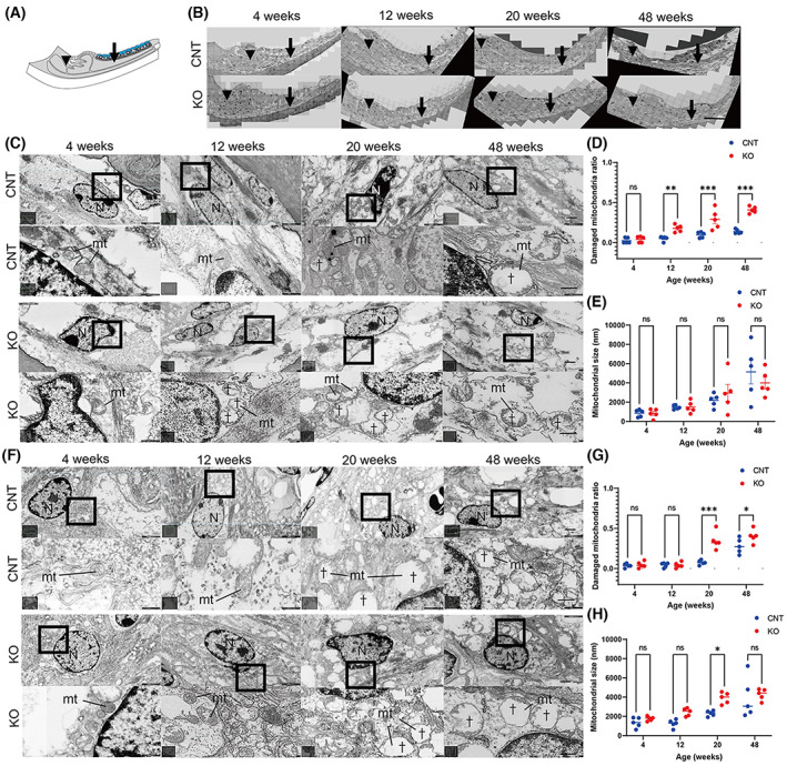
Previous studies have revealed that age-related hearing loss (AHL) in Cdk5 regulatory subunit-associated protein 1 (Cdk5rap1)-knockout mice is associated with pathology in the cochlea. Here, we aimed to identify mitochondrial alterations in the cochlea of Cdk5rap1-knockout mice with AHL. Mitochondria in the spiral ganglion neurons (SGNs) and hair cells (HCs) were normal despite senescence; however, the mitochondria of types I, II, and IV spiral ligament fibrocytes were ballooned, damaged, and ballooned, respectively, in the stria vascularis. Our results suggest that the accumulation of dysfunctional mitochondria in the lateral wall, rather than the loss of HCs and SGNs, leads to the onset of AHL. Our results provide valuable information regarding the underlying mechanisms of AHL and the relationship between aberrant tRNA modification-induced hearing loss and mitochondrial dysfunction.
Keywords: Cdk5rap1; age-related hearing loss; microstructural findings; mitochondria; mitochondrial tRNA; transmission electron microscopy.
© 2023 The Authors. FEBS Open Bio published by John Wiley & Sons Ltd on behalf of Federation of European Biochemical Societies.
Conflict of interest statement
The authors declare no conflict of interest.
Figures
Similar articles
-
Cdk5 regulatory subunit-associated protein 1 knockout mice show hearing loss phenotypically similar to age-related hearing loss.Mol Brain. 2021 May 17;14(1):82. doi: 10.1186/s13041-021-00791-w. Mol Brain. 2021. PMID: 34001214 Free PMC article.
-
Downregulation of REST in the cochlea contributes to age-related hearing loss via the p53 apoptosis pathway.Cell Death Dis. 2022 Apr 13;13(4):343. doi: 10.1038/s41419-022-04774-0. Cell Death Dis. 2022. PMID: 35418568 Free PMC article.
-
Oxidative stress and ROS metabolism via down-regulation of sirtuin 3 expression in Cmah-null mice affect hearing loss.Aging (Albany NY). 2015 Aug;7(8):579-94. doi: 10.18632/aging.100800. Aging (Albany NY). 2015. PMID: 26319214 Free PMC article.
-
[Molecular mechanism of age-related hearing loss: toward its prevention].Nihon Jibiinkoka Gakkai Kaiho. 2009 May;112(5):414-21. doi: 10.3950/jibiinkoka.112.414. Nihon Jibiinkoka Gakkai Kaiho. 2009. PMID: 19517797 Review. Japanese.
-
Role of mitochondrial dysfunction and mitochondrial DNA mutations in age-related hearing loss.Hear Res. 2007 Apr;226(1-2):185-93. doi: 10.1016/j.heares.2006.06.004. Epub 2006 Jul 25. Hear Res. 2007. PMID: 16870370 Review.
Cited by
-
Mitochonic acid 5 mitigates age-related hearing loss progression by targeting defective 2-methylthiolation in mitochondrial transfer RNAs.Front Cell Neurosci. 2025 Apr 7;19:1541347. doi: 10.3389/fncel.2025.1541347. eCollection 2025. Front Cell Neurosci. 2025. PMID: 40260078 Free PMC article.
-
Pathophysiological insights and therapeutic developments in age-related hearing loss: a narrative review.Front Aging Neurosci. 2025 Aug 20;17:1657603. doi: 10.3389/fnagi.2025.1657603. eCollection 2025. Front Aging Neurosci. 2025. PMID: 40908956 Free PMC article. Review.
-
Translational response to mitochondrial stresses is orchestrated by tRNA modifications.bioRxiv [Preprint]. 2024 Feb 14:2024.02.14.580389. doi: 10.1101/2024.02.14.580389. bioRxiv. 2024. PMID: 38405984 Free PMC article. Preprint.
-
Mitochondrial Hearing Loss: Genetic Variants and Clinical Progression.Cureus. 2025 Jun 12;17(6):e85825. doi: 10.7759/cureus.85825. eCollection 2025 Jun. Cureus. 2025. PMID: 40656278 Free PMC article.
References
-
- Pickles JO (2004) Mutation in mitochondrial DNA as a cause of presbyacusis. Audiol Neurootol 9, 23–33. - PubMed
-
- Seidman MD, Bai U, Khan M and Quirk WS (1997) Mitochondrial DNA deletions associated with aging and presbyacusis. Arch Otolaryngol Head Neck Surg 123, 1039–1045. - PubMed
-
- Fischel‐Ghodsian N, Prezant TR, Chaltraw WE, Wendt KA, Nelson RA, Amos KS and Falk RE (1997) Mitochondrial gene mutation is a significant predisposing factor in aminoglycoside ototoxicity. Am J Otolaryngol 18, 173–178. - PubMed
Publication types
MeSH terms
Substances
LinkOut - more resources
Full Text Sources
Molecular Biology Databases
Research Materials
