Multicenter Study
. 2023 May 31;13(1):8834.
doi: 10.1038/s41598-023-33723-w.

Automated LVO detection and collateral scoring on CTA using a 3D self-configuring object detection network: a multi-center study

Affiliations
Multicenter Study

Automated LVO detection and collateral scoring on CTA using a 3D self-configuring object detection network: a multi-center study

Omer Bagcilar et al. Sci Rep. .

Abstract

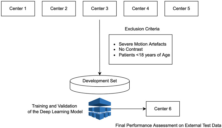
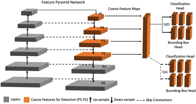
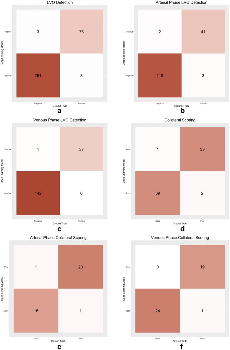
The use of deep learning (DL) techniques for automated diagnosis of large vessel occlusion (LVO) and collateral scoring on computed tomography angiography (CTA) is gaining attention. In this study, a state-of-the-art self-configuring object detection network called nnDetection was used to detect LVO and assess collateralization on CTA scans using a multi-task 3D object detection approach. The model was trained on single-phase CTA scans of 2425 patients at five centers, and its performance was evaluated on an external test set of 345 patients from another center. Ground-truth labels for the presence of LVO and collateral scores were provided by three radiologists. The nnDetection model achieved a diagnostic accuracy of 98.26% (95% CI 96.25-99.36%) in identifying LVO, correctly classifying 339 out of 345 CTA scans in the external test set. The DL-based collateral scores had a kappa of 0.80, indicating good agreement with the consensus of the radiologists. These results demonstrate that the self-configuring 3D nnDetection model can accurately detect LVO on single-phase CTA scans and provide semi-quantitative collateral scores, offering a comprehensive approach for automated stroke diagnostics in patients with LVO.


Conflict of interest statement

D.A. is the CEO and co-founder of Hevi AI Health Tech. M.Y. is the chief AI scientist and co-founder of Hevi AI Health Tech. Other authors declare no interest.

Figures

Figure 1
Figure 1
The study sample selection along with the model training and selection processes. We collected brain computed tomography angiography scans from Centers 1–5 and excluded patients following the exclusion criteria. The development set had 2425 patients with 435 large vessel occlusion positive scans. We train our nnDetection model on the development set and tested the model’s performance on the test data from Center 6.
Figure 2
Figure 2
The representative diagram of the nnDetection Model. The backbone of nnDetection is a pyramid-like network with bottom-up (left) and top-down (right) pathways with interconnected layers. The upper layers have a lower spatial resolution yet have representative features for the task at hand. The classification and regression (i.e., bounding boxes) were carried out on the representative averaged feature maps. On the bottom-up path the spatial resolution of the feature maps decreases while getting richer and denser information, while the top-down recovers the spatial dimension. The skip connection between the pathways facilitates information flow. The orange-colored features maps (P5–P2) are used for the object detection and classification. In this study, there was a primary task used in every scan: (1) the identification of the zone of the middle cerebral artery (regression) and the large vessel occlusion (classification); and one auxiliary task used in patients with large vessel occlusion: the branch for the identification of the brain hemisphere (regression) where the collateral scoring will be made by the classification branch.
Figure 3
Figure 3
The examples of correct predictions by the deep learning model. The predictions of the model are shown with a dashed box, while the ground-truth box with continuous lines. The red colors indicate the side with pathology while the green colors indicate the normal side. (a) The deep learning model correctly identified the left middle cerebral artery M1 segment occlusion. (b) The deep learning model correctly assigned collateral status as poor in a patient with right middle cerebral artery M1 segment occlusion. (c) The deep learning model correctly assigned collateral status as good in a patient with left middle cerebral artery M1 segment occlusion.
Figure 4
Figure 4
Examples of false predictions by the deep learning model. (a) The deep learning model incorrectly identified LVO in the right middle cerebral artery M1 segment. In this patient, a large intraparenchymal hematoma (arrow) displaces the middle cerebral artery medially and upwards. Hence, the model probably failed to identify the normal contrast filling of the artery and made an incorrect prediction. (b) The deep learning model failed to identify the left middle cerebral artery proximal M2 segment occlusion. (c) The deep learning model incorrectly assigned collateral status as good in a patient with left middle cerebral artery M1 segment occlusion. The experts assigned collateral status as poor in this patient.
Figure 5
Figure 5
Confusion matrices displaying the deep learning model's predictions compared to the ground-truth labels. The figure illustrates the model's performance in detecting large vessel occlusion on the overall test set (a), arterial-phase scans (b), and venous-phase scans (c). Additionally, the model's performance in collateral scoring is presented for the test set (d), arterial-phase scans (e), and venous-phase scans (f).

References

    1. WHO EMRO|Stroke, Cerebrovascular accident|Health topics. World Health Organization-Regional Office for the Eastern Mediterranean.http://www.emro.who.int/health-topics/stroke-cerebrovascular-accident/in....
    1. Smith WS, et al. Significance of large vessel intracranial occlusion causing acute ischemic stroke and TIA. Stroke. 2009;40:3834–3840. doi: 10.1161/STROKEAHA.109.561787. - DOI - PMC - PubMed
    1. Jovin TG, et al. Thrombectomy within 8 hours after symptom onset in ischemic stroke. N. Engl. J. Med. 2015;372:2296–2306. doi: 10.1056/NEJMoa1503780. - DOI - PubMed
    1. Berkhemer OA, et al. Collateral status on baseline computed tomographic angiography and intra-arterial treatment effect in patients with proximal anterior circulation stroke. Stroke. 2016;47:768–776. doi: 10.1161/STROKEAHA.115.011788. - DOI - PubMed
    1. Saver JL, et al. Time to treatment with endovascular thrombectomy and outcomes from ischemic stroke: A meta-analysis. JAMA. 2016;316:1279–1288. doi: 10.1001/jama.2016.13647. - DOI - PubMed

Publication types