. 2023 May 31;14(1):3166.
doi: 10.1038/s41467-023-38698-w.

Cryo-EM structure of the folded-back state of human β-cardiac myosin

Affiliations

Cryo-EM structure of the folded-back state of human β-cardiac myosin

Alessandro Grinzato et al. Nat Commun. .

Abstract

To save energy and precisely regulate cardiac contractility, cardiac muscle myosin heads are sequestered in an 'off' state that can be converted to an 'on' state when exertion is increased. The 'off' state is equated with a folded-back structure known as the interacting-heads motif (IHM), which is a regulatory feature of all class-2 muscle and non-muscle myosins. We report here the human β-cardiac myosin IHM structure determined by cryo-electron microscopy to 3.6 Å resolution, providing details of all the interfaces stabilizing the 'off' state. The structure shows that these interfaces are hot spots of hypertrophic cardiomyopathy mutations that are thought to cause hypercontractility by destabilizing the 'off' state. Importantly, the cardiac and smooth muscle myosin IHM structures dramatically differ, providing structural evidence for the divergent physiological regulation of these muscle types. The cardiac IHM structure will facilitate development of clinically useful new molecules that modulate IHM stability.


Conflict of interest statement

The authors declare no competing interests.

Figures
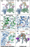
Fig. 1
Fig. 1. Cryo-EM density map of the IHM of β-cardiac myosin.
a Complete map of the interacting heads motif (IHM) at a resolution of 3.6 Å (map 1). The blocked head (BH) is colored in green, the free head (FH) is colored blue, the essential light chain (ELC) is colored in pink and the regulatory light chain (RLC) is colored in light orange. b Map masked on the two heads improved the resolution to 3.2 Å (map 2). c, d map 2 allows visualization of the side chains of the active site, the ADP, the hydrolyzed phosphate and the magnesium. The map is represented as mesh for clarity. e Interface between the two heads of the IHM (map 1). f Light chains and N-terminal extensions (Nterm represented as spheres, NtermELC is colored in deep purple and NtermRLC is colored in orange) and coiled-coils are seen without ambiguity in map 1. Black arrows point to the N-terminal extensions.
Fig. 2
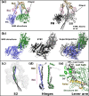
Fig. 2. The IHM structure strongly differs from homology models.
a Overall view of the cryo-EM structure of the β-cardiac myosin IHM (left) and of the homology model built from low-resolution maps of relaxed muscle thick filaments (PDB code 5TBY). BH: blocked head; FH: free head; ELC: essential light chain; RLC: regulatory light chain. b The relative orientation of the two heads varies between the structure and the model. The two IHMs are superimposed on the BH motor domain (green), the difference in the FH head position (blue for the structure (left and right), black lines for the model (center and right) are highlighted with arrows. c S2 interacts with the BH differently in the structure as compared to the model. d The lever arm conformations in the structure and the model are compared for the FH and the BH respectively, with omission of the light chains for clarity. The regions where the ELC and the RLC bind are indicated. e The conformation and position of the Relay and Converter greatly differ in the structure compared to the 5TBY model (grey). In (c), the structures are superimposed on both heads (which includes both Motor domains and the lever arms with bound LCs), in (d) the structures are superimposed on the Converter (residues 708-777), in (e), the structures are superimposed on the N-term subdomain (residues 3-202). See also Supplementary Movie 3.
Fig. 3
Fig. 3. The IHM is stabilized by multiple interfaces.
a Footprint of the interfaces stabilizing the sequestered state. The subdomains involved in the interaction are colored and the contours of interface surfaces indicate which region of the IHM is involved in recognizing this surface: L50 (wheat); U50 (marine blue); Relay (yellow); Converter (green); coiled-coil (black); HCM-loop (dark red); Loop-4 (cyan). The large flat surface called “Mesa” is delimited by magenta dotted lines. The primary head/head interaction site (PHHIS) regroups BH surface residues involved in binding the FH head next to the Mesa (see b). b Cartoon representation of the head-head interfaces. Key-residues involved in the interactions are represented as sticks. Red dotted lines represent the main interactions mediated by the BH HCM-loop, the PHHIS and Loop-4 residues (sticks. Key-interactions mediated by the HCM-loop through R403 are contoured in a black circle. The connectors and elements involved in the interface are shown and named in Supplementary Fig. 6. c Coiled-coil/BH interface. d ELC/RLC interface on the BH, the differences with the FH are illustrated in Supplementary Fig. 8. e RLC/RLC interface involving the N-terminal extension of the RLC from the FH (FH-RLCNterm ext).
Fig. 4
Fig. 4. The sequestered states of β-cardiac myosin and smooth muscle myosin (SmMyo2) differ greatly.
a Relative orientation of the BH and FH heads in SmIHM (PDB code 7MF3,) and CarIHM. SmIHM and CarIHMs are aligned on the BH (partly shown in green), the CarFH (blue) is rotated 22° anticlockwise compared to the SmFH (beige), see also cartoon providing the contour of these FH heads. b Comparison of the head-head interfaces in SmIHM and CarIHM, two views are represented. On the left, IHMs are aligned on the FH motor domain; on the right, they are aligned on the BH motor domain. The CarIHM is represented in transparency. Note the difference in the HCM-loop on the left, and on the FHRelay (yellow) and FHConverter positions (Blue) on the right. The red dashed line circle shows the HD-linker. c Sequence alignment of different structural elements involved in IHM interactions showing the divergence: Card (Homo sapiens β-cardiac myosin); Sm (Gallus gallus smooth muscle myosin); Sk (Homo sapiens fast skeletal muscle myosin); NM2a (Homo sapiens nonmuscle myosin 2a); Tar (Aphonopelma tarantula striated muscle myosin 2). Regions of interactions are in colored box: conserved in red, divergent in green. d Comparison of the position of the coiled-coils on the surface of SmIHM heads and the CarIHM. e Comparison of the angles at the hinges of flexibility: (left) pliant region shown by an alignment on the Converter; (right) ELC/RLC interface in the FH and BH lever arms, shown by an alignment on IQ1. f Comparison of the RLC-RLC interface, the models are aligned on the IQ2 of the FH. A sequence alignment of the N-term extension of the RLCs is shown with conserved regions between smooth muscle RLC (SmRLC) and cardiac RLC (CarRLC) boxed in red. The phosphorylation sites (S19 for SmRLC, S15 for CarRLC) are indicated as a red “S”.
Fig. 5
Fig. 5. Consequences of the differences between tarantula muscle myosin and cardiac myosin IHMs in thick filament regulation.
The relative orientation of the two heads in tarantula IHM (TarBH in brown and TarFH in dark grey) is compared to CarIHM (CarBH in green and CarFH in blue) (a) and SmIHM (SmBH in pink and SmFH in gold yellow) (b). Interestingly, the relative orientation of the heads in TarIHM is closer to SmIHM. c Shows how CarIHM and TarIHM are oriented relative to the core of the thick filament (dark green) and the actin (thin) filament (light red). The differences in the relative orientation of the heads would induce different interfaces. A black line shows the difference in the orientation of the FH in CarIHM and TarIHM.

Update of

Similar articles

Cited by

References

    1. Robert-Paganin J, Pylypenko O, Kikuti C, Sweeney HL, Houdusse A. Force generation by myosin motors: a structural perspective. Chem. Rev. 2020;120:5–35. doi: 10.1021/acs.chemrev.9b00264. - DOI - PubMed
    1. Janssen I, Heymsfield SB, Wang ZM, Ross R. Skeletal muscle mass and distribution in 468 men and women aged 18-88 yr. J. Appl. Physiol. 2000;89:81–88. doi: 10.1152/jappl.2000.89.1.81. - DOI - PubMed
    1. Stewart MA, Franks-Skiba K, Chen S, Cooke R. Myosin ATP turnover rate is a mechanism involved in thermogenesis in resting skeletal muscle fibers. Proc. Natl Acad. Sci. 2010;107:430–435. doi: 10.1073/pnas.0909468107. - DOI - PMC - PubMed
    1. Naber N, Cooke R, Pate E. Slow myosin ATP turnover in the super-relaxed state in tarantula muscle. J. Mol. Biol. 2011;411:943–950. doi: 10.1016/j.jmb.2011.06.051. - DOI - PMC - PubMed
    1. Hooijman P, Stewart MA, Cooke R. A new state of cardiac myosin with very slow ATP turnover: a potential cardioprotective mechanism in the heart. Biophys. J. 2011;100:1969–1976. doi: 10.1016/j.bpj.2011.02.061. - DOI - PMC - PubMed

Publication types