. 2023 May 31;13(1):8870.
doi: 10.1038/s41598-023-36073-9.

Effects of three microtubule-associated proteins (MAP2, MAP4, and Tau) on microtubules' physical properties and neurite morphology

Affiliations

Effects of three microtubule-associated proteins (MAP2, MAP4, and Tau) on microtubules' physical properties and neurite morphology

Kohei Nishida et al. Sci Rep. .

Abstract

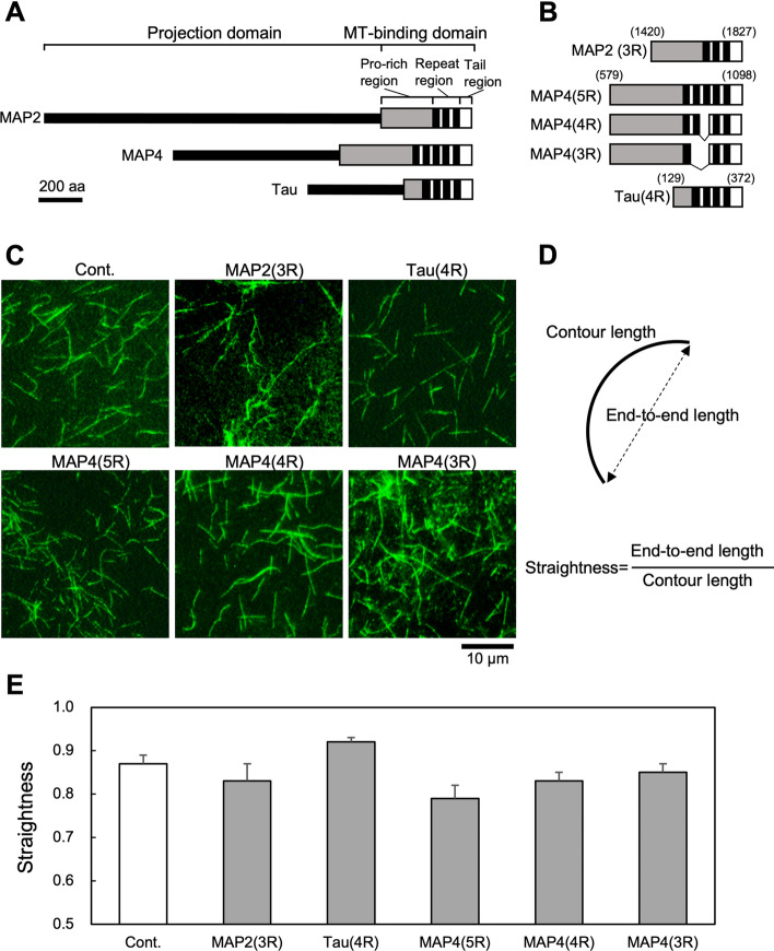
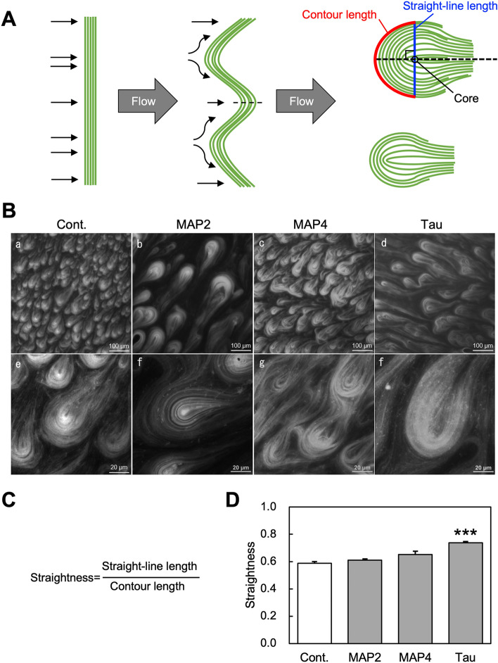
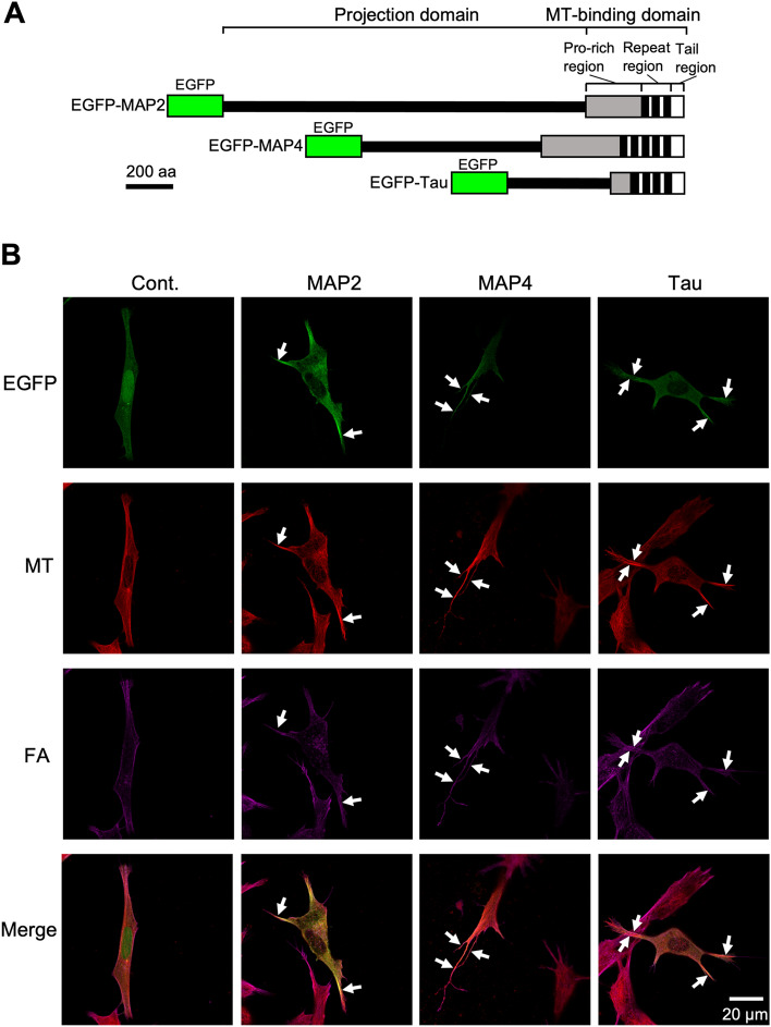
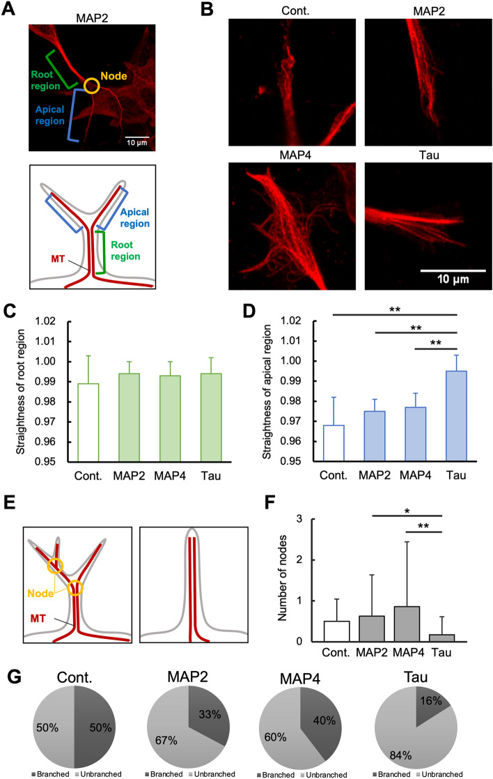
The physical properties of cytoskeletal microtubules have a multifaceted effect on the expression of their cellular functions. A superfamily of microtubule-associated proteins, MAP2, MAP4, and tau, promote the polymerization of microtubules, stabilize the formed microtubules, and affect the physical properties of microtubules. Here, we show differences in the effects of these three MAPs on the physical properties of microtubules. When microtubule-binding domain fragments of MAP2, tau, and three MAP4 isoforms were added to microtubules in vitro and observed by fluorescence microscopy, tau-bound microtubules showed a straighter morphology than the microtubules bound by MAP2 and the three MAP4 isoforms. Flexural rigidity was evaluated by the shape of the teardrop pattern formed when microtubules were placed in a hydrodynamic flow, revealing that tau-bound microtubules were the least flexible. When full-length MAPs fused with EGFP were expressed in human neuroblastoma (SH-SY5Y) cells, the microtubules in apical regions of protrusions expressing tau were straighter than in cells expressing MAP2 and MAP4. On the other hand, the protrusions of tau-expressing cells had the fewest branches. These results suggest that the properties of microtubules, which are regulated by MAPs, contribute to the morphogenesis of neurites.


Conflict of interest statement

The authors declare no competing interests.

Figures

Figure 1
Figure 1
Schematic illustration of MBD fragments of MAPs used in this study and estimation of rigidities of the MAPs-bound microtubules. (A) Schematic structures of MAP2, MAP4, and tau superfamily proteins. The structures are classified into a projection domain and a microtubule-binding domain (MBD). MBDs are further separated into Pro-rich, repeat, and tail regions. The repeat region consists of tandemly repeated AP sequences shown as black boxes. (B) Structures of MBD fragments of MAPs used in this study. The numbers indicate amino acid residue number. Three isoforms of MAP4 with different repeat numbers were also used. The number before R in parentheses indicates the number of repeats. (C) Fluorescence microscopic observation of microtubules bound to MBD fragments of MAPs. 500 nM tubulin dimers labeled with DyLight488 was mixed with 20 nM of MBD fragments of MAPs in the presence of 15 µM taxol, and incubated for 60 min at 37 °C. Samples were observed by fluorescence microscopy using a 100 × objective lens. (D) Definition of straightness. The straightness of microtubules was defined as end-to-end length/contour length. If microtubules are straight, then straightness = 1. (E) Straightness of microtubules (n = 50) without (Cont.) or with MAP fragments.
Figure 2
Figure 2
Evaluation of the effect of MAPs on the flexural rigidity of microtubules by analyzing their teardrop pattern. (A) A schematic model of teardrop pattern formation. The straightness of the head region of teardrop patterns was measured from randomly selected independent teardrops. Blue and red lines indicate straight-line length through teardrop core and contour length, respectively (top right teardrop). (B) Fluorescence microscopic images of teardrop patterns formed in the presence or absence (Cont.) of microtubule-binding domain (MBD) fragments of MAPs. 135 µM of taxol-stabilized microtubules labeled with DyLight488 was mixed with 13.5 µM of MBD fragments of MAPs, forming teardrop patterns. MAP2, MAP4, and tau are MAP2(3R), MAP4(5R), and tau(4R), respectively in Fig. 1B. The upper row (a–d) and lower row (e–f) are low-magnification and high-magnification microscopic images, respectively. (C) Definition of straightness. In this case, the straightness of microtubules was defined as straight-line length/contour length. (D) Relationship between types of MAPs added and straightness (n = 10). *** denote P < 0.001 for Cont., as determined by a Mann–Whitney U test.
Figure 3
Figure 3
Measurement of tensile strength of MAPs-bound microtubules using a mechanical chamber. (A) Schematic diagram of the mechanical chamber. Microtubules composed of tubulin dimers equivalent to 200 nM were immobilized on PDMS bound to the anti-GFP antibody via GFP-fused kinesin, and then PDMS were stretched after the addition of 10 nM MBD fragments of MAPs. MAP2, MAP4, and tau are MAP2(3R), MAP4(5R), and tau(4R), respectively in Fig. 1B. (B) Typical fluorescence microscopic images of MAPs-bound microtubules before stretching (left), after stretching 500 µm (middle), and after stretching 7000 µm (right). Orange arrowheads indicate cracks caused by stretching. (C) Number of cracks per microtubule after stretching 7000 µm (n = 10).
Figure 4
Figure 4
Confocal microscopic images of paraformaldehyde-fixed SH-SY5Y cells expressing EGFP-MAPs. (A) Schematic structures of EGFP-MAPs. EGFP was fused to the N-terminal region of each full-length MAP. (B) SH-SY5Y cells were transfected with EGFP (Cont.) or EGFP-MAPs (MAP2, MAP, and tau) constructs, fixed with paraformaldehyde, then observed by confocal microscopy using a 100 × objective lens. From top to bottom, EGFP, microtubules (MT), actin filaments (FA), and their merged images are shown. White arrows indicate protrusions that enclose thick microtubule bundles.
Figure 5
Figure 5
Microtubule bundles present in the protrusions of SH-SY5Y cells expressing EGFP-tau have straighter microtubules in the apical region and fewer branches than cells expressing EGFP-MAP2 and EGFP-MAP4. (A) An image of a cell protrusion expressing EGFP-MAP2. In this study, the tip of the branch (node) is defined as the apical region, and the part from the cell body to the first branch is defined as the root region. (B) Images of typical apical regions of a cell protrusion expressing EGFP or EGFP-MAPs (MAP2, MAP4, and tau). (C, D) Rigidities of the root region (C) and apical region (D) of a cell protrusion expressing each EGFP-MAP. Rigidities were calculated as: straight line length at the edges of region/length along the region of protrusions containing microtubule bundles. (E) Quantification of cell branching. The schematic diagrams show that the number of nodes is 2 (left) or 0 (right). (F) Node number of cell protrusions expressing each EGFP-MAP. (G) The ratio of branched protrusion and unbranched protrusion per cell. Number of cells used for evaluation: EGFP (n = 6), EGFP-MAP2 (n = 12), EGFP-MAP4 (n = 24), EGFP-tau (n = 24). * and ** denote 0.01 < P < 0.05 and 0.001 < P < 0.01, respectively, as determined by a Mann–Whitney U test.

References

    1. Alberts B, et al. Molecular Biology of the Cell. 6. Garland Science; 2014.
    1. Tirnauer JS, Bierer BE. EB1 proteins regulate microtubule dynamics, cell polarity, and chromosome stability. J. Cell Biol. 2000;149:761–766. - PMC - PubMed
    1. McNally FJ, Vale RD. Identification of katanin, an ATPase that severs and disassembles stable microtubules. Cell. 1993;75:419–429. - PubMed
    1. Murphy DB, Borisy GG. Association of high-molecular-weight proteins with microtubules and their role in microtubule assembly in vitro. Proc. Natl. Acad. Sci. USA. 1975;72:2696–2700. - PMC - PubMed
    1. Weingarten MD, Lockwood AH, Hwo SY, Kirschner MW. A protein factor essential for microtubule assembly. Proc. Natl. Acad. Sci. USA. 1975;72:1858–1862. - PMC - PubMed

Publication types