. 2023 Jun;618(7965):625-633.
doi: 10.1038/s41586-023-06140-2. Epub 2023 May 31.

Axonemal structures reveal mechanoregulatory and disease mechanisms

Affiliations

Axonemal structures reveal mechanoregulatory and disease mechanisms

Travis Walton et al. Nature. 2023 Jun.

Abstract

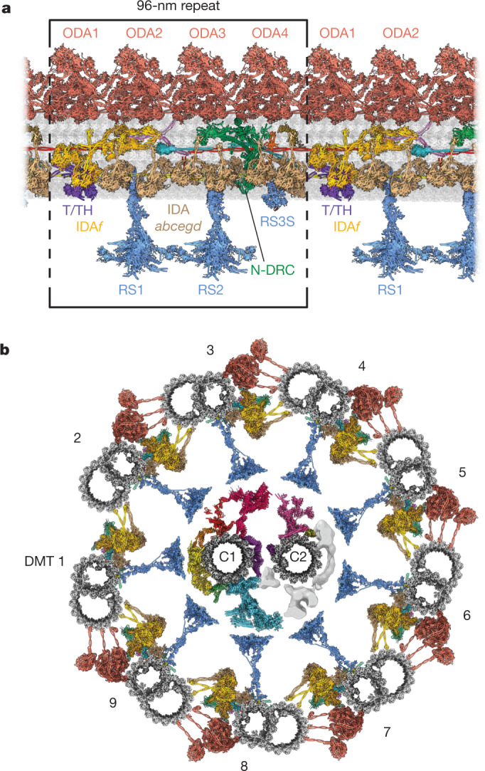
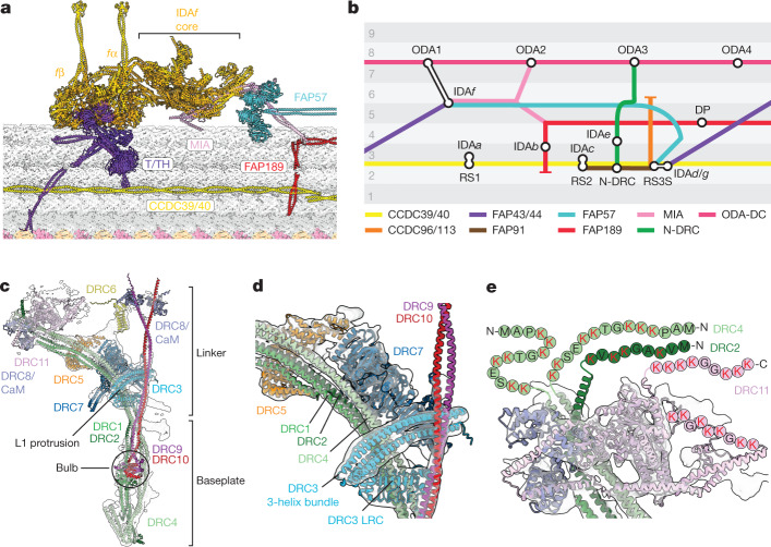
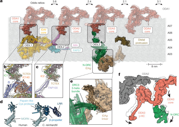
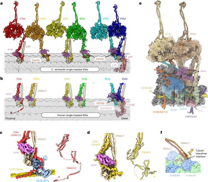
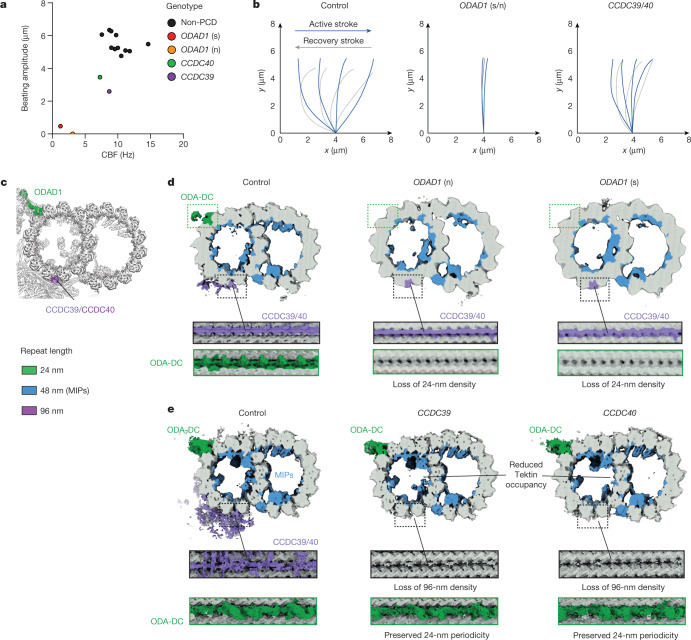
Motile cilia and flagella beat rhythmically on the surface of cells to power the flow of fluid and to enable spermatozoa and unicellular eukaryotes to swim. In humans, defective ciliary motility can lead to male infertility and a congenital disorder called primary ciliary dyskinesia (PCD), in which impaired clearance of mucus by the cilia causes chronic respiratory infections1. Ciliary movement is generated by the axoneme, a molecular machine consisting of microtubules, ATP-powered dynein motors and regulatory complexes2. The size and complexity of the axoneme has so far prevented the development of an atomic model, hindering efforts to understand how it functions. Here we capitalize on recent developments in artificial intelligence-enabled structure prediction and cryo-electron microscopy (cryo-EM) to determine the structure of the 96-nm modular repeats of axonemes from the flagella of the alga Chlamydomonas reinhardtii and human respiratory cilia. Our atomic models provide insights into the conservation and specialization of axonemes, the interconnectivity between dyneins and their regulators, and the mechanisms that maintain axonemal periodicity. Correlated conformational changes in mechanoregulatory complexes with their associated axonemal dynein motors provide a mechanism for the long-hypothesized mechanotransduction pathway to regulate ciliary motility. Structures of respiratory-cilia doublet microtubules from four individuals with PCD reveal how the loss of individual docking factors can selectively eradicate periodically repeating structures.


Conflict of interest statement

The authors declare no competing interests.

Figures

Fig. 1
Fig. 1. Atomic model of the modular repeat of the C. reinhardtii axoneme.
a, A longitudinal section of an atomic model of a fully decorated DMT showing the 96-nm repeat boxed. b, Cross-sectional model of the C.reinhardtii axoneme composed of nine copies of the 96-nm repeat revealed by our study and the C1 and C2 microtubules of the central apparatus (PDB: 7SQC and 7SOM). The model combines data from multiple sources and provides a consensus view that does not fully capture the asymmetries observed in vivo.
Fig. 2
Fig. 2. Structures of mechanoregulatory complexes from the C. reinhardtii axoneme.
a, Atomic model depicting the multipartite tethering of IDAf by the MIA complex (FAP73/100), a FAP57 homodimer and T/TH. Coiled coils of CCDC39/40 and FAP189 interact with T/TH and MIA, respectively. b, A conceptual schematic, in the style of a London Underground map, showing the interconnectivity between axonemal complexes in a 96-nm repeat. Circles represent axonemal complexes, coloured lines represent the path of docking factors and alternating grey zones represent protofilaments. DP, distal protrusion. c, Composite cryo-EM map and model of the N-DRC. CaM, calmodulin. d, The position of DRC3 at a fork in the N-DRC structure. e, DRC2, DRC4 and DRC11 position flexible, lysine-rich regions towards the neighbouring DMT. Individual letters indicate amino acids.
Fig. 3
Fig. 3. Regulatory complexes modulate inter-dynein interactions.
a, Selected atomic models and their cryo-EM densities showing the longitudinal arrangement of ODA1–ODA4 and their adjacent complexes on the axoneme. Each ODA exists in open and closed conformations, as revealed by 3D classification of the cryo-EM dataset. Odds ratios for the co-occurrence of open and closed states between ODA pairs are shown above the bidirectional arrows. Odds ratios were calculated using 160,444 particles for ODA1, 162,179 particles for ODA2, 214,203 particles for ODA3 and 177,762 particles for ODA4. Scale bar, 8 nm (the length of a tubulin heterodimer) with microtubule polarity indicated. b, OIDL1 between the TCTEX of ODA1 and IDAf subunits IC140 and fβ. c, OIDL2 between the TCTEX of ODA2 and MIA subunit FAP100. d, Models of human (left) and C.reinhardtii (right) DRC7 with the domains coloured in different shades of blue. Human DRC7 lacks the β-propeller and LRR domains that mediate OIDL3 in C.reinhardtii. MORN, membrane occupation and recognition nexus. e, Interaction between the three-helix bundle of DRC3 and the linker of IDAg. f, Overlay of the two major conformational states of N-DRC. Arrows denote movement from the closed to the open state.
Fig. 4
Fig. 4. Atomic model of the 96-nm repeat of a human axoneme.
a, Longitudinal section of the 96-nm repeat of a human respiratory-cilia DMT. b, Cross-section of a human DMT. c, Atomic model of human RSs coloured by subunit. The inset shows the symmetric head of RS1 viewed from above. Only the composition of the base of RS3 is known; the rest of the complex is shown as a density map in a and c.
Fig. 5
Fig. 5. Species-specific compositions and positions of single-headed IDAs.
a, Atomic model of the six single-headed IDAs as they appear on the C.reinhardtii DMT. Each IDA is associated with actin and either centrin or a p28 homodimer; p38 and p44 are specific docking factors for IDAd. b, Cropped image showing the DMT-bound bases of the human single-headed IDAs. Compared with those of C.reinhardtii, IDAb and IDAe have different subunit compositions and docking positions. c,d, Cryo-EM density of the dynein tail domains was used to identify the HC of IDAa as DNAH12 (c) and that of IDAc as DNAH3 (d). e, Cryo-EM map and model showing the molecular environment of IDAg and IDAd and its connection with RS3S in C.reinhardtii. A similar arrangement is found at the base of human RS3. f, The DNALI1 homodimer of human IDAe interacts directly with the tubulin interdimer interface. Similar interactions with tubulin are observed for human IDAa, IDAb and IDAc and their equivalents in C.reinhardtii (through the equivalent p28 homodimer).
Fig. 6
Fig. 6. Structures of DMTs from individuals with PCD.
a, Beating amplitude is plotted against ciliary beat frequency (CBF) for primary brushing samples from human nasal epithelia for 11 people without PCD and patients with PCD carrying biallelic ODAD1 (splice site (s) and nonsense (n)), CCDC39 and CCDC40 mutations. b, Schematic models of the corresponding ciliary waveforms observed in primary nasal brushings of patients with PCD and people without PCD (control). c, Cross-section of the atomic model of the human 96-nm DMT repeat depicting the position of ODAD1 and the CCDC39/40 coiled coil. The proteins are coloured according to their periodicity in the axoneme. d,e, Cross and longitudinal sections through cryo-EM maps reconstructed from DMTs isolated from pseudostratified respiratory epithelia cultured from either a healthy individual (control) or nasal brushings from patients with PCD. Map densities are coloured according to the repeat length with tubulin shown in grey. d, Mutations in ODAD1 cause complete loss of the ODA-DC (green), which repeats with 24-nm periodicity. e, Mutations in CCDC39 and CCDC40 cause complete loss of the 96-nm repeat (mauve). Apart from tektins, which had reduced occupancy, most MIPs (blue) are unaffected. Control maps are reconstructed from similar numbers of particles at a similar resolution and periodicity to the mutant maps for direct comparison.
Extended Data Fig. 1
Extended Data Fig. 1. Structural comparison of inner dynein arm f (IDAf) and outer dynein arm (ODA) from C. reinhardtii.
a, Atomic model showing outer dynein arm 1 (ODA1) and inner dynein arm f (IDAf) bound to a doublet microtubule (DMT, gray). Dynein subunits are colored based upon their structural or functional similarity and listed in the table. Non-shared subunits are labeled. All other external complexes have been removed for clarity. b, Comparison of the cores of ODA (left) and IDAf (right). Paralogous subunits in these two structures occupy similar positions. IDAf-specific subunits IC97 and FAP120 bind the LC tower, whereas the ODA-specific LC4 binds γ-HC. c, Comparison of motor domain orientation in IDAf and ODA. The ODA, faded and outlined by a dashed line, is overlaid by IDAf. The atomic models were aligned by superposing their cores. α-HC of ODA is omitted for clarity. Compared to ODA, the helical bundles of IDAf between the core region and the motors are less curled, resulting in rotation of the motors by 45–65° and displacement by 10 nm. These conformations differ from the active state of cytoplasmic dynein, suggesting that the connection between the core and the motor may help specialize each dynein for distinct roles.
Extended Data Fig. 2
Extended Data Fig. 2. The C. reinhardtii MIA complex contributes to the docking of inner dynein arm f (IDAf) to the doublet microtubule (DMT).
a, MIA (a heterodimer of FAP73/FAP100) interacts with protofilament A05 of the DMT through a C-terminal helix of FAP100 beneath the fβ heavy chain tail. Unmodeled density of FAP100 is also shown. b, The N-terminus of the MIA coiled coil interacts with a FAP189 family member near the site of its 90° bend on the DMT surface. c, Comparison of the binding of docking complex subunits DC1/2 to ODA and MIA to IDAf. Both coiled coils bisect the helical bundles of the dynein heavy chains within the dynein cores. d, Cryo-EM map showing the interaction between the fα tail, MIA, and the β-propeller domains of a FAP57 homodimer.
Extended Data Fig. 3
Extended Data Fig. 3. Structure of the C. reinhardtii tether/tetherhead (T/TH) complex.
a, Composite cryo-EM map colored by T/TH subunit and the motor domains of inner dynein arm (IDA) fα and fβ. The doublet microtubule surface is shown in gray. The N-terminal pair of β-propellers of FAP44 interact with the AAA1, AAA6, and C-terminal domains of the fα motor. b, Domain organization of T/TH subunits FAP43, FAP44, and MOT7. Both FAP43 and FAP44 contain an N-terminal pair of β-propellers, followed by a helical stellate structure, and a microtubule-associated coiled coil (CC). MOT7 contains a BLUF domain that is proposed to sense blue light. c, Schematic illustrating the six-armed stellate structure of T/TH with MOT7 at the center. d, Reverse side of the stellate showing the position of MOT7 and its BLUF domain (bright red) within the cryo-EM map. The resolution is too low to observe any flavin cofactors. On the left, the N-terminal pair of β-propellers from FAP43 bind arm 2 of the stellate. e, Interaction of the FAP43/44 coiled coil with the inner junction proteins FAP20 and PACRG. Bulky sidechain residues used for identification are labeled.
Extended Data Fig. 4
Extended Data Fig. 4. Integrative modeling of the nexin-dynein regulatory complex (N-DRC) from C. reinhardtii and H. sapiens.
a, Domain architecture of C. reinhardtii N-DRC subunits. b, Architecture of the N-DRC “bulb” in C. reinhardtii. c, Model of the N-DRC linker in the cryo-ET subtomogram average of the wildtype C. reinhartii axoneme (EMD-20338). Small differences between our model and the cryo-ET map are likely due to the loss of interactions with the neighboring doublet microtubule (DMT) in our splayed axoneme cryo-EM sample. d, Model of the N-DRC linker in the subtomogram average of the drc7 mutant axoneme (EMD-20339). The DRC7 subunit resides outside of the cryo-ET density, consistent with the loss of DRC7 in the mutant axonemes. Reduced density of the distal lobe, containing the C-termini of DRC9 and 10, suggest an altered or weakened interaction between DRC9/10 and the neighboring DMT in this mutant. e, Model of the N-DRC linker in the subtomogram average of the drc11 mutant axoneme (EMD-20340). Reduced density of the proximal lobe is consistent with the position of DRC11. f, Orthogonal views of the atomic model of human N-DRC. The bulb of human N-DRC (DRC1/9/10) is uncoupled from the baseplate unlike in C. reinhardtii. g, Interaction between the bulb and the N-terminal tail domain of DNAH7 (IDAe). h, Interaction between the 3-helix bundle of DRC3 and the linker of DNAH6 (IDAg).
Extended Data Fig. 5
Extended Data Fig. 5. Identification of FAP78 as the distal protrusion (DP) of the C. reinhardtii axoneme.
a, Domain architecture of FAP78. FAP78 contains 16 armadillo (ARM) repeats interrupted with a helical/unstructured region. The C-terminal half contains a microtubule (MT) binding domain and an EF-hand like domain, which is not resolved in our cryo-EM map. b, Cryo-EM map of the DP colored by domain. c, Atomic model of FAP78. d, Sidechain density of a section of FAP78 boxed in panel c. e, The FAP78 model positioned within the subtomogram average of the C. reinhardtii axoneme (EMD-6872). The height of the DP is 155 Å in this map. FAP78 is located within 3–5 nm of IDAf and ODA4, suggesting a possible role in dynein regulation, although we do not observe direct interactions. f, The DP is absent from axonemes from human respiratory cilia (EMD-5950). The expected location is denoted by a dashed oval. The subtomogram averages in panels e and f are colored according to the color scheme established in Fig. 1. g, Phylogenetic distribution of FAP78 is limited to green algae and other single-celled ciliates. The homolog in T. thermophila only aligns to the C-terminal half of FAP78 and does not contain ARM repeats, which likely explains why a distal protrusion is not observed in subtomogram averages of T. thermophila axonemes (EMD-12119).
Extended Data Fig. 6
Extended Data Fig. 6. Radial spoke 3 (RS3).
a, Model and cryo-EM map showing the base of human RS3 and its association with IDAd/g. Coiled coils from CCDC96/113 and CFAP57 are docking factors for RS3. CFAP43/44 (components of the T/TH complex) are docking factors for IDAd/g. b, Model and cryo-EM map of C. reinhardtii RS3S. c, Domain organization of RS3S subunits. The boxes on FAP91 indicate consecutive binding sites for RS2, the N-DRC and RS3S. Both the oxidoreductase-like domain of FAP61 and the calmodulin (CaM)-like domain of FAP251 interact with FAP91. d, Atomic model of the docking factors for RS3S in C. reinhardtii.
Extended Data Fig. 7
Extended Data Fig. 7. Primary ciliary dyskinesia (PCD) patient information.
a, Integrative Genomics Viewer (IGV) snapshot illustrating the homozygous exon 1–3 deletion in the CCDC40 gene of patient ID03. b, Analysis of high-speed video data from PCD patients showing defects in epithelia consistency, cilia beat frequency, beat angle, and amplitude compared to healthy, non-PCD controls. The box represents the interquartile range, the center represents the median, and the whiskers represent minimum and maximum measurements. Statistical significance was determined by one-way ANOVA (non-parametric Kruskal-Wallis), with * P ≤ 0.05, ** P ≤ 0.01, *** P ≤ 0.001 and **** P ≤ 0.0001. The number of movies used for the analysis were 104 (control); 3 (ODAD1 s); 8 (ODAD1 n); 8 (CCDC40), and 6 (CCDC39). For each mutation, the movies came from the same patient. ODAD1 (s) refers to individual ID01 with a splice mutation, and ODAD1 (n) refers to individual ID02 with a nonsense mutation in ODAD1. c, Representative clinical transmission electron microscopy (TEM) images selected from a minimum of >100 screened cross-sections showing axoneme cross sections from a non-PCD control (with axonemal complexes labeled), two individuals with mutations in ODAD1 (ID01 and ID02), an individual with a mutation in CCDC40 (ID03), and an individual with a mutation in CCDC39 (ID04). Red arrows mark the absence of outer dynein arms (ODAs). Orange arrows mark microtubule (MT) transposition. Yellow arrows mark the absence of inner dynein arms (IDAs). Purple arrows mark an abnormal central apparatus (CA). d, Compared to a non-PCD control, radial spokes (blue arrows) attached to doublet microtubules in individual ID03 (with a CCDC40 mutation) are intermittently bound, consistent with the loss of strict 96-nm periodicity. These micrographs sections were selected from a total of 37,071 non-PCD control micrographs, and 3,483 CCDC40 micrographs. Source data
Extended Data Fig. 8
Extended Data Fig. 8. Coiled-coil docking factors potentially contribute to axonemal asymmetry.
a, Atomic models of the FAP189 and FAP57 homodimers. Other axonemal complexes are omitted for clarity. FAP189 forms an L-shaped coiled coil that occupies the interprotofilament cleft between protofilaments A04 and A05. b, Fit of FAP189 atomic model into the cryo-EM map. Labeled amino acids are denoted below by bold font in the multiple sequence alignment of FAP189 with its paralogs FAP58 and MBO2. The conserved sequence ALSXXLENP, highlighted in red, corresponds to residues that interact with FAP100 of the MIA complex (see Extended Data Fig. 2b). c, Agreement of cryo-EM sidechain density with the atomic model of FAP57. Residues labeled in the model are denoted by bold font in the multiple sequence alignment below. d, Domain architecture of FAP189, FAP58, and MBO2. e, Phylogenetic tree showing the relationship between FAP189, FAP58, and MBO2. FAP189 and FAP58 share 80% sequence identity. f, Domain architecture of FAP57, FBB7 and FAP331, which are composed of tandem N-terminal β-propellers and a long, helical C-terminal region. g, Phylogenetic tree showing the relationship between FAP57, FBB7, and FAP331.

References

    1. Legendre M, Zaragosi L-E, Mitchison HM. Motile cilia and airway disease. Semin. Cell Dev. Biol. 2021;110:19–33. doi: 10.1016/j.semcdb.2020.11.007. - DOI - PubMed
    1. Ishikawa T. Axoneme structure from motile cilia. Cold Spring Harb. Perspect. Biol. 2017;9:a028076. doi: 10.1101/cshperspect.a028076. - DOI - PMC - PubMed
    1. Bui KH, Yagi T, Yamamoto R, Kamiya R, Ishikawa T. Polarity and asymmetry in the arrangement of dynein and related structures in the Chlamydomonas axoneme. J. Cell Biol. 2012;198:913–925. doi: 10.1083/jcb.201201120. - DOI - PMC - PubMed
    1. Kubo S, et al. Remodeling and activation mechanisms of outer arm dyneins revealed by cryo-EM. EMBO Rep. 2021;22:e52911. doi: 10.15252/embr.202152911. - DOI - PMC - PubMed
    1. Walton T, Wu H, Brown A. Structure of a microtubule-bound axonemal dynein. Nat. Commun. 2021;12:477. doi: 10.1038/s41467-020-20735-7. - DOI - PMC - PubMed

Publication types

MeSH terms

Substances

LinkOut - more resources