. 2023 May 31;11(1):86.
doi: 10.1186/s40478-023-01579-w.

Astrocytic pathology in Alpers' syndrome

Affiliations

Astrocytic pathology in Alpers' syndrome

Laura A Smith et al. Acta Neuropathol Commun. .

Abstract

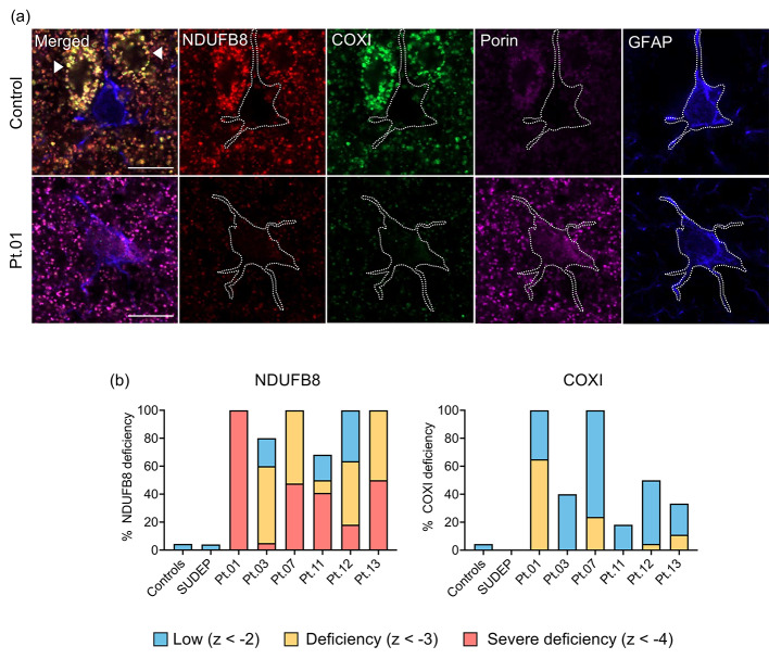
Refractory epilepsy is the main neurological manifestation of Alpers' syndrome, a severe childhood-onset mitochondrial disease caused by bi-allelic pathogenic variants in the mitochondrial DNA (mtDNA) polymerase gamma gene (POLG). The pathophysiological mechanisms underpinning neuronal hyperexcitabilty leading to seizures in Alpers' syndrome remain unknown. However, pathological changes to reactive astrocytes are hypothesised to exacerbate neural dysfunction and seizure-associated cortical activity in POLG-related disease. Therefore, we sought to phenotypically characterise astrocytic pathology in Alpers' syndrome. We performed a detailed quantitative investigation of reactive astrocytes in post-mortem neocortical tissues from thirteen patients with Alpers' syndrome, eight neurologically normal controls and five sudden unexpected death in epilepsy (SUDEP) patients, to control for generalised epilepsy-associated astrocytic pathology. Immunohistochemistry to identify glial fibrillary acidic protein (GFAP)-reactive astrocytes revealed striking reactive astrogliosis localised to the primary visual cortex of Alpers' syndrome tissues, characterised by abnormal-appearing hypertrophic astrocytes. Phenotypic characterisation of individual GFAP-reactive astrocytes demonstrated decreased abundance of mitochondrial oxidative phosphorylation (OXPHOS) proteins and altered expression of key astrocytic proteins including Kir4.1 (subunit of the inwardly rectifying K+ ion channel), AQP4 (astrocytic water channel) and glutamine synthetase (enzyme that metabolises glutamate). These phenotypic astrocytic changes were typically different from the pathology observed in SUDEP tissues, suggesting alternative mechanisms of astrocytic dysfunction between these epilepsies. Crucially, our findings provide further evidence of occipital lobe involvement in Alpers' syndrome and support the involvement of reactive astrocytes in the pathogenesis of POLG-related disease.

Keywords: Alpers’ syndrome; Aquaporin 4 (AQP4); GFAP; Glutamine synthetase (GS); Kir4.1; Mitochondrial Epilepsy; POLG; Reactive astrogliosis.


Conflict of interest statement

The authors declare that they have no competing interests.

Figures

Fig. 1
Fig. 1
Reactive astrogliosis in Alpers’ syndrome. (a) Increased intensity and distribution of glial fibrillary acidic protein (GFAP) + labelling in occipital cortex tissues from patients with Alpers’ syndrome (Pt.11 and Pt.13) relative to controls and SUDEP patients. GFAP + labelling is intense in the necrotic lesion of Pt.11 (red arrow head), and shows selective cortical layer involvement in adjacent preserved tissue (blue arrow head). Hypertrophic ameboid GFAP + astrocytes are abundant across all cortical layers of Pt.13. Scale bars = 100 μm. (b) Quantitative analysis of the percentage area of cortical GFAP + staining revealed significantly increased GFAP + labelling in multiple Alpers’ syndrome patient tissues compared to controls (N = 8) [40]. Data presented as mean + SD. Multiple comparison analyses relative to control data: ** P < 0.01, *** P < 0.001. (c) Analyses assessing the correlation between the percentage area of GFAP + labelling with the density of interneuron subtypes (parvalbumin+, calretinin+, calbindin + and somatostatin+) and pyramidal neurons (SMI-32+), expressed as z-scores, using our published neuronal density data set [19]. Legend = Spearman-rank r value. * P < 0.05, ** P < 0.01
Fig. 2
Fig. 2
Hypertrophic astrocytes in Alpers’ syndrome. (a) The mean (+ SD) area (µm2) and volume (µm3) of individual two-dimensional (2-D) and three-dimensional (3-D) GFAP + astrocytes, respectively, are increased in occipital cortex tissues from patients with Alpers’ syndrome relative to control astrocytes (N = 5 control cases). Dotted line indicates the mean area and volume of control astrocytes. Random individual 2-D and 3-D astrocytes were imaged for analysis. Multiple comparison analyses relative to control data: * P < 0.05, ** P < 0.01, **** P < 0.0001. (b) Hypertrophic astrocytes identified based on GFAP + staining of Patient 13 are presented. Scale bars = 10 μm
Fig. 3
Fig. 3
Oxidative phosphorylation protein deficiencies in Alpers’ syndrome patient astrocytes. (a) Quadruple immunofluorescence assay revealed decreased protein abundance of NDUFB8 (complex I subunit) and COXI (complex IV subunit) within GFAP + astrocytes in occipital cortex tissues from patients with Alpers’ syndrome relative to control astrocytes, despite increased porin signal (mitochondrial marker). Note astrocytes demonstrate weak immunoreactivity of all mitochondrial markers compared to adjacent neurons (arrow head). Scale bars = 10 μm. (b) Graphs demonstrate the percentage of GFAP + astrocytes with decreased mean optical intensity of NDUFB8 and COXI normalised to porin, relative to control astrocytes. Levels of deficiencies are based on standard deviation limits of control data: z-score < -2 = low expression; z-score < -3 = deficient; z-score < -4 = severely deficient [45]. N = 5 Controls, N = 5 SUDEP cases
Fig. 4
Fig. 4
Altered expression of Kir4.1, AQP4 and glutamine synthetase in Alpers’ syndrome patient astrocytes. (a) Immunofluorescence to identify Kir4.1 (purple), (b) AQP4 (orange) and (c) glutamine synthetase (red), using GFAP+ (green) and Hoescht (blue, nuclear marker) to identify individual reactive astrocytes in the occipital cortex. Scale bars = 10 μm. The mean optical intensity of Kir4.1, AQP4 and glutamine synthetase within individual GFAP + astrocytes are presented as circles. For Pt.11 – Pt.13 occipital cortex data, orange circles denote astrocytes imaged in lesioned cortex, and red circles denote astrocytes imaged in adjacent non-lesioned cortex. Box plots demonstrate the median, upper/lower quartiles and range of data. Multiple comparison analyses relative to control data: *P < 0.05, ** P < 0.01, *** P < 0.001, **** P < 0.0001. Note: Kir4.1 intensity was selectively increased in astrocytes from lesioned cortex of Pt.13 (P < 0.0001). N = 5 controls, N = 5 SUDEP

Similar articles

Cited by

References

    1. Ropp PA, Copeland WC. Cloning and characterization of the human mitochondrial DNA polymerase, DNA polymerase gamma. Genomics. 1996;36(3):449–458. - PubMed
    1. Naviaux RK, Nguyen KV. POLG mutations associated with Alpers syndrome and mitochondrial DNA depletion. Ann Neurol. 2004;55(5):706–712. - PubMed
    1. Lim A, Thomas RH. The mitochondrial epilepsies. Eur J Paediatr Neurol. 2020;24:47–52. - PubMed
    1. Copeland WC, Human DNA Polymerase Gamma Mutation Database [Available from: https://tools.niehs.nih.gov/polg/index.cfm/main/home/limitresults/false
    1. Naviaux RK, Nyhan WL, Barshop BA, Poulton J, Markusic D, Karpinski NC, Haas RH. Mitochondrial DNA polymerase Gamma Deficiency and mtDNA Depletion in a child with Alpers’ syndrome. Annu Neurol. 1999;45:54–58. - PubMed

Publication types

MeSH terms

Substances