

Association of perivascular fat attenuation on computed tomography and heart failure with preserved ejection fraction
- PMID: 37259241
- PMCID: PMC10375150
- DOI: 10.1002/ehf2.14419
Association of perivascular fat attenuation on computed tomography and heart failure with preserved ejection fraction
Abstract
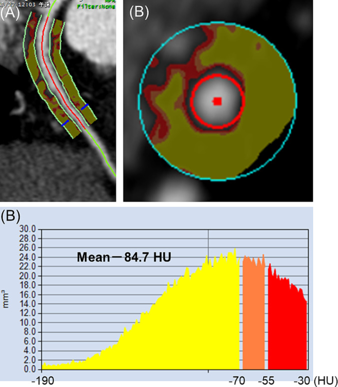
Aims: Heart failure with a preserved ejection fraction (HFpEF) is associated with chronic inflammation. We aimed to investigate the association between pericoronary adipose tissue attenuation (PCATA) on coronary computed tomography angiography as a novel noninvasive marker of pericoronary inflammation and the presence of HFpEF.
Methods and results: This retrospective study included 607 outpatients (median age, 65 years; 50% male) who underwent both echocardiography and coronary computed tomography angiography. Patients with obstructive coronary artery disease were excluded from this study. PCATA was compared between patients with and without HFpEF, which was diagnosed according to the Heart Failure Association (HFA)-PEFF score. PCATA was assessed at the proximal 40-mm segments of all three major coronary arteries on coronary computed tomography angiography. Patients with HFpEF had higher PCATA in all coronary arteries compared to the control participants: left anterior descending artery (LAD), -65.2 ± 6.9 Hounsfield units (HU) vs. -68.1 ± 6.7 HU; left circumflex artery (LCX), -62.7 ± 6.8 HU vs. -65.4 ± 6.6 HU; and right coronary artery (RCA), -63.6 ± 8.5 HU vs. -65.5 ± 7.7 HU (P < 0.01). Multivariate logistic regression analysis, including conventional risk factors, revealed that PCATA per standard deviation in the LAD (odds ratio [OR], 1.449; 95% confidence interval [CI], 1.152-1.823), LCX (OR, 1.634; 95% CI, 1.283-2.081), and RCA (OR, 1.388; 95% CI, 1.107-1.740) were independently associated with HFpEF. The association between PCATA and HFpEF was mostly consistent across various patient clinical characteristics. The left ventricular mass and left atrial volume index showed a mild correlation with LAD-PCATA (ρ = 0.13 [P < 0.01] and ρ = 0.24 [P < 0.01]) and LCX-PCATA (ρ = 0.16 [P < 0.01] and ρ = 0.23 [P < 0.01]).
Conclusions: High PCATA score was significantly associated with the presence of HFpEF. Our results suggest that inflammation in the pericoronary artery adipose tissue is one of the underlying mechanisms of HFpEF.
Keywords: Adipose tissue; Computed tomography; Coronary artery; Heart failure; Inflammation.
© 2023 The Authors. ESC Heart Failure published by John Wiley & Sons Ltd on behalf of European Society of Cardiology.
Conflict of interest statement
None declared.
Figures


Similar articles
-
The prognostic value of epicardial and pericoronary adipose tissue in heart failure with preserved ejection fraction using coronary computed tomography angiography.Br J Radiol. 2025 Feb 1;98(1166):229-236. doi: 10.1093/bjr/tqae216. Br J Radiol. 2025. PMID: 39454024
-
Pericoronary adipose tissue attenuation in patients with acute aortic dissection based on coronary computed tomography angiography.Quant Imaging Med Surg. 2024 Jan 3;14(1):31-42. doi: 10.21037/qims-23-253. Epub 2023 Nov 23. Quant Imaging Med Surg. 2024. PMID: 38223036 Free PMC article.
-
Determinants of Pericoronary Adipose Tissue Attenuation on Computed Tomography Angiography in Coronary Artery Disease.J Am Heart Assoc. 2020 Aug 4;9(15):e016202. doi: 10.1161/JAHA.120.016202. Epub 2020 Jul 30. J Am Heart Assoc. 2020. PMID: 32750306 Free PMC article.
-
Drugs That Ameliorate Epicardial Adipose Tissue Inflammation May Have Discordant Effects in Heart Failure With a Preserved Ejection Fraction as Compared With a Reduced Ejection Fraction.J Card Fail. 2019 Dec;25(12):986-1003. doi: 10.1016/j.cardfail.2019.09.002. Epub 2019 Sep 18. J Card Fail. 2019. PMID: 31541742 Review.
-
Menopause, epicardial adiposity and preserved ejection fraction heart failure.Int J Cardiol. 2024 Nov 15;415:132478. doi: 10.1016/j.ijcard.2024.132478. Epub 2024 Aug 22. Int J Cardiol. 2024. PMID: 39179034 Review.
Cited by
-
Impact of pericoronary adipose tissue attenuation and coronary microvascular dysfunction on cardiac remodeling and dysfunction.Heart Vessels. 2025 Jul 14. doi: 10.1007/s00380-025-02576-w. Online ahead of print. Heart Vessels. 2025. PMID: 40659864
-
Lower estimates of myocardial perfusion are associated with greater aortic perivascular adipose tissue density in humans independent of aortic stiffness.Am J Physiol Heart Circ Physiol. 2024 Oct 1;327(4):H927-H934. doi: 10.1152/ajpheart.00436.2024. Epub 2024 Aug 16. Am J Physiol Heart Circ Physiol. 2024. PMID: 39150391
-
Computed tomography-based assessment of pericoronary adipose tissue in cardiovascular diseases: Diagnostic and prognostic implications.World J Radiol. 2025 Jun 28;17(6):107281. doi: 10.4329/wjr.v17.i6.107281. World J Radiol. 2025. PMID: 40606053 Free PMC article. Review.
-
Coronary computed tomography angiography for clinical practice.Jpn J Radiol. 2024 Jun;42(6):555-580. doi: 10.1007/s11604-024-01543-1. Epub 2024 Mar 8. Jpn J Radiol. 2024. PMID: 38453814 Free PMC article. Review.
-
Association of mean pericoronary adipose tissue attenuation with different demographic factors in a subgroup of patients without coronary artery disease stratified by sex, body mass index, and age.Quant Imaging Med Surg. 2024 Jan 3;14(1):503-513. doi: 10.21037/qims-23-951. Epub 2024 Jan 2. Quant Imaging Med Surg. 2024. PMID: 38223068 Free PMC article.
References
Publication types
MeSH terms
LinkOut - more resources
Full Text Sources
Medical
