. 2023 Jan 23;16(2):164.
doi: 10.3390/ph16020164.

Topical Administration of a Nanoformulation of Chitosan-Hyaluronic Acid-Epoetin Beta in a Rat Model of Glaucoma

Affiliations

Topical Administration of a Nanoformulation of Chitosan-Hyaluronic Acid-Epoetin Beta in a Rat Model of Glaucoma

Beatriz Silva et al. Pharmaceuticals (Basel). .

Abstract

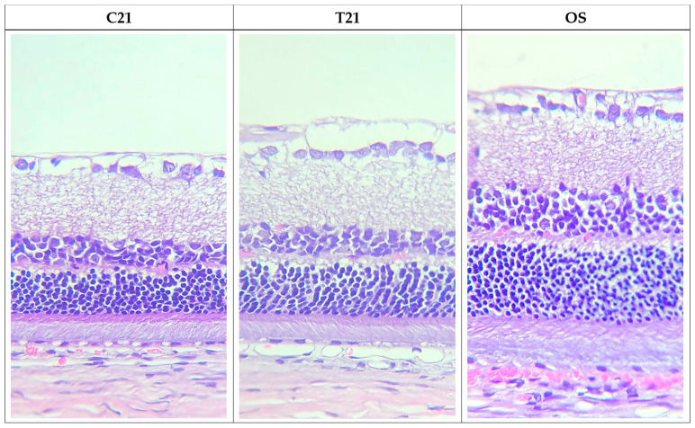
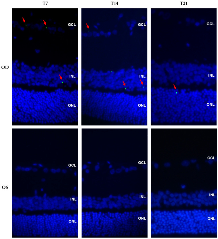
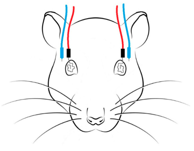
The present work investigates the effects of chitosan-hyaluronic acid-epoetin beta (CS/HA-EPOβ) nanoparticles after topical ocular administration in a rat glaucoma model. Wistar Hannover rats (n = 24) were submitted to a complete ophthalmological examination and electroretinography, followed by glaucoma induction in their right eye on day 1 of the study. Treatment group (T) received CS/HA-EPOβ nanocarriers (n = 12), while the control group (C) received only empty ones. Electroretinography was repeated on day 3 (n = 24) and before euthanasia on day 7 (n = 8), 14 (n = 8), and 21 (n = 8), followed by bilateral enucleation and histological assessment. The animals showed good tolerance to the nanoformulation. Maximum IOP values on the right eye occurred shortly after glaucoma induction (T = 62.6 ± 8.3 mmHg; C = 63.6 ± 7.9 mmHg). Animals from the treated group presented a tendency for faster recovery of retinal electrical activity (p > 0.05). EPOβ was detected on the retina of all treated eyes using immunofluorescence. Control animals presented with thinner retinas compared to the treated ones (p < 0.05). Therefore, topical ocular administration of CS/HA-EPOβ nanoparticles enabled EPOβ delivery to the retina of glaucomatous rats and promoted an earlier retinal recovery, confirming EPOβ's neuroprotective effects. The encouraging results of this preclinical study pave the way for new strategies for topical ocular administration of neuroprotective compounds.

Keywords: chitosan; epoetin beta; glaucoma; hyaluronic acid; nanoparticles; neuroprotection; ocular delivery.


Conflict of interest statement

The authors declare no conflict of interest.

Figures

Figure 1
Figure 1
Pictures of a rat 1 h (a) and 3 days (b) after the cauterization of the 3 episcleral veins of the OD. The animal shows symmetry between both ocular globes and no signs of ocular discomfort.
Figure 2
Figure 2
Mean IOP from the OD (orange) and from the OS (blue) since the day of glaucoma induction (t = 0) until 21 days later, in the treatment (a) and the control (b) groups. Time is presented in days in the X axis and IOP is presented in mmHg in the Y axis.
Figure 2
Figure 2
Mean IOP from the OD (orange) and from the OS (blue) since the day of glaucoma induction (t = 0) until 21 days later, in the treatment (a) and the control (b) groups. Time is presented in days in the X axis and IOP is presented in mmHg in the Y axis.
Figure 3
Figure 3
Example of ERG waveforms recorded from the OD at the beginning of the study (black) and at day 3 after the glaucoma induction (orange) in an animal from the control group. a, a-wave; b, b-wave; N, zero/basis; SLR, scotopic luminescence response; PA, photopic adaptation; PLR, photopic luminescence response; PF, photopic flicker; SA, scotopic adaptation; dB, decibel; min, minutes.
Figure 4
Figure 4
ERG waveforms of the OD (orange) and the OS (black) recorded at day 3 and day 21 after the glaucoma induction, from the treatment group. SLR, scotopic luminescence response; PF, photopic flicker; SA, scotopic adaptation; OD, right eye; OS, left eye.
Figure 5
Figure 5
Photomicrographs of retinal sections from the OD, 21 days after glaucoma induction, from the control group (C21) and treated group (T21), and from a non-glaucomatous eye (OS; n = 4). We can observe that the improvement in retinal thickness was more pronounced in the treated group. Hematoxylin and eosin staining, 40× magnification.
Figure 6
Figure 6
Immunofluorescence photomicrographs showing cross sections of the OD (treated eye) and OS (control eye) in the treatment groups T7, T14, and T21 (magnification 40×). Images show the merging of green and blue filters. Red arrows indicate EPOβ stained in green and cell nuclei are stained in blue with DAPI. GCL, ganglion cell layer; INL, inner nuclear layer; ONL, outer nuclear layer.
Figure 7
Figure 7
Representation of the ERG setup: active electrodes (red) with silver tips were placed on both corneas, and reference electrodes (blue) were placed subcutaneously between the ears and the lateral canthus ipsilateral to the tested eye.
Figure 8
Figure 8
Representation of a rat ocular globe: yellow arrows indicate the cauterized episcleral veins; (1) lateral vein, (2) dorso-lateral vein, (3) dorso-medial vein. Extraocular muscles: SR, superior rectus; SO, superior oblique; MR, medial rectus; IR, inferior rectus; IO, inferior oblique; LR, lateral rectus.
Figure 9
Figure 9
Photo of a rat ocular globe during the microsurgical procedure: (a) yellow arrow indicates a dorsal episcleral vein before conjunctiva incision; (b) yellow arrow indicates a lateral episcleral vein after the conjunctival incision.
Figure 10
Figure 10
Photo of a rat ocular globe during the microsurgical procedure: (a) before the cauterization of the dorsal episcleral vein, showing normal iris vascularization; (b) after the cauterization of the dorsal episcleral vein, showing an evident generalized iridal congestion (green arrow).
Figure 11
Figure 11
Representation of a rat ocular globe painted with tissue dyes: (a) frontal view; (b) caudal view. Photographs of a rat ocular globe painted with tissue dyes: (c) lateral view; (d) caudal view. In green we can identify the transected optic nerve. ON, optic nerve (previously published by our group in Silva et al., 2022 [11]).

Similar articles

Cited by

References

    1. Vidal-Sanz M., Salinas-Navarro M., Nadal-Nicolás F.M., Alarcón-Martínez L., Valiente-Soriano F.J., Miralles de Imperial J., Avilés-Trigueros M., Agudo-Barriuso M., Villegas-Pérez M.P. Understanding glaucomatous damage: Anatomical and functional data from ocular hypertensive rodent retinas. Prog. Retin. Eye Res. 2012;31:1–27. doi: 10.1016/j.preteyeres.2011.08.001. - DOI - PubMed
    1. Ghate D., Edelhauser H.F. Barriers to glaucoma drug delivery. J. Glaucoma. 2008;17:147–156. doi: 10.1097/IJG.0b013e31814b990d. - DOI - PubMed
    1. Pardue M.T., Allen R.S. Neuroprotective strategies for retinal disease. Prog. Retin. Eye Res. 2018;65:50–76. doi: 10.1016/j.preteyeres.2018.02.002. - DOI - PMC - PubMed
    1. Huang W., Gao F., Hu F., Huang J., Wang M., Xu P., Zhang R., Chen J., Sun X., Zhang S., et al. Asiatic acid prevents retinal ganglion cell apoptosis in a rat model of glaucoma. Front. Neurosci. 2018;12:489. doi: 10.3389/fnins.2018.00489. - DOI - PMC - PubMed
    1. Rhee J., Shih K.C. Use of gene therapy in retinal ganglion cell neuroprotection: Current concepts and future directions. Biomolecules. 2021;11:581. doi: 10.3390/biom11040581. - DOI - PMC - PubMed

LinkOut - more resources