Review
. 2023 Feb 7;16(2):249.
doi: 10.3390/ph16020249.

Smart Biomimetic Nanozymes for Precise Molecular Imaging: Application and Challenges

Affiliations
Review

Smart Biomimetic Nanozymes for Precise Molecular Imaging: Application and Challenges

Qiao Luo et al. Pharmaceuticals (Basel). .

Abstract

New nanotechnologies for imaging molecules are widely being applied to visualize the expression of specific molecules (e.g., ions, biomarkers) for disease diagnosis. Among various nanoplatforms, nanozymes, which exhibit enzyme-like catalytic activities in vivo, have gained tremendously increasing attention in molecular imaging due to their unique properties such as diverse enzyme-mimicking activities, excellent biocompatibility, ease of surface tenability, and low cost. In addition, by integrating different nanoparticles with superparamagnetic, photoacoustic, fluorescence, and photothermal properties, the nanoenzymes are able to increase the imaging sensitivity and accuracy for better understanding the complexity and the biological process of disease. Moreover, these functions encourage the utilization of nanozymes as therapeutic agents to assist in treatment. In this review, we focus on the applications of nanozymes in molecular imaging and discuss the use of peroxidase (POD), oxidase (OXD), catalase (CAT), and superoxide dismutase (SOD) with different imaging modalities. Further, the applications of nanozymes for cancer treatment, bacterial infection, and inflammation image-guided therapy are discussed. Overall, this review aims to provide a complete reference for research in the interdisciplinary fields of nanotechnology and molecular imaging to promote the advancement and clinical translation of novel biomimetic nanozymes.

Keywords: magnetic resonance imaging; molecular imaging; multimodal imaging; nanozymes; photoacoustic imaging; positron emission tomography.


Conflict of interest statement

The authors declare no conflict of interest.

Figures

Scheme 1
Scheme 1
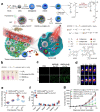
Schematic illustration of biomimetic nanozymes for precise molecular imaging.
Figure 1
Figure 1
Fe5C2@Fe3O4 NPs for MRI. (a) Schematic illustration of Fe5C2@Fe3O4 NPs with Ph-responsive Fe2+ release, ROS generation, and T2/T1 signal-conversion abilities. (b,c) pH-dependent MRI mode switching of PEG/Fe5C2@Fe3O4 nanoparticles. (d) Representative T2—weighted MR images. (e) Representative T1—weighted MR images. Reproduced with permission from Ref. [99]. Copyright © 2019 American Chemical Society. ** p<0.05
Figure 2
Figure 2
APMN NPs for MRI. (a) Schematic illustration of the main synthetic process of APMN NPs and the mechanism of this biomimetic theranostic nanoplatform for TME-responsive MR imaging and efficient induction of multimodal synergistic treatments. (be) MR images of APMN NPs under different conditions. (f) T1—weighted MR images of APMN NPs in cells. (g) T1—weighted MR images of MCF-7 tumor-bearing nude mice. Reproduced with permission from Ref. [100]. Copyright © 2022 Acta Materialia Inc. Published by Elsevier Ltd. All rights reserved.
Figure 3
Figure 3
Pt-CuS Janus for PAI. (a) Schematic illustration of the main synthesis procedures and antitumor mechanism of PCPT. (b) Photothermal stability of the Pt-CuS solution. (c) The production of 1O2 in the presence of PCPT (50 ppm) under different conditions. (d) The viability of CT26 cells after different treatments. (e) In vivo PA imaging of tumor-bearing mice. (f) In vitro PA imaging of different concentrations of Pt-CuS NPs. (g) Biodistribution of Pt in the tumor and main organs after injection of PCPT. (h) Tumor volume change curves during therapy. Reproduced with permission from Ref. [104]. Copyright © 2019 American Chemical Society.
Figure 4
Figure 4
AMP NRs for PAI. (a) Preparation procedure and schematic illustration of AMP NRs for tumor-microenvironment-activated nanozyme-mediated theranostics. (b) PAI signal intensity of AMP NRs with different concentrations of H2O2. (c) PAI signal intensity of AMP NRs at different pH values in the presence of 100 × 10−6 M H2O2. (d) Detection of ROS levels in 4T1 cells treated with different formulations. (e) CLSM images of 4T1 cells treated with AMP NPs (B@MIL-100/PVP). (f) Representative PAI images of tumors after different treatments. Reproduced with permission from Ref. [106]. Copyright © 2019 WILEY-VCH Verlag GmbH & Co. KGaA, Weinheim.
Figure 5
Figure 5
HSC-2 for NIR-II PA/NIR-II FL imaging. (a) Schematic illustration of adjustable photoacoustic/fluorescence image-guided photothermal/catalytic therapy in the NIR-II window. (b) POD-like activity and (c) CAT-like activity of HSC-2 in different systems over time. (d) PA images of tumors in mice at different time points after intratumoral injection of HSC-2 (808 nm laser (0.3 W cm−2) irradiation; 1064 nm). (e) NIR-II FL images of 4T1 tumor-bearing mice after systemic administration of HSC-2 (1000 LP and 100 ms). Reproduced with permission from Ref. [107]. Copyright © 2021 Wiley-VCH GmbH.
Figure 6
Figure 6
GNPs for FLI and infrared thermography. (a) Fabrication process of GNPs. (b) Schematic illustration of the catalytic activity of GNPs. (c,d) H2O2 content in tumor tissue determined with an H2O2 detection kit. Flow cytometry images of the pH probes BCECF-AM © and 2-NBDG (d) to determine the cellular pH value and glucose consumption, respectively. (e) Ex vivo fluorescence images of RC, IR@RC, and IRG@RC with/without laser irradiation. (f) In vivo fluorescence images of tumor-bearing mice. (g) Thermal imaging at the tumor site after laser irradiation by in vivo infrared thermography. Reproduced with permission from Ref. [108]. Copyright © 2022 The Authors. Published by Elsevier Ltd.
Figure 7
Figure 7
HMONs-Au-Col@64Cu-TA-PVP for PET. (a) Schematic showing the process of preparing the in situ polymerized hollow mesoporous organosilica biocatalytic nanoreactor for synergistic PDT/CDT. (b) Detection of H2O2 production after the indicated treatment detected with a hydrogen peroxide assay kit. (c) Fluorescence images of ROS generation by BxPC-3 cells after the indicated treatment in the culture medium with glucose. (d) Representative in vivo PET imaging of tumor-bearing mice. (e) Quantification of HMON-Au@64Cu-TA-PVP and HMON-Au-Col@64Cu-TA-PVP tumor uptake. (f) Quantification of the PET signal intensities in the main organs of tumor-bearing mice. (g) The average tumor growth curves after treatment. Reproduced with permission from Ref. [111]. Copyright © 2019 WILEY-VCH Verlag GmbH & Co. KGaA, Weinheim.
Figure 8
Figure 8
FeTIR for multimodal imaging. (a) Schematic illustration showing the preparation of oxygen-deficient bimetallic oxide FeWOX nanosheets as peroxidase-like nanozymes for sensing tumors via multimodal imaging. (b) Comparison of TMB oxidation by FeWOX-PEG, H2O2, and FeWOX-PEG/H2O2. (c) Fluorescence spectra obtained from different treatment groups containing H2O2, FeWOX-PEG, and FeWOX-PEG/H2O2. (d) In vitro ratiometric PA imaging at different concentrations of H2O2 (0–200 × 10−6 M) with the FeTIR nanoprobe. (e) In vivo ratiometric PA imaging of muscle and 4T1 tumor tissues of mice after injection of the FeTIR nanoprobe. (f). In vivo ratiometric PA imaging of inflammation sites in mice after FeTIR nanoprobe injection. Reproduced with permission from Ref. [115]. Copyright © 2020 Wiley-VCH GmbH.

References

    1. Massoud T.F., Gambhir S.S. Molecular Imaging in Living Subjects: Seeing Fundamental Biological Processes in a New Light. Genes Dev. 2003;17:545–580. doi: 10.1101/gad.1047403. - DOI - PubMed
    1. James M.L., Gambhir S.S. A Molecular Imaging Primer: Modalities, Imaging Agents, and Applications. Physiol. Rev. 2012;92:897–965. doi: 10.1152/physrev.00049.2010. - DOI - PubMed
    1. Böhmer V.I., Szymanski W., Feringa B.L., Elsinga P.H. Multivalent Probes in Molecular Imaging: Reality or Future? Trends Mol. Med. 2021;27:379–393. doi: 10.1016/j.molmed.2020.12.006. - DOI - PubMed
    1. van Duijnhoven S.M.J., Robillard M.S., Langereis S., Grüll H. Bioresponsive Probes for Molecular Imaging: Concepts and in Vivo Applications: Bioresponsive Molecular Imaging Probes. Contrast Media Mol. Imaging. 2015;10:282–308. doi: 10.1002/cmmi.1636. - DOI - PubMed
    1. Reynolds F., Kelly K.A. Techniques for Molecular Imaging Probe Design. Mol. Imaging. 2011;10:407–419. doi: 10.2310/7290.2011.00003. - DOI - PMC - PubMed

LinkOut - more resources