. 2023 Jul;43(7):1234-1250.
doi: 10.1161/ATVBAHA.122.318904. Epub 2023 Jun 1.

CD47 Activation by Thrombospondin-1 in Lymphatic Endothelial Cells Suppresses Lymphangiogenesis and Promotes Atherosclerosis

Affiliations

CD47 Activation by Thrombospondin-1 in Lymphatic Endothelial Cells Suppresses Lymphangiogenesis and Promotes Atherosclerosis

Bhupesh Singla et al. Arterioscler Thromb Vasc Biol. 2023 Jul.

Abstract

Background: TSP1 (thrombospondin-1)-a well-known angiogenesis inhibitor-mediates differential effects via interacting with cell surface receptors including CD36 (cluster of differentiation) and CD47. However, the role of TSP1 in regulating lymphangiogenesis is not clear. Our previous study suggested the importance of cell-specific CD47 blockade in limiting atherosclerosis. Further, our experiments revealed CD47 as a dominant TSP1 receptor in lymphatic endothelial cells (LECs). As the lymphatic vasculature is functionally linked to atherosclerosis, we aimed to investigate the effects of LEC TSP1-CD47 signaling inhibition on lymphangiogenesis and atherosclerosis.

Methods: Murine atherosclerotic and nonatherosclerotic arteries were utilized to investigate TSP1 expression using Western blotting and immunostaining. LEC-specific knockout mice were used to determine the in vivo role of LEC Cd47 in lymphangiogenesis and atherosclerosis. Various in vitro cell-based assays, in vivo Matrigel plug implantation, molecular biological techniques, and immunohistological approaches were used to evaluate the underlying signaling mechanisms.

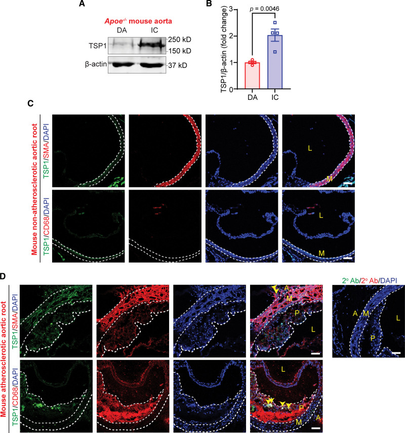
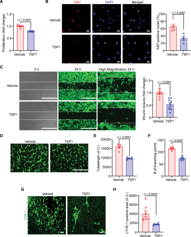
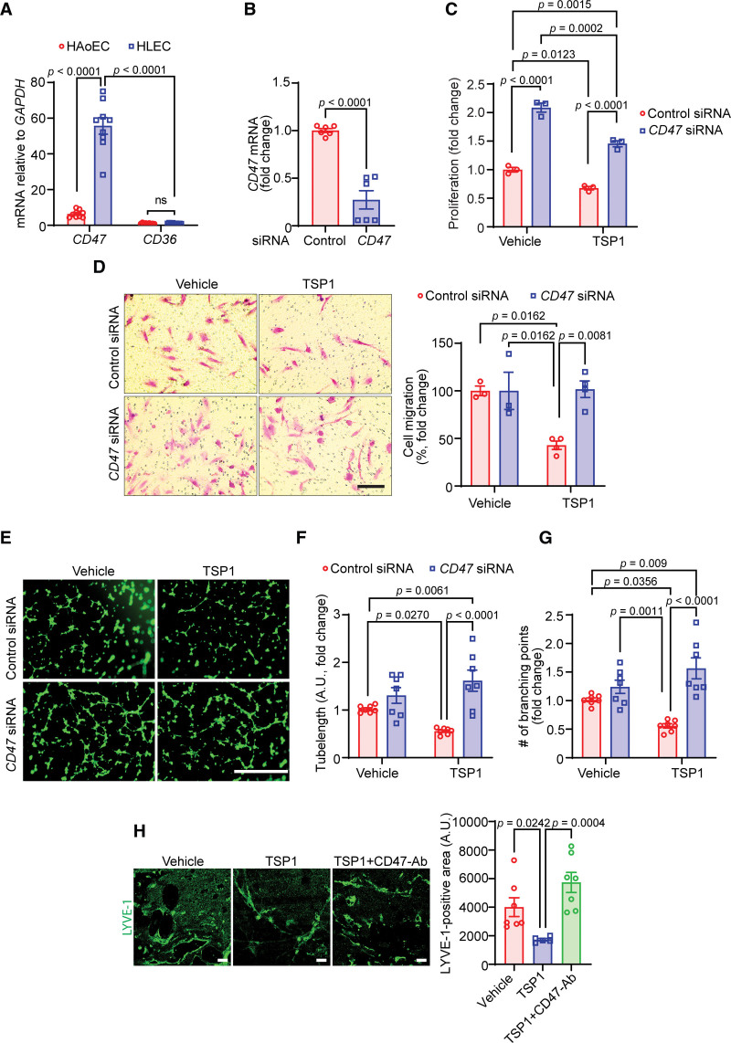
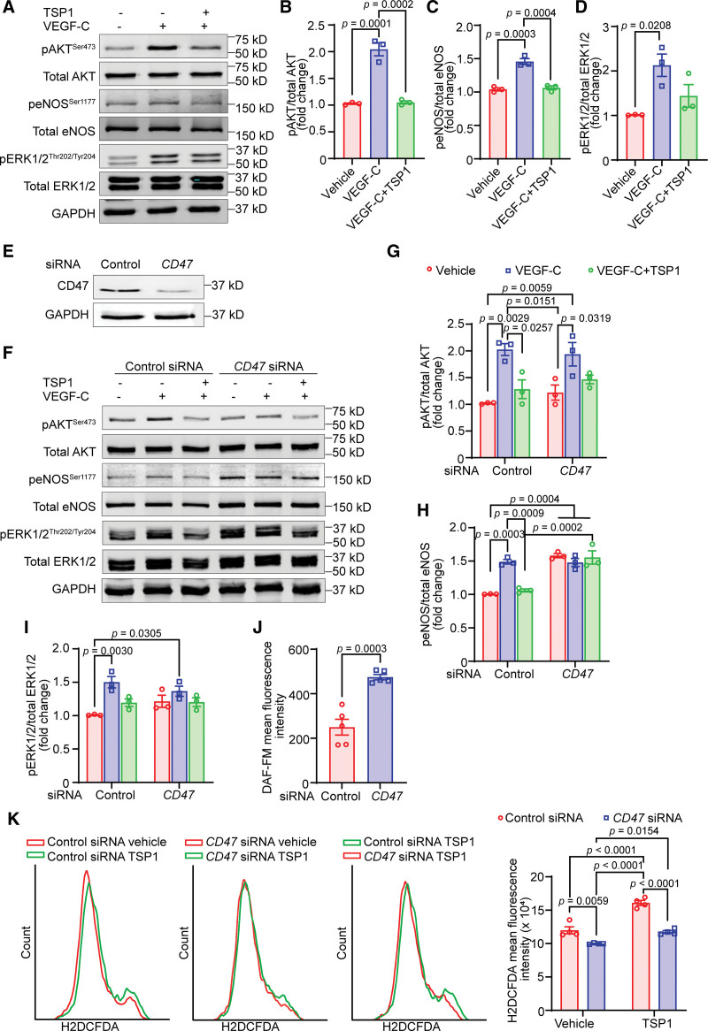
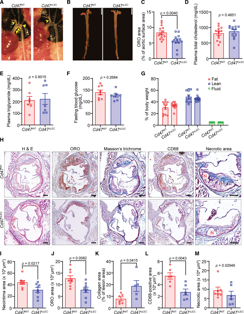
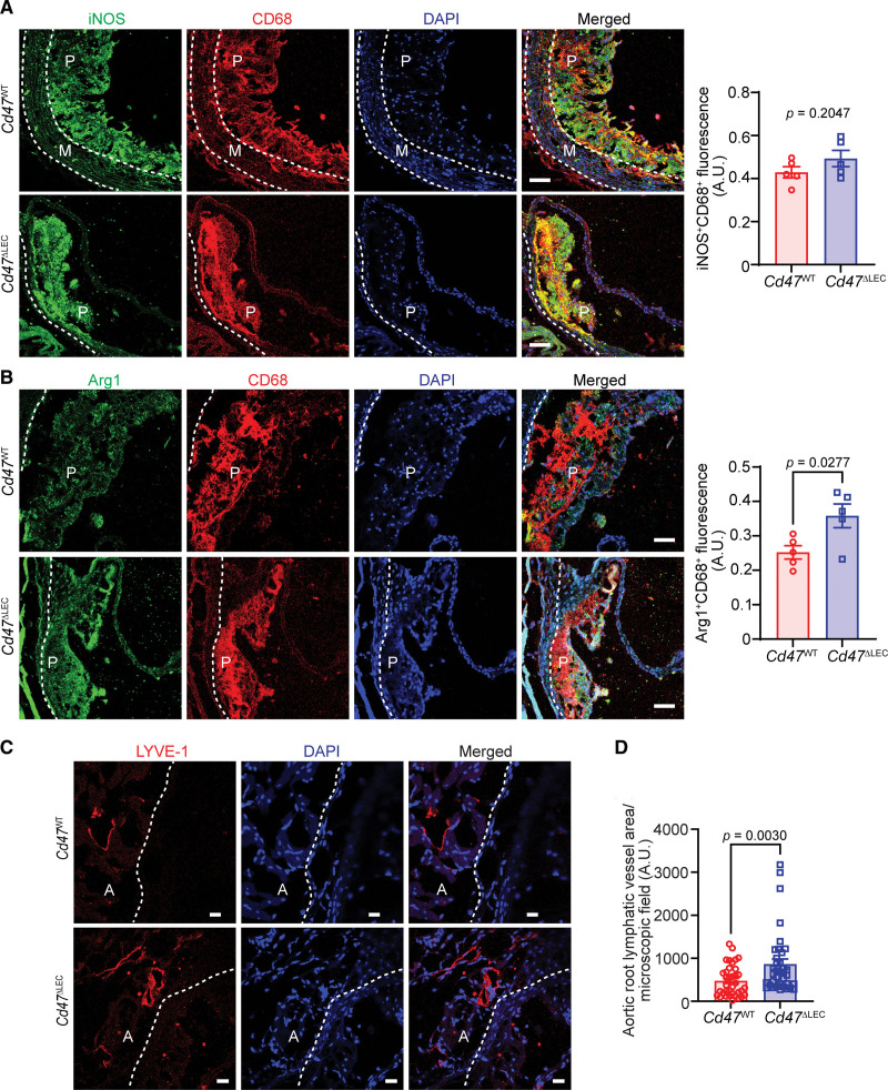
Results: Elevated TSP1 expression was observed in mouse atherosclerotic aortic tissue compared with nonatherosclerotic control tissue. TSP1 at pathological concentrations suppressed both in vitro and in vivo lymphangiogenesis. Mechanistically, TSP1 inhibited VEGF (vascular endothelial growth factor)-C-induced AKT and eNOS activation in LEC and attenuated NO (nitric oxide) production. Further, CD47 silencing in LEC prevented the effects of TSP1 on lymphangiogenic AKT-eNOS signaling and lymphangiogenesis. Atheroprone AAV (adeno-associated virus) 8-PCSK9-injected LEC-specific Cd47 knockout mice (Cd47ΔLEC) had reduced atherosclerosis in both aorta and aortic root compared with control mice (Cd47ΔWT). However, no differences in metabolic parameters including body weight, plasma total cholesterol levels, and fasting blood glucose were observed. Additional immunostaining experiments performed on aortic root cross-sections indicated higher lymphatic vessel density in Cd47ΔLEC mice in comparison to controls.

Conclusions: These findings demonstrate that TSP1 inhibits lymphangiogenesis via activation of CD47 in LEC, and loss of LEC Cd47 attenuates atherosclerotic lesion formation. Collectively, these results identify LEC CD47 as a potential therapeutic target in atherosclerosis.

Keywords: aorta; atherosclerosis; endothelial cells; fasting; lymphangiogenesis.


Conflict of interest statement

Disclosures None.

Figures

Figure 1.
Figure 1.
TSP1 (thrombospondin-1) protein levels are upregulated in murine atherosclerotic arteries. A, Representative Western blot images of TSP1 and β-actin expression in plaque (P)-free descending aorta (DA) segments and atherosclerotic inner curvature (IC) of male Apoe−/− mice fed with a Western diet for 12 weeks. B, Bar graph shows mean TSP1 protein levels (n=4). C and D, Wild-type and Apoe−/− mice (12-wk Western diet) aortic sinus cross-sections were immunostained to investigate TSP1 (green), SMA (smooth muscle cell actin; red), and CD68 (cluster of differentiation; macrophage marker, red) expression. Yellow arrowheads indicate TSP1 expression in adventitia (A) and intraplaque areas (n=3). Scale bar, 50 µm. Statistical analyses were performed using a 2-tailed unpaired Student t test (B). Data represent mean±SEM. L indicates lumen; and M, media.
Figure 2.
Figure 2.
TSP1 (thrombospondin-1) suppresses both in vitro and in vivo lymphangiogenesis. A, Vehicle- and TSP1-pretreated (22 nM, 4 h) human lymphatic endothelial cells (LECs) were stimulated with VEGF (vascular endothelial growth factor)-C (100 ng/mL) and proliferation investigated after 48 hours using the WST (water-soluble tetrazolium)-1 assay. Data are representative of 3 independent experiments performed at least in duplicate. B, LECs plated on coverslips were pretreated and stimulated as in A for 24 hours. Cells were immunostained for Ki67 (red) and nuclei counterstained with DAPI (4’,6-diamidino-2-phenylindole; blue). Images of at least 4 random microscopic fields were captured. Representative images are shown. Scale bar, 20 µm. Bar graph represents the percentage of Ki67-positive nuclei (n=4–5). C, LEC migration in response to vehicle (VEGF-C) and TSP1 (VEGF-C+TSP1) was investigated after 24 hours using Culture-Insert 2 Well 24 (ibidi USA). Representative images of wounds at 0 and 24 hours are shown. Scale bar, 1000 μm. Bar diagram shows quantification of wound closure (n=9). D, Vehicle- or TSP1-pretreated LECs were seeded in wells of a Matrigel-coated plate in basal medium containing VEGF-C±TSP1 and tube formation determined after 6 hours. Representative images of tube formation are shown. Scale bar, 1000 µm. Images of random fields were captured and tube length (E) and number of branching points (F) quantified (n=6). G and H, Wild-type male mice were injected SC with Matrigel solutions premixed with either VEGF-C or VEGF-C+TSP1. Plugs were isolated after 10 days, sectioned, and immunostained for LYVE-1 (lymphatic vessel endothelial hyaluronan receptor-1). G, Representative images of LYVE-1 staining of cross-sections of the Matrigel plugs are shown. Scale bar, 20 μm. H, Quantification of LYVE-1–positive area (n=5–7). Statistical analyses were performed using a 2-tailed unpaired Student t test (A–C, E, and F) and a Mann-Whitney U test (H). Data represent mean±SEM.
Figure 3.
Figure 3.
CD47 (cluster of differentiation) mediates TSP1 (thrombospondin-1)-induced inhibition of lymphangiogenesis. A, Quantitative real-time polymerase chain reaction (qRT-PCR) was performed to determine the relative mRNA levels of CD47 and CD36 in human lymphatic endothelial cells (HLECs) and human aortic endothelial cells (HAoEC). Bar graph represents mRNA levels in comparison to CD36 (n=9). B, Control and CD47 siRNA-treated lymphatic endothelial cells (LECs; 48 h) were utilized to quantify CD47 transcript expression using qRT-PCR (n=6). C, WST (water-soluble tetrazolium)-1 assay was conducted to investigate the effects of CD47 silencing on LEC proliferation in response to TSP1 treatment as described in Figure 2A. Data are representative of 3 independent experiments performed at least in quadruplicate. D, Control and CD47-silenced LECs were used to evaluate cell migration. Scale bar, 200 µm. xBar graph represents the percentage of migrated cells (n=3–4). E through G, Control and CD47-silenced cells were pretreated as in Figure 2D and seeded in wells of a Matrigel-coated plate in basal medium containing VEGF (vascular endothelial growth factor)-C±TSP1 and tube formation determined. Representative images of tube formation are shown (E). Scale bar, 1000 µm. Tube length (F) and number of branching points (G) quantified (n=7). H, Wild-type male mice were injected SC with Matrigel solutions premixed with either VEGF-C, VEGF-C+TSP1+IgG, or VEGF-C+TSP1+CD47-blocking antibody. Plugs were isolated after 10 days, sectioned, and immunostained for LYVE-1 (lymphatic vessel endothelial hyaluronan receptor-1). Representative images of LYVE-1 staining of the cross-sections of the Matrigel plugs and quantification of LYVE-1–positive area are shown (n=5–7). Scale bar, 20 μm. Statistical analyses were performed using 2-way ANOVA (A, C, D, F, and G) with Bonferroni (A), Tukey (C, F, and G), and Sidak (D) multiple comparisons test, 2-tailed unpaired Student t test (B), and Kruskal-Wallis test for multiple comparisons (H). Data represent mean±SEM.
Figure 4.
Figure 4.
TSP1 (thrombospondin-1)-induced CD47 (cluster of differentiation) activation blocks VEGF (vascular endothelial growth factor)-C–stimulated lymphangiogenic signaling. A through D, Lymphatic endothelial cells (LECs) were pretreated with TSP1 (22 nM, 16 h) in 0.5% fetal bovine serum (FBS) containing basal media MV2, stimulated with VEGF-C (15 min), and cell lysates subjected to Western blot analysis. A, Representative Western blot images are shown. B through D, Bar diagrams represent mean protein levels expressed as a ratio of phospho-to-total proteins, AKT (B), eNOS (C), and ERK1/2 (D; n=3). E, LECs were treated with control or CD47-siRNA (48 h) and immunoblotting done to determine CD47 expression (ab175388). F through I, Control or CD47-siRNA–treated cells were treated as in Figure 4A and Western blot experiments executed. F, Representative Western blot images are shown. G through I, Bar diagrams represent pAKT/total AKT (G), peNOS/total eNOS (H), and pERK1/2/total ERK1/2 (I; n=3). J, Control or CD47-silenced LECs were pretreated with TSP1 (22 nM, 4 h), stimulated with VEGF-C (100 ng/mL, 1 h) and analyzed for NO (nitric oxide) production using DAF-FM diacetate. K, Control or CD47-silenced LECs were pretreated with vehicle or TSP1 (1 h), incubated with H2DCFDA solution, and fluorescence analyzed using flow cytometry. Statistical analyses were performed using 1-way ANOVA (B–D), 2-way ANOVA with Tukey test for multiple comparisons (G–I and K), and 2-tailed unpaired Student t test (J). Data represent mean±SEM.
Figure 5.
Figure 5.
Lymphatic endothelial cell (LEC)–specific Cd47 deficiency reduces atherosclerotic lesion formation. A through M, Male Cd47WT and Cd47ΔLEC mice were injected with AAV (adeno-associated virus) 8-PCSK9 IP, fed a Western diet for 16 weeks and atherosclerosis analyzed. A, Representative in situ images of aortic arch (yellow arrowheads point to atherosclerotic lesions). Scale bar, 2 mm. B, Representative en face oil red O (ORO) staining of aorta. Scale bar, 5 mm. C, Quantification of plaque area in aorta (n=13–14). D through G, Bar diagrams show plasma total cholesterol (D), plasma triglycerides (E), fasting blood glucose levels (F), and body composition (fat, lean, and fluid mass; G; n=8–13). H, Representative images of aortic root cross-sections stained with H&E (neointima area), ORO (lipid accumulation), Masson trichrome (collagen content), CD68 (cluster of differentiation; macrophage burden), and necrotic area (encircled in red). Scale bar, 200 µm. I through M, Bar diagrams show neointima area (I), lipid deposition (J), collagen content (K), macrophage accumulation (L), and necrotic area (M) in aortic root sections (n=5–9). Statistical analyses were performed using a 2-tailed unpaired Mann-Whitney U test (C, E, and M), 2-tailed unpaired Student t test (D, F, and I–L), and 2-way ANOVA followed by Sidak post hoc test (G). Data represent mean±SEM.
Figure 6.
Figure 6.
Lymphatic endothelial cell (LEC)–specific deletion of Cd47 (cluster of differentiation) in mice increases arterial lymphatic vessel (LV) density. A through D, Aortic root cross-sections from male AAV (adeno-associated virus) 8-PCSK9–injected Cd47WT and Cd47ΔLEC mice (16-wk Western diet) were immunostained for CD68, iNOS (inducible NO [nitric oxide] synthase), Arg1 (arginase 1), and LYVE-1 (lymphatic vessel endothelial hyaluronan receptor-1). Nuclei were counterstained with DAPI (4’,6-diamidino-2-phenylindole; blue). Representative confocal images of iNOS (green; A) and CD68 (red); Arg1 (green; B) and CD68 (red; scale bar, 50 µm); and LYVE-1 staining (red; C; scale bar, 20 µm) are shown (n=5–6). Statistical analyses were performed using a 2-tailed unpaired Student t test (A and B) and a Mann-Whitney U test (C). Data represent mean±SEM. A indicates adventitia; M, media; and P, plaque.

References

    1. Lawton JS, Tamis-Holland JE, Bangalore S, Bates ER, Beckie TM, Bischoff JM, Bittl JA, Cohen MG, DiMaio JM, Don CW, et al. . 2021 ACC/AHA/SCAI guideline for coronary artery revascularization: executive summary: a report of the American College of Cardiology/American Heart Association joint committee on clinical practice guidelines. Circulation. 2022;145:e4–e17. doi: 10.1161/CIR.0000000000001039 - PubMed
    1. Martel C, Li W, Fulp B, Platt AM, Gautier EL, Westerterp M, Bittman R, Tall AR, Chen SH, Thomas MJ, et al. . Lymphatic vasculature mediates macrophage reverse cholesterol transport in mice. J Clin Invest. 2013;123:1571–1579. doi: 10.1172/JCI63685 - PMC - PubMed
    1. Yeo KP, Lim HY, Thiam CH, Azhar SH, Tan C, Tang Y, See WQ, Koh XH, Zhao MH, Phua ML, et al. . Efficient aortic lymphatic drainage is necessary for atherosclerosis regression induced by ezetimibe. Sci Adv. 2020;6:eabc2697. doi: 10.1126/sciadv.abc2697 - PMC - PubMed
    1. Rademakers T, van der Vorst EP, Daissormont IT, Otten JJ, Theodorou K, Theelen TL, Gijbels M, Anisimov A, Nurmi H, Lindeman JH, et al. . Adventitial lymphatic capillary expansion impacts on plaque T cell accumulation in atherosclerosis. Sci Rep. 2017;7:45263. doi: 10.1038/srep45263 - PMC - PubMed
    1. Vuorio T, Nurmi H, Moulton K, Kurkipuro J, Robciuc MR, Ohman M, Heinonen SE, Samaranayake H, Heikura T, Alitalo K, et al. . Lymphatic vessel insufficiency in hypercholesterolemic mice alters lipoprotein levels and promotes atherogenesis. Arterioscler Thromb Vasc Biol. 2014;34:1162–1170. doi: 10.1161/ATVBAHA.114.302528 - PMC - PubMed

Publication types

MeSH terms