Review
. 2023 Dec 12;11(1):eesp00112023.
doi: 10.1128/ecosalplus.esp-0011-2023. Epub 2023 Jun 1.

Structure, Assembly, and Function of Flagella Responsible for Bacterial Locomotion

Affiliations
Review

Structure, Assembly, and Function of Flagella Responsible for Bacterial Locomotion

Tohru Minamino et al. EcoSal Plus. .

Abstract

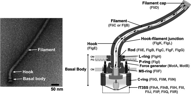
Many motile bacteria use flagella for locomotion under a variety of environmental conditions. Because bacterial flagella are under the control of sensory signal transduction pathways, each cell is able to autonomously control its flagellum-driven locomotion and move to an environment favorable for survival. The flagellum of Salmonella enterica serovar Typhimurium is a supramolecular assembly consisting of at least three distinct functional parts: a basal body that acts as a bidirectional rotary motor together with multiple force generators, each of which serves as a transmembrane proton channel to couple the proton flow through the channel with torque generation; a filament that functions as a helical propeller that produces propulsion; and a hook that works as a universal joint that transmits the torque produced by the rotary motor to the helical propeller. At the base of the flagellum is a type III secretion system that transports flagellar structural subunits from the cytoplasm to the distal end of the growing flagellar structure, where assembly takes place. In recent years, high-resolution cryo-electron microscopy (cryoEM) image analysis has revealed the overall structure of the flagellum, and this structural information has made it possible to discuss flagellar assembly and function at the atomic level. In this article, we describe what is known about the structure, assembly, and function of Salmonella flagella.

Keywords: bacterial flagellum; chemotaxis; cryoEM image analysis; energy coupling; flagellar assembly; flagellar gene regulation; motility; torque generation; transmembrane proton channel; type III secretion system.


Figures

FIG 1
FIG 1
The Salmonella flagellum. An electron micrograph of the flagellum isolated from Salmonella (left) and its schematic diagram (right) are shown. The flagellum is divided into at least three distinct functional parts: a basal body that is a bidirectional rotary motor, a hook that functions as a universal joint, and a filament that acts as a helical propeller. A hook-filament junction connects the hook and filament. A filament cap is located at the tip of the filament and facilitates filament assembly. To construct the flagellum beyond the cytoplasmic membrane, flagellar structural subunits are translocated via a type III secretion system (fT3SS) that is located at the base of the flagellum and assemble at the tip of the growing structure. Several force generators surround the basal body. OM, outer membrane; PG, peptidoglycan layer; CM, cytoplasmic membrane.
FIG 2
FIG 2
CryoEM structure of the Salmonella hook-basal body. Atomic model of the Salmonella native hook-basal body structure in Cα ribbon representation (PDB accession number 7CGO). The atomic model of the basal body consists of the MS ring (FliF) (slate gray); the P ring (FlgI) (medium purple); the L ring (FlgH) (purple); the polypeptide channel complex formed by FliP (cornflower blue), FliQ (light blue), and FliR (navy blue); and the rod (light gray). The hook (FlgE) (light green) is connected directly to the rod.
FIG 3
FIG 3
Rotational symmetry of basal body rings. (A) Vertical section of the atomic model of the S ring in Cα ribbon representation (PDB accession number 7CGO). FliF self-assembles into the MS ring with 34-fold rotational symmetry. The polypeptide channel complex with a stoichiometry of 5 FliP subunits, 4 FliQ subunits, and 1 FliR subunit is located inside the MS ring. FliP (cornflower blue) and FliR (navy blue) assemble into a helical structure inside the MS ring, and the central pore of the FliP5-FliR1 complex serves as a polypeptide channel that allows export substrates to be translocated across the cytoplasmic membrane. (B) Vertical section of the atomic model of the LP ring in Cα ribbon representation (PDB accession number 7CGO). The P ring (FlgI) (medium purple) and the L ring (FlgH) (purple) together form a very strong and stable ring complex with 26-fold rotational symmetry, and the LP ring acts as a molecular bushing for high-speed rod rotation inside it. The central channel of the axial structure is a physical pathway for the diffusion of the flagellar structural subunits in an unfolded conformation to the distal end of the growing flagellar structure.
FIG 4
FIG 4
Structure of the C ring. (A) CryoEM image of the Salmonella basal body and its schematic diagram. The cryoEM image of the basal body in the left panel is modified from references and and is used here to provide a visual reference for the schematic diagram shown in the right panel. The basal body consists of the C ring, the MS ring, the P ring, the L ring, and the rod. There is no MotA5-MotB2 complex in the purified basal body (left), but multiple MotA5-MotB2 complexes are present around the MS-ring–C-ring complex and function as force generators for the flagellar motor (right). The C ring is composed of FliG, FliM, and FliN. The N-terminal domain of FliG (FliGN) binds to the C-terminal cytoplasmic domain of FliF (FliFC) to form the FliG ring on the cytoplasmic face of the MS ring. The C-terminal domain of FliG (FliGC) is located in the upper part of the C ring. The middle domain of FliM (FliMM) is located between the middle domain of FliG (FliGM) and FliN and forms a continuous wall of the C ring. The continuous spiral density at the bottom edge of the C ring is made of the C-terminal domain of FliM (FliMC) and FliN. OM, outer membrane; PG, peptidoglycan layer; CM, cytoplasmic membrane. (B) Homology model of Salmonella FliG built based on the crystal structure of FliG derived from Aquifex aeolicus (PDB accession number 3HJL). The Cα backbone is color-coded from blue to red, going through the rainbow colors from the N to the C termini. FliG consists of three distinct FliGN, FliGM, and FliGC domains and two helix linkers named HelixNM and HelixMC. FliGC is divided into two distinct FliGCN and FliGCC subdomains. The highly conserved Arg281 and Asp289 residues in FliGCC are responsible for interactions with the cytoplasmic domain of the MotA5-MotB2 complex. The conserved MFXF motif allows FliGCC to rotate 180° relative to FliGCN. Thr103 in HelixNM interacts with Pro169 and Ala173 in HelixMC. In contrast to the wild-type motor that is placed in a default CCW state, the in-frame deletion of three residues, Pro169, Ala170, and Ala171, locks the flagellar motor in the CW state. (C) Two distinct conformations of the FliGC domain. Two distinct homology models of Salmonella FliGC were built based on the structures of Helicobacter pylori FliGC under PDB accession numbers 3USY (green) and 3USW (cyan). Conformational rearrangements of the conserved MFXF motif induce a 180° rotation of FliGCC relative to FliGCN to reorient the Arg281 and Asp289 residues. (D) Structural comparisons between the wild-type FliGMC fragment composed of FliGM, HelixMC, and FliGC (green) and its CW-locked variant (cyan). Homology models of the wild-type FliGMC-FliMM complex and the CW-locked form of FliGMC were built based on the FliGMC/FliMM complex (PDB accession number 4FHR) and the CW-locked FliGMC variant (PDB accession number 3AJC) derived from Thermotoga maritima, respectively. The FliGM domain of the CW-locked variant is superimposed onto that of the FliGMC/FliMM complex. HelixMC (magenta) is located at the interface between FliGM and FliMM (dark gray). In contrast, the CW-locked deletion not only induces a distinct orientation of HelixMC relative to the FliGM-FliMM interface but also goes through a 90° rotation of FliGCC.
FIG 5
FIG 5
Common structural features of the flagellar axial structure. (A) Atomic model of the FlgE subunit in the hook structure (PDB accession number 7CGO). The Cα backbone is color-coded from blue to red, going through the rainbow colors from the N to the C termini. The N-terminal and C-terminal regions of FlgE are intrinsically disordered in its monomeric state, but when incorporated into the hook structure, they form an α-helical coiled coil in the inner-core D0 domain. (B) Arrangement of FlgE subunits in the hook. Major helical lines are indicated by arrows. The FlgE subunits along the 11-start helical line comprise the protofilament. The number labeled on the subunits represents the number of the subunit starting from the central subunit (subunit 0) along the 1-start helical line. The number also shows the direction of the helical line. FlgE monomers are sequentially assembled along the 1-start helix.
FIG 6
FIG 6
CryoEM structure of the filament cap. Shown are side (left) and top (right) views of the atomic model of the filament cap in Cα ribbon representation (PDB accession number 6SIH). Five FliD subunits self-assemble into the filament cap structure consisting of a pentagonal plate and five extended leg-like domains.
FIG 7
FIG 7
CryoEM structure of the rod. (A) Atomic model of the rod in Cα ribbon representation (PDB accession number 7CGO). The rod consists of 6 FliE (gold), 5 FlgB (aquamarine), 6 FlgC (pink), 5 FlgF (dark green), and 24 FlgG (light gray) subunits and is connected directly to the polypeptide channel complex consisting of 5 FliP subunits (cornflower blue), 4 FliQ subunits (light blue), and 1 FliR subunit (navy blue). (B) Interactions of FliE with the MS ring, FliP, and FliR. FliE consists of three α-helices, α1, α2, and α3. Helix α1 binds to the inner wall of the MS ring. Helices α2 and α3 of FliE, which form domain D0, bind to either the FliP or the FliR subunit of the polypeptide channel complex to form the most proximal part of the rod inside the MS ring. Helix α2 is invisible when FliE binds to FliR.
FIG 8
FIG 8
Structural comparison of the distal rod and hook. (A) Atomic model of the distal rod in Cα ribbon representation (PDB accession number 7CGO). FlgG is composed of three domains, D0, Dc, and D1, that are arranged from the inner to the outer parts of the distal rod. The L stretch of domain Dc of FlgG makes the rod straight and rigid. (B) Atomic model of the native supercoiled hook in Cα ribbon representation (PDB accession number 7CGO). FlgE is composed of four domains, D0, Dc, D1, and D2, that are arranged from the inner to the outer parts of the hook. Gaps between FlgE subunits are responsible for the bending flexibility of the hook structure.
FIG 9
FIG 9
Structure of the fT3SS. Shown are atomic models of the FlhB1-FliP5-FliQ4-FliR1 complex (PDB accession number 6S3L), the C-terminal cytoplasmic domain of FlhB (FlhBC) (PDB accession number 3B0Z), the C-terminal cytoplasmic domain of FlhA (FlhAC) (PDB accession number 3A5I), the FliHC2-FliI complex (PDB accession number 5B0O), and FliJ (PDB accession number 3AJW) in Cα ribbon representation. There is no atomic model for the N-terminal transmembrane domain of FlhA with eight transmembrane helices (FlhATM) and the N-terminal domain of FliH (FliHN). Because the electron density corresponding to FlhBC in the cryoEM structure of the FlhB1-FliP5-FliQ4-FliR1 complex is very poor, the position of FlhBC relative to FlhBTM remains unknown. The flagellar type III secretion system consists of a transmembrane export gate complex with a stoichiometry of 9 FlhA subunits (magenta), 1 FlhB subunit (plum), 5 FliP subunits (cornflower blue), 4 FliQ subunits (light blue), and 1 FliR subunit (navy blue) and a cytoplasmic ATPase ring complex with a stoichiometry of 12 FliH subunits (brown), 6 FliI subunits (khaki), and 1 FliJ subunit (dark olive green). The transmembrane export complex is located within the MS ring. FliJ binds to the center of the FliI6 ring, and two C-terminal domains of the FliH dimer (FliHC2) bind to different regions of the N-terminal domain of each FliI subunit of the FliI6 ring. The FliI6-FliJ ring complex is tightly bound to the C ring by the interaction between FliHN2 and FliN in the C ring. CM, cytoplasmic membrane.
FIG 10
FIG 10
Structure of the MotA5-MotB2 complex. (A) Model for the inactive and active forms of the MotA5-MotB2 complex. The MotA5-MotB2 complex is divided into four distinct functional parts: a cytoplasmic domain responsible for the interaction with the rotor protein FliG, a transmembrane proton (H+) channel domain involved in inward-directed H+ flow, a peptidoglycan (PG)-binding domain (PGB) responsible for binding to the PG layer (PDB accession number 2ZVY), and a flexible linker region containing a plug helix. (Left) When the PGB domain adopts a compact conformation, the plug helix binds to the H+ channel domain to suppress massive H+ flow. (Right) When the inactive MotA5-MotB2 complex, which is freely diffusing across the cytoplasmic membrane, encounters a rotor, interactions between its cytoplasmic domain and FliG are postulated to trigger the dissociation of the plug segment from the H+ channel, followed by the unfolding of the N-terminal α-helix of the PGB domain, which extends 5 nm and binds the PG layer. As a result, the MotA5-MotB2 complex becomes a force generator to drive flagellar motor rotation. PG, peptidoglycan layer; Peri, periplasmic space; CM, cytoplasmic membrane; Cyto, cytoplasm. (B) CryoEM structure of the MotA5-MotB2 complex. Shown is an atomic model of the purified MotA5-MotB2 complex in Cα ribbon representation (PDB accession number 6YKM). The five MotA subunits (goldenrod) form a pentameric ring structure, with two transmembrane helices of the MotB dimer (red and orchid) bound to the center of the MotA ring. Each plug helix of the MotB dimer binds to the periplasmic loop of MotA.
FIG 11
FIG 11
Load-dependent stator assembly mechanism. (Right) At very low loads, a few stator units surround the rotor. (Left) As the external load increases, the number of active stator units increases to a maximum of about 10. The association rates of the stator unit are essentially the same for both low and high loads, but the dissociation rate is much higher at low loads than at high loads.
FIG 12
FIG 12
Hierarchical flagellar gene expression in Salmonella. Flagellar assembly begins with the basal body, followed by the hook and, finally, the filament. FlhD and FlhC form the FlhD4-FlhC2 complex that induces transcription from class 2 promoters, thereby producing several hook-basal bodies (HBBs) on the Salmonella cell surface. During HBB assembly, FlgM binds to FliA to inhibit transcription from class 3 promoters. When the hook length reaches its mature length of 55 nm, FlgM is secreted via the fT3SS into the culture medium. As a result, FliA can act as a flagellum-specific sigma factor (σ28), allowing RNA polymerase to transcribe class 3 genes encoding proteins required for filament formation, motility, and chemotaxis. The flgKL, flgMN, fliAZY, and fliDST operons highlighted in magenta are transcribed from both class 2 and class 3 promoters. OM, outer membrane; PG, peptidoglycan layer; CM, cytoplasmic membrane.
FIG 13
FIG 13
Model for the substrate specificity switching of the fT3SS. The fT3SS transports the hook-type export substrates FlgD, FlgE, and FliK during hook assembly but does not transport the filament-type substrates FlgK/L, FlgM, and FliD, which form a complex with FlgN, FliA, and FliT in the cytoplasm, respectively. At that point, no flagellin molecules (FliC and FljB) are expressed. When the hook length reaches about 55 nm, the C-terminal domain of FliK (FliKC) binds to FlhBCC to induce a conformational change of a cleaved form of FlhBC. Next, the FliKC-FlhBC complex binds to FlhAC to induce the remodeling of the FlhAC ring structure, allowing the fT3SS to terminate the export of hook-type proteins and initiate the export of filament-type proteins.

Similar articles

Cited by

References

    1. Minamino T, Imada K. 2015. The bacterial flagellar motor and its structural diversity. Trends Microbiol 23:267–274. doi:10.1016/j.tim.2014.12.011. - DOI - PubMed
    1. Terashima H, Kawamoto A, Morimoto YV, Imada K, Minamino T. 2017. Structural differences in the bacterial flagellar motor among bacterial species. Biophys Physicobiol 14:191–198. doi:10.2142/biophysico.14.0_191. - DOI - PMC - PubMed
    1. Macnab RM. 1996. Flagella and motility, p 123–145. In Neidhardt FC, Curtiss R, III, Ingraham JL, Lin ECC, Low KB, Magasanik B, Reznikoff WS, Riley M, Schaechter M, Umbarger HE (ed), Escherichia coli and Salmonella typhimurium: cellular and molecular biology, 2nd ed. ASM Press, Washington, DC.
    1. Macnab RM. 2003. How bacteria assemble flagella. Annu Rev Microbiol 57:77–100. doi:10.1146/annurev.micro.57.030502.090832. - DOI - PubMed
    1. Minamino T, Namba K. 2004. Self-assembly and type III protein export of the bacterial flagellum. J Mol Microbiol Biotechnol 7:5–17. doi:10.1159/000077865. - DOI - PubMed

LinkOut - more resources