. 2023 May 1;5(11):3018-3032.
doi: 10.1039/d3na00105a. eCollection 2023 May 30.

Dual imaging agent for magnetic particle imaging and computed tomography

Affiliations

Dual imaging agent for magnetic particle imaging and computed tomography

Sitong Liu et al. Nanoscale Adv. .

Abstract

Magnetic particle imaging (MPI) is a novel biomedical imaging modality that allows non-invasive, tomographic, and quantitative tracking of the distribution of superparamagnetic iron oxide nanoparticle (SPION) tracers. While MPI possesses high sensitivity, detecting nanograms of iron, it does not provide anatomical information. Computed tomography (CT) is a widely used biomedical imaging modality that yields anatomical information at high resolution. A multimodal imaging agent combining the benefits of MPI and CT imaging would be of interest. Here we combine MPI-tailored SPIONs with CT-contrast hafnium oxide (hafnia) nanoparticles using flash nanoprecipitation to obtain dual-imaging MPI/CT agents. Co-encapsulation of iron oxide and hafnia in the composite nanoparticles was confirmed via transmission electron microscopy and elemental mapping. Equilibrium and dynamic magnetic characterization show a reduction in effective magnetic diameter and changes in dynamic magnetic susceptibility spectra at high oscillating field frequencies, suggesting magnetic interactions within the composite dual imaging tracers. The MPI performance of the dual imaging agent was evaluated and compared to the commercial tracer ferucarbotran. The dual-imaging agent has MPI sensitivity that is ∼3× better than this commercial tracer. However, worsening of MPI resolution was observed in the composite tracer when compared to individually coated SPIONs. This worsening resolution could result from magnetic dipolar interactions within the composite dual imaging tracer. The CT performance of the dual imaging agent was evaluated in a pre-clinical animal scanner and a clinical scanner, revealing better contrast compared to a commercial iodine-based contrast agent. We demonstrate that the dual imaging agent can be differentiated from the commercial iodine contrast agent using dual energy CT (DECT) imaging. Furthermore, the dual imaging agent displayed energy-dependent CT contrast arising from the combination of SPION and hafnia, making it potentially suitable for virtual monochromatic imaging of the contrast agent distribution using DECT.


Conflict of interest statement

The authors have declared that no competing interest exists.

Figures
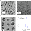
Fig. 1
Fig. 1. Nanoparticles and nanoclusters evaluated via transmission electron microscopy (TEM) and dynamic light scattering (DLS). (A) TEM images of SPIONs. (B) TEM images of hafnium oxide nanoparticles. (C) Representative TEM images of dual imaging agents. (D) Representative DLS analysis of dual imaging agents.
Fig. 2
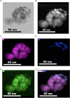
Fig. 2. Bright-field TEM image, HADDF STEM image, and elemental mapping images of dual imaging agents. (A) Bright-field TEM. (B) HADDF STEM image. (C) Elemental mapping showing iron content. (D) Elemental mapping showing hafnium content. (E) Elemental mapping showing oxygen content. (F) Superposition of iron, hafnium, and HADDF STEM images.
Fig. 3
Fig. 3. Magnetization versus magnetic field curves obtained using a SQUID magnetometer. (A) SPIONs singly coated with PEG-silane. (B) Dual imaging agents. (C) Histogram of magnetic diameters for nanoparticles singly coated with PEG (single particles with a polymer corona) and dual imaging agents (composite nanoparticles).
Fig. 4
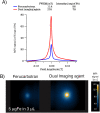
Fig. 4. (A) PSF for ferucarbotran and the dual imaging agents. (B) Side by side phantoms 2D isotropic images for ferucarbotran and dual imaging agents.
Fig. 5
Fig. 5. Evaluation of CT performance of the dual imaging agents and Omnipaque at various concentrations in different scanners IVIS® Spectrum In Vivo Imaging System data is shown in (A) and (C) while Aquilion ONE GENESIS SCT data is shown in (B) and (D). Representative grayscale images for the dual imaging agents and Omnipaque at various concentrations were presented in (A) and (B), while quantitative data are presented in (C) and (D).
Fig. 6
Fig. 6. Evaluation of CT performance in Aquilion ONE GENESIS scanner using a dual-energy CT. Representative grayscale CT images (A), CT performance at various concentrations (B), and CNR (C) of the dual imaging agents and Omnipaque.

References

    1. Gleich B. Weizenecker R. Tomographic imaging using the nonlinear response of magnetic particles. Nature. 2005;435:1214–1217. doi: 10.1038/nature03808. doi: 10.1038/nature03808. - DOI - DOI - PubMed
    1. Zheng B. et al., Quantitative Magnetic Particle Imaging Monitors the Transplantation, Biodistribution, and Clearance of Stem Cells In Vivo. Theranostics. 2016;6:291–301. doi: 10.7150/thno.13728. doi: 10.7150/thno.13728. - DOI - DOI - PMC - PubMed
    1. Zheng B. et al., Magnetic Particle Imaging tracks the long-term fate of in vivo neural cell implants with high image contrast. Sci. Rep. 2015;5:14055. doi: 10.1038/srep14055. doi: 10.1038/srep14055. - DOI - DOI - PMC - PubMed
    1. Yu E. Y. et al., Magnetic Particle Imaging for Highly Sensitive, Quantitative, and Safe in Vivo Gut Bleed Detection in a Murine Model. ACS Nano. 2017;11:12067–12076. doi: 10.1021/acsnano.7b04844. doi: 10.1021/acsnano.7b04844. - DOI - DOI - PMC - PubMed
    1. Fuller E. G. et al., Theranostic nanocarriers combining high drug loading and magnetic particle imaging. Int. J. Pharm. 2019;572:118796. doi: 10.1016/j.ijpharm.2019.118796. doi: 10.1016/j.ijpharm.2019.118796. - DOI - DOI - PubMed