

Relationship between morphometric and mechanical properties of superficial lumbosacral soft tissue layers in healthy young adults
- PMID: 37260591
- PMCID: PMC10228649
- DOI: 10.3389/fphys.2023.1175035
Relationship between morphometric and mechanical properties of superficial lumbosacral soft tissue layers in healthy young adults
Abstract
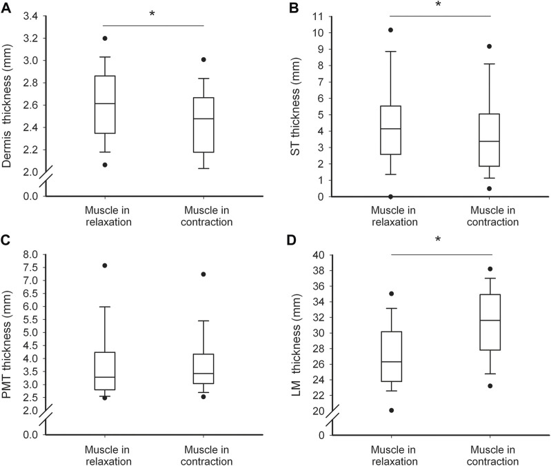
Introduction: It is commonly considered that myotonometry is a non-invasive method capable of quantifying linear elastic and viscoelastic properties of the myofascial tissue through the application of a weak mechanical impulse to the surface of the skin. However, before the impulse can reach the myofascial tissue, it must cross more superficial tissues such as the skin and subcutaneous tissue (ST). All these superficial tissues have different distributions and organizations of structural components. Therefore, the study aimed to examine the potential relationships between the mechanical and morphometric properties of various superficial soft tissues surrounding the lumbar multifidus muscle (LM). Methods: Myotonometric measurements of dynamic stiffness, logarithmic decrement, and creep, and ultrasonographic measurements of thickness and echogenicity of cutaneous, subcutaneous, perimuscular tissue, and LM were obtained from 50 healthy individuals in the resting prone position and during contralateral arm lift. Results: The most important findings were that in both the relaxed and contracted LM state, the dynamic stiffness strongly negatively (r = -0.69; p < 0.001 in relaxation, r = -0.83; p < 0.001 in contraction) and creep strongly positively (r = 0.79; p < 0.001 in relaxation, r = 0.85; p < 0.001 in contraction) correlated with the thicknesses of the ST. Similar but weaker correlations were noticed between both these measures and the perimuscular tissue thickness. Elasticity was uncorrelated to the thicknesses of the tissues. With LM contraction (change from the relaxed to contracted state), the relative increase in dynamic stiffness was correlated with the relative decrease in dermis (r = -0.51; p < 0.001) and ST (r = -0.47; p = 0.001) thickness, and with the relative increase in LM (r = 0.36; p = 0.010) thickness. Moreover, the relative decrease (thinning) in the ST thickness was correlated with the relative increase in logarithmic decrement (i.e., decrease in soft tissue elasticity, r = -0.37, p = 0.011). The mechanical properties of the soft tissues were not related to their echogenicity. Discussion: In conclusion, the thicker the subcutaneous and perimuscular layers, the lesser the stiffness and the greater the time-dependent deformation to the external force of the tissues surrounding the LM during its relaxation and isometric contraction. Moreover, the greater the thinning of the ST and the thickening of the LM during its contraction, the higher the increase in lumbosacral tissue stiffness and the decrease in elasticity. Therefore, one should consider the thickness of the ST before planning or analyzing the outcomes of myotonometric or other external biomechanical measurements to avoid drawing the wrong conclusions about the mechanical properties of the myofascial tissue.
Keywords: low back; lumbar myofascial; plastic surgery; rehabilitative ultrasound imaging (RUSI); spinal surgery.
Copyright © 2023 Grześkowiak, Kocur and Łochyński.
Conflict of interest statement
The authors declare that the research was conducted in the absence of any commercial or financial relationships that could be construed as a potential conflict of interest.
Figures


References
-
- Agyapong-Badu S., Warner M., Samuel D., Stokes M. (2018). Practical considerations for standardized recording of muscle mechanical properties using a myometric device: Recording site, muscle length, state of contraction and prior activity. J. Musculoskelet. Res. 21, 2. 10.1142/s0218957718500100 - DOI
-
- Andonian B. J., Masi A. T., Aldag J. C., Barry A. J., Coates B. A., Emrich K., et al. (2015). Greater resting lumbar extensor myofascial stiffness in younger ankylosing spondylitis patients than age-comparable healthy volunteers quantified by myotonometry. Arch. Phys. Med. Rehabil. 96, 2041–2047. 10.1016/j.apmr.2015.07.014 - DOI - PubMed
LinkOut - more resources
Full Text Sources