. 2023 May 13;26(6):106861.
doi: 10.1016/j.isci.2023.106861. eCollection 2023 Jun 16.

Anomalous diffusion of nanoparticles in the spatially heterogeneous biofilm environment

Affiliations

Anomalous diffusion of nanoparticles in the spatially heterogeneous biofilm environment

Bart Coppens et al. iScience. .

Abstract

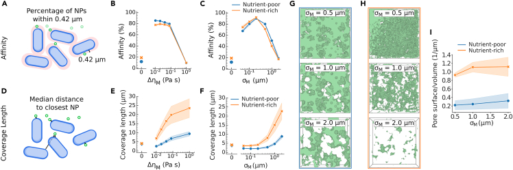
Biofilms contain extracellular polymeric substances (EPS) that provide structural support and restrict penetration of antimicrobial treatment. To overcome limited penetration, functionalized nanoparticles (NPs) have been suggested as carriers for antimicrobial delivery. Using microscopy, we evaluate the diffusion of nanoparticles in function of the structure of Salmonella biofilms. We observe anomalous diffusion and heterogeneous mobility of NPs resulting in distinct NPs distribution that depended on biofilm structure. Through Brownian dynamics modeling with spatially varying viscosity around bacteria, we demonstrated that spatial gradients in diffusivity generate viscous sinks that trap NPs near bacteria. This model replicates the characteristic diffusion signature and vertical distribution of NPs in the biofilm. From a treatment perspective, our work indicates that both biofilm structure and the level of EPS can impact NP drug delivery, where low levels of EPS might benefit delivery by immobilizing NPs closer to bacteria and higher levels hamper delivery due to shielding effects.

Keywords: Nanoparticles; Nanoscience; Nanotechnology.


Conflict of interest statement

The authors declare no competing interests.

Figures

None
Graphical abstract
Figure 1
Figure 1
Structural quantification of Salmonella biofilms grown in rich and poor nutrient conditions (A–H)Maximum intensity orthogonal projections of fluorescent blue Salmonella enterica Typhimurium biofilm, incubated in nutrient-poor a or nutrient-rich b conditions. Contrast was enhanced equally in both conditions for visualization purposes. Comparison between nutrient-poor and nutrient-rich grown biofilms of c mean thickness (p = 0.01), d mean pairwise distance from each bacteria to its 20 nearest neighbors (p = 0.00036),e nematic order of each bacteria relative to its 20 nearest neighbors (p = 8.3e-6), f mean angle of each bacteria relative to the substrate (p = 3.3e-5), g biofilm volume relative to substrate area (p = 0.18), h volume density of bacteria with respect to 3D image volume (p = 0.42). Error bars indicate standard deviation over the mean of 3 biological repeats. Measures for each of the three independent biofilm repeats are shown as points. All significance levels were obtained via pairwise t-tests, without multiple test correction.
Figure 2
Figure 2
Nanoparticle penetration in structurally different biofilms (A and B) Mean height distribution (perpendicular to the glass surface) of NPs (green) and bacteria (blue), 1 h after NP addition to nutrient-poor and nutrient-rich Salmonella enterica Typhimurium biofilms. Distribution of b NP displacement tracks (colored relative to track length as a measure for mobility) over 1 min, 20 min after NP addition in a nutrient-poor biofilm (bacteria shown in blue). (C) Ensemble probability distribution of displacements with a lag time of Δt= 0.1 s. The dashed line shows the slope for a Laplacian fit, estimated via non-linear least squares on the displacement curves on log scale. (D) Distribution of the diffusion coefficients D, estimated via linear least squares on the time-averaged mean squared displacement (TAMSD) as x¯2(Δ)DΔ. A break was included in the y axis to show the distribution of larger D as well. (E) Distribution of anomalous diffusion exponents α, estimated via non-linear least squares on the TAMSD.
Figure 3
Figure 3
Brownian dynamics modeling in experimentally observed biofilm structures (A) Schematic of Brownian dynamics model, which shows a Gaussian decay in viscosity with increasing distance to the surface of the bacteria. Viscosity is assumed to be additive, and hence further slowing down diffusion when in the vicinity of multiple bacteria. (B) CLSM images of fluorescently stained EPS, using EbbaBiolight 680, for nutrient-poor (left) and nutrient-rich (right) biofilms. (C) Normalized EPS intensity in function of the distance from the surface of segmented bacteria fitted with non-linear least squares as a Gaussian decay I=I0exp((x/σM)2). (D) Fitted Gaussian decay length σM for nutrient-poor and nutrient-rich conditions. Error bars indicate standard deviation over three biological repeats, and measures for each of the three independent biofilm repeats are shown as points. A t-test resulted in a p value of 0.85. (E) Simulated NPs (red) in nutrient-poor and nutrient-rich biofilms acquired through segmentation of the bacterial channel of the experimental CLSM images. Bacteria are colored relative to their height in the biofilm. (F) Ensemble displacement distributions for both nutrient-poor (top) and nutrient-rich biofilm (bottom) structures with a lag time of Δt=0.1s. The filled area denotes the standard deviation over three biological repeats. (G) Height distribution of the simulated NPs for varying EPS viscosity ΔηM. Height distributions were obtained by simulated NP positions and empirical bacterial positions from three biological repeats.
Figure 4
Figure 4
The effect of EPS on NP penetration in experimentally observed biofilms (A) Schematic of affinity, which is defined as the fraction of NP within the average experimentally fitted EPS length scale σM=0.42μm. (B) Affinity in function of EPS viscosity ΔηM for nutrient-poor and nutrient-rich biofilms, where ΔηM indicates homogeneous bulk viscosity η0. EPS length scale σM was kept constant at 0.42 μm. (C) Affinity in function of EPS length scale σM for nutrient-poor and nutrient-rich biofilms. EPS viscosity ΔηM was kept constant at 0.01 Pas. (D) Schematic of coverage length, which is defined as the median distance to the closest NP over all bacteria. (E) Coverage length in function of EPS viscosity ΔηM for nutrient-poor and nutrient-rich biofilms after 10 min simulation time. EPS length scale was kept constant at 0.42 μm. (F–H) Coverage length in function of EPS length scale σM for nutrient-poor and nutrient-rich biofilms after 10 min simulation time. EPS viscosity ΔηM was kept constant at 0.01 Pas. Pore volume, where the color intensity scales according to log(1/η(x)), such that the green volume shows where NPs can diffuse freely, in the empirical nutrient-poor g and nutrient-rich h biofilms in function of EPS length-scale σM with ΔηM=0.01Pas. (I) Pore surface area to volume ratio for nutrient-rich and nutrient-poor biofilms, for varying σM. Curves and filled area respectively indicate the mean and standard deviation of three biological repeats.
Figure 5
Figure 5
Virtual biofilm structure generation allows control of bacterial organization (A) Bacterial organization in virtual biofilms for various characteristic lengths Lt. (B) Side view of pore volumes (shown in green), where NPs can diffuse freely (η=η0), in virtual biofilms of characteristic length Lt=13.6μm and Lt=34.0μm. Pore volume, where the color intensity scales according to log(1/η(x)), such that the green volume shows where NPs can diffuse freely, is shown for EPS length-scale σM=0.5μm and σM=1.0μm. For visualization purposes, we visualized only a slice of 20 μm, at the center of the biofilm. (C) Affinity (threshold kept constant at 0.42 μm) in function of characteristic length Lt and EPS length-scale σM, at constant EPS viscosity ΔηM=0.01Pas. (D) Coverage length in function of characteristic length Lt and EPS length-scale σM, at constant EPS viscosity ΔηM=0.01Pas.
Figure 6
Figure 6
Conceptual representation of diffusion through the biofilm EPS (A) The conventional depiction of the biofilm with bacteria (blue) submerged in a nearly uniform EPS material (red). Increased viscosity only provides a transient barrier to penetration of particles (green). At long timescale, specific immobilization is required to maintain a concentration gradient. (B) EPS is distributed in a strongly heterogeneous viscosity landscape that provides sharp, local diffusivity gradients. These generate viscous sinks that establish equilibrium concentration gradients and explain both retention and shielding of particles in the biofilm.

Similar articles

Cited by

References

    1. Flemming H.C., Wingender J. The biofilm matrix. Nat. Rev. Microbiol. 2010;8:623–633. doi: 10.1038/nrmicro2415. - DOI - PubMed
    1. Flemming H.C., Wuertz S. Bacteria and archaea on Earth and their abundance in biofilms. Nat. Rev. Microbiol. 2019;17:247–260. doi: 10.1038/s41579-019-0158-9. - DOI - PubMed
    1. Karygianni L., Ren Z., Koo H., Thurnheer T. Biofilm matrixome: extracellular components in structured microbial communities. Trends Microbiol. 2020;28:668–681. doi: 10.1016/j.tim.2020.03.016. - DOI - PubMed
    1. Guzmán-Soto I., McTiernan C., Gonzalez-Gomez M., Ross A., Gupta K., Suuronen E.J., Mah T.F., Griffith M., Alarcon E.I. Mimicking biofilm formation and development: recent progress in in vitro and in vivo biofilm models. iScience. 2021;24:102443. doi: 10.1016/j.isci.2021.102443. - DOI - PMC - PubMed
    1. Xiu W., Shan J., Yang K., Xiao H., Yuwen L., Wang L. Recent development of nanomedicine for the treatment of bacterial biofilm infections. View. 2021;2:20200065. doi: 10.1002/viw.20200065. - DOI

LinkOut - more resources