. 2023 May 26:17:1593-1609.
doi: 10.2147/DDDT.S397541. eCollection 2023.

0.5-5% Supramolecular Salicylic Acid Hydrogel is Safe for Long-Term Topical Application and Improves the Expression of Genes Related to Skin Barrier Homeostasis in Mice Models

Affiliations

0.5-5% Supramolecular Salicylic Acid Hydrogel is Safe for Long-Term Topical Application and Improves the Expression of Genes Related to Skin Barrier Homeostasis in Mice Models

Can Zhou et al. Drug Des Devel Ther. .

Abstract

Background: As a keratolytic, salicylic acid (SA) can be topically applied in various formulations and doses in dermatology. Supramolecular SA hydrogel, a new SA formulation with higher bioavailability, is developed and commercially available nowadays. However, there still remain concerns that the long-term and continual application of SA at low concentrations may jeopardize the cutaneous barrier properties.

Aim of the study: To reveal the long-term effects of 0.5-5% supramolecular SA hydrogel on the skin barrier in normal mice models.

Materials and methods: The 0.5%, 1%, 2%, and 5% supramolecular SA hydrogel or hydrogel vehicle without SA was applied to mice's shaved dorsal skin once per day respectively. Tissue samples of the dorsal skin were harvested on day 14 and 28 of the serial application of SA for histopathological observation and transcriptomic analysis.

Results: Following topical supramolecular SA hydrogel therapy with various concentrations of SA (0.5%, 1%, 2%, and 5%) for 14 days and 28 days, there were no obvious macroscopic signs of impaired cutaneous health and no inflammatory or degenerative abnormalities were observed in histological results. Additionally, the transcriptomic analysis revealed that on day 14, SA dramatically altered the expression of genes related to the extracellular matrix structural constituent. And on day 28, SA regulated gene expression profiles of keratinization, cornified envelope, and lipid metabolism remarkably. Furthermore, the expression of skin barrier related genes was significantly elevated after the application of SA based on RNA-seq results, and this is likely to be associated with the PPAR signaling pathway according to the enrichment analysis.

Conclusion: Our findings demonstrated that the sustained topical administration of the 0.5-5% supramolecular SA hydrogel for up to 28 days did no harm to normal murine skin and upregulated the expression of genes related to the epidermal barrier.

Keywords: PPAR signaling pathway; skin barrier; supramolecular salicylic acid hydrogel; transcriptomic analysis.


Conflict of interest statement

The authors declare no conflicts of interest in this work.

Figures

Figure 1
Figure 1
Effects of SA with different concentrations on the skin morphology of mice. (A) Protocol for evaluating the effect of SA on the dorsal skin of normal murine. The murine skin was treated with vehicle control or 0.5%, 1%, 2%, and 5% SA for 28 consecutive days. At the end of day 14 and 28, the samples were harvested and subsequently analyzed. (B) H&E staining of the back skin on day 14 and day 28. Each picture is representative of data from three different individuals. Scale Bars= 50µm. (C) Quantitative evaluation of epidermal thickness in mice skin specimens treated with SA for 14 days and 28 days. Values are expressed as the mean ± SD (n = 3). (D) Immunohistochemistry for Ki67 in dorsal skin sections obtained from the control group and 5%SA group on day 14 and 28.
Figure 2
Figure 2
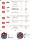
Co-expression patterns of genes regulated by SA on day 14 and 28. (A and B) The three most significant co-expression modules on day 14 (A) and day 28 (B) and their respective enriched GO terms. The profile number on the top left corner of each red box was assigned by STEM. The black lines in the red profile boxes depict the gene expression patterns in dose size order. The p-value of each profile is above the white profile box. Each line in the white box represents the expression pattern of a gene. Enriched GO terms for each module, the number of genes for the term, and the corresponding FDR (False discovery rate) are shown at the right. (C and D) Chord diagram of enriched GO terms and their genes in profile 40 on (C) day 14 and (D) day 28.
Figure 3
Figure 3
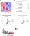
5% SA mainly regulates the gene expression profiles of extracellular matrix on day 14. (A) Heatmap of DEGs in 5%SA group (left) and control group (right) on day 14. (B) The most significant top 20 GO terms of DEGs. The dot size indicates the number of DEGs enriched in the corresponding GO term. Different p-values are indicated by dot colors. The rich factor is the ratio of the DEG number to the total number of genes in the GO term. (C and D) Volcano plots of genes included in GO terms for (C) collagen-containing extracellular matrix and (D) extracellular matrix structural constituent. (E) Representation of DEGs related to extracellular matrix structural constituent conferring tensile strength.
Figure 4
Figure 4
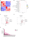
5% SA upregulates genes associated with keratin filament and keratinization on day 28. (A) Heatmap of DEGs in 5%SA group (left) and control group (right) on day 28. (B) The most significant top 20 GO terms of DEGs. The dot size indicates the number of DEGs enriched in the corresponding GO term. Different p-values are indicated by dot colors. The rich factor is the ratio of the DEG number to the total number of genes in the GO term. (C and D) Volcano plots of genes included in GO terms for (C) keratin filament and (D) keratinization. (E) Representation of DEGs related to the late cornified envelope.
Figure 5
Figure 5
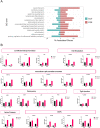
SA upregulates genes associated with the skin barrier. (A) Barrier-related significantly enriched GO terms between the 5%SA group and the control group. Green bars represent day 14, and red bars represent day 28. (B) Barrier biomarkers show greater differential expression across 5% SA vs the control group.
Figure 6
Figure 6
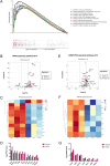
KEGG pathway analysis on day 14. (A) Multiple GSEA analysis of the KEGG pathway which achieved the top 10 highest enrichment scores. (B) The volcano plot shows the expression of PPAR signaling pathway associated genes. (C) Heatmap of PPAR signaling pathway associated genes between 5%SA and vehicle on day 14. (D) The FPKM values of genes associated with the PPAR signaling pathway. (E) The volcano plot shows the expression of cGMP-PKG signaling pathway associated genes. (F) Heatmap of cGMP-PKG signaling pathway associated genes between 5%SA and vehicle on day 14. (G) The FPKM values of genes associated with the cGMP-PKG signaling pathway.
Figure 7
Figure 7
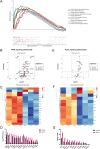
KEGG pathway analysis on day 28. (A) Multiple GSEA analysis of the KEGG pathway which achieved the top 10 highest enrichment scores. (B) The volcano plot shows the expression of PPAR signaling pathway associated genes. (C) Heatmap of PPAR signaling pathway associated genes between 5%SA and vehicle on day 28. (D) The FPKM values of genes associated with the PPAR signaling pathway. (E) The volcano plot shows the expression of NOTCH signaling pathway associated genes. (F) Heatmap of NOTCH signaling pathway associated genes between 5%SA and vehicle on day 28. (G) The FPKM values of genes associated with the NOTCH signaling pathway.

References

    1. Natsuga K. Epidermal barriers. Cold Spring Harb Perspect Med. 2014;4(4):a018218. doi:10.1101/cshperspect.a018218 - DOI - PMC - PubMed
    1. Lee AY. Molecular mechanism of epidermal barrier dysfunction as primary abnormalities. Int J Mol Sci. 2020;21(4):1. - PMC - PubMed
    1. Egawa G, Kabashima K. Barrier dysfunction in the skin allergy. Allergol Int. 2018;67(1):3–11. doi:10.1016/j.alit.2017.10.002 - DOI - PubMed
    1. Lefevre-Utile A, Braun C, Haftek M, Aubin F. Five functional aspects of the epidermal barrier. Int J Mol Sci. 2021;22(21):11676. doi:10.3390/ijms222111676 - DOI - PMC - PubMed
    1. Eyerich S, Eyerich K, Traidl-Hoffmann C, Biedermann T. Cutaneous barriers and skin immunity: differentiating a connected network. Trends Immunol. 2018;39(4):315–327. doi:10.1016/j.it.2018.02.004 - DOI - PubMed