On the Analyses of Medical Images Using Traditional Machine Learning Techniques and Convolutional Neural Networks
- PMID: 37260910
- PMCID: PMC10071480
- DOI: 10.1007/s11831-023-09899-9
On the Analyses of Medical Images Using Traditional Machine Learning Techniques and Convolutional Neural Networks
Abstract
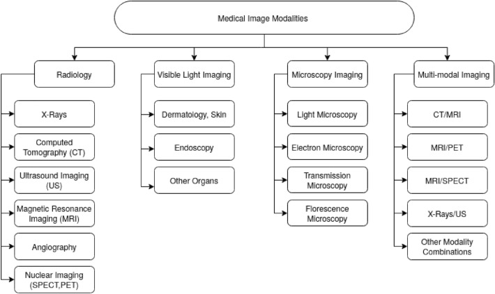
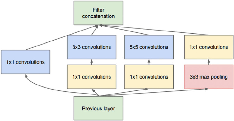
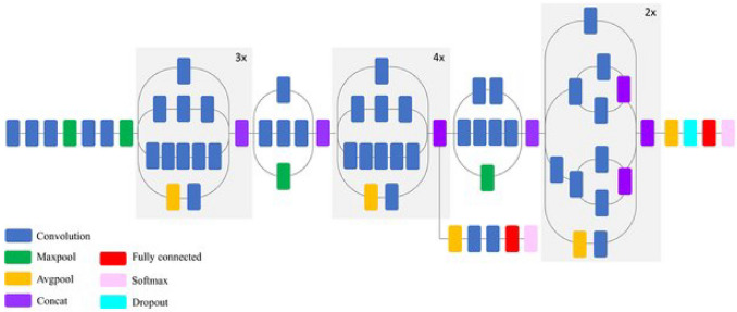
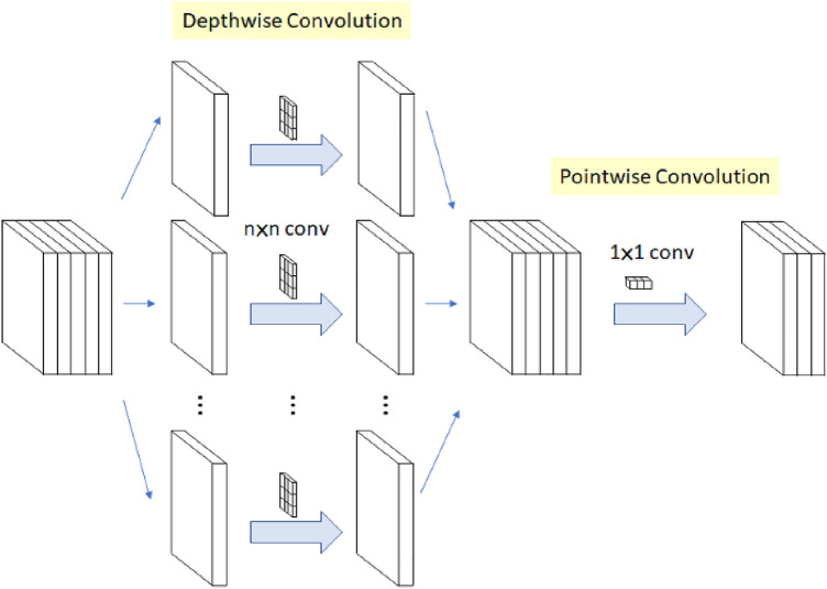
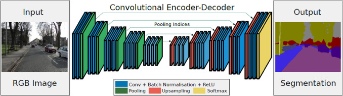
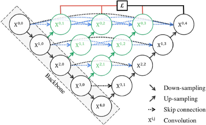
Convolutional neural network (CNN) has shown dissuasive accomplishment on different areas especially Object Detection, Segmentation, Reconstruction (2D and 3D), Information Retrieval, Medical Image Registration, Multi-lingual translation, Local language Processing, Anomaly Detection on video and Speech Recognition. CNN is a special type of Neural Network, which has compelling and effective learning ability to learn features at several steps during augmentation of the data. Recently, different interesting and inspiring ideas of Deep Learning (DL) such as different activation functions, hyperparameter optimization, regularization, momentum and loss functions has improved the performance, operation and execution of CNN Different internal architecture innovation of CNN and different representational style of CNN has significantly improved the performance. This survey focuses on internal taxonomy of deep learning, different models of vonvolutional neural network, especially depth and width of models and in addition CNN components, applications and current challenges of deep learning.
© The Author(s) 2023.
Conflict of interest statement
Conflict of interestThe authors have no competing interests to declare that are relevant to the content of this article.
Figures


Similar articles
-
Language Processing Model Construction and Simulation Based on Hybrid CNN and LSTM.Comput Intell Neurosci. 2021 Jul 6;2021:2578422. doi: 10.1155/2021/2578422. eCollection 2021. Comput Intell Neurosci. 2021. Retraction in: Comput Intell Neurosci. 2023 Aug 2;2023:9757696. doi: 10.1155/2023/9757696. PMID: 34306049 Free PMC article. Retracted.
-
Combined Transfer Learning and Test-Time Augmentation Improves Convolutional Neural Network-Based Semantic Segmentation of Prostate Cancer from Multi-Parametric MR Images.Comput Methods Programs Biomed. 2021 Oct;210:106375. doi: 10.1016/j.cmpb.2021.106375. Epub 2021 Aug 28. Comput Methods Programs Biomed. 2021. PMID: 34500139
-
MRI-based brain tumor detection using convolutional deep learning methods and chosen machine learning techniques.BMC Med Inform Decis Mak. 2023 Jan 23;23(1):16. doi: 10.1186/s12911-023-02114-6. BMC Med Inform Decis Mak. 2023. PMID: 36691030 Free PMC article.
-
3D Deep Learning on Medical Images: A Review.Sensors (Basel). 2020 Sep 7;20(18):5097. doi: 10.3390/s20185097. Sensors (Basel). 2020. PMID: 32906819 Free PMC article. Review.
-
Application of Convolutional Neural Network-Based Detection Methods in Fresh Fruit Production: A Comprehensive Review.Front Plant Sci. 2022 May 16;13:868745. doi: 10.3389/fpls.2022.868745. eCollection 2022. Front Plant Sci. 2022. PMID: 35651761 Free PMC article. Review.
Cited by
-
Deep Learning Approaches for Medical Image Analysis and Diagnosis.Cureus. 2024 May 2;16(5):e59507. doi: 10.7759/cureus.59507. eCollection 2024 May. Cureus. 2024. PMID: 38826977 Free PMC article. Review.
-
Deep learning-based anatomical position recognition for gastroscopic examination.Technol Health Care. 2024;32(S1):39-48. doi: 10.3233/THC-248004. Technol Health Care. 2024. PMID: 38669495 Free PMC article.
-
HTRecNet: a deep learning study for efficient and accurate diagnosis of hepatocellular carcinoma and cholangiocarcinoma.Front Cell Dev Biol. 2025 Mar 24;13:1549811. doi: 10.3389/fcell.2025.1549811. eCollection 2025. Front Cell Dev Biol. 2025. PMID: 40196844 Free PMC article.
-
A Novel Heteromorphous Convolutional Neural Network for Automated Assessment of Tumors in Colon and Lung Histopathology Images.Biomimetics (Basel). 2023 Aug 16;8(4):370. doi: 10.3390/biomimetics8040370. Biomimetics (Basel). 2023. PMID: 37622975 Free PMC article.
-
Robust Bi-CBMSegNet framework for advancing breast mass segmentation in mammography with a dual module encoder-decoder approach.Sci Rep. 2025 Jul 8;15(1):24434. doi: 10.1038/s41598-025-09775-5. Sci Rep. 2025. PMID: 40628947 Free PMC article.
References
-
- Winsberg F, Elkin M, Jr Macy J, Bordaz V, Weymouth W. Detection of radiographic abnormalities in mammograms by means of optical scanning and computer analysis. Radiology. 1967;89(2):211–215. doi: 10.1148/89.2.211. - DOI
-
- Kimme C, O’Loughlin BJ, Sklansky J (1977) Automatic detection of suspicious abnormalities in breast radiographs. In: Data structures, computer graphics, and pattern recognition, pp 427–447. Elsevier
-
- Ishida M, Kato H, Doi K, Frank PH (1982) Development of a new digital radiographic image processing system. In: Application of optical instrumentation in medicine X, vol 347, pp 42–48. SPIE
-
- Chen CM, Chou YH, Tagawa N, Do Y (2013) Computer-aided detection and diagnosis in medical imaging
LinkOut - more resources
Full Text Sources