

Systematic Review of Artificial Intelligence for Abnormality Detection in High-volume Neuroimaging and Subgroup Meta-analysis for Intracranial Hemorrhage Detection
- PMID: 37261453
- PMCID: PMC10233528
- DOI: 10.1007/s00062-023-01291-1
Systematic Review of Artificial Intelligence for Abnormality Detection in High-volume Neuroimaging and Subgroup Meta-analysis for Intracranial Hemorrhage Detection
Abstract
Purpose: Most studies evaluating artificial intelligence (AI) models that detect abnormalities in neuroimaging are either tested on unrepresentative patient cohorts or are insufficiently well-validated, leading to poor generalisability to real-world tasks. The aim was to determine the diagnostic test accuracy and summarise the evidence supporting the use of AI models performing first-line, high-volume neuroimaging tasks.
Methods: Medline, Embase, Cochrane library and Web of Science were searched until September 2021 for studies that temporally or externally validated AI capable of detecting abnormalities in first-line computed tomography (CT) or magnetic resonance (MR) neuroimaging. A bivariate random effects model was used for meta-analysis where appropriate. This study was registered on PROSPERO as CRD42021269563.
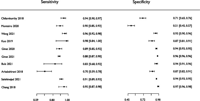
Results: Out of 42,870 records screened, and 5734 potentially eligible full texts, only 16 studies were eligible for inclusion. Included studies were not compromised by unrepresentative datasets or inadequate validation methodology. Direct comparison with radiologists was available in 4/16 studies and 15/16 had a high risk of bias. Meta-analysis was only suitable for intracranial hemorrhage detection in CT imaging (10/16 studies), where AI systems had a pooled sensitivity and specificity 0.90 (95% confidence interval [CI] 0.85-0.94) and 0.90 (95% CI 0.83-0.95), respectively. Other AI studies using CT and MRI detected target conditions other than hemorrhage (2/16), or multiple target conditions (4/16). Only 3/16 studies implemented AI in clinical pathways, either for pre-read triage or as post-read discrepancy identifiers.
Conclusion: The paucity of eligible studies reflects that most abnormality detection AI studies were not adequately validated in representative clinical cohorts. The few studies describing how abnormality detection AI could impact patients and clinicians did not explore the full ramifications of clinical implementation.
Keywords: Anomaly detection; Brain MRI; Clinical validation; Deep learning; Machine learning.
© 2023. The Author(s).
Conflict of interest statement
S. Agarwal, D. Wood, M. Grzeda, C. Suresh, M. Din, J. Cole, M. Modat and T.C. Booth declare that they have no competing interests.
Figures


References
-
- Dixon S. Diagnostic imaging dataset annual statistical release 2020/21. 2021. https://www.england.nhs.uk/statistics/statistical-work-areas/diagnostic-.... Accessed 20 Mar 2023.
-
- The Royal College of Radiologists London. Clinical radiology UK workforce census 2020 report. 2020. https://www.rcr.ac.uk/system/files/publication/field_publication_files/c.... Accessed 20 Mar 2023.
-
- World Health Organization. Cancer control: early detection. WHO Guide for effective programmes. 2007. http://apps.who.int/iris/bitstream/10665/43743/1/9241547338_eng. Accessed 15 Feb 2022.
Publication types
MeSH terms
Grants and funding
LinkOut - more resources
Full Text Sources
Medical
