. 2023 Jun 27;42(6):112591.
doi: 10.1016/j.celrep.2023.112591. Epub 2023 May 31.

Btbd11 supports cell-type-specific synaptic function

Affiliations

Btbd11 supports cell-type-specific synaptic function

Alexei M Bygrave et al. Cell Rep. .

Abstract

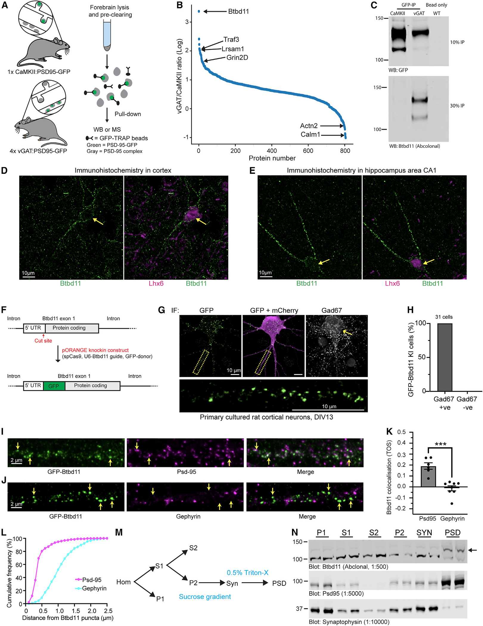
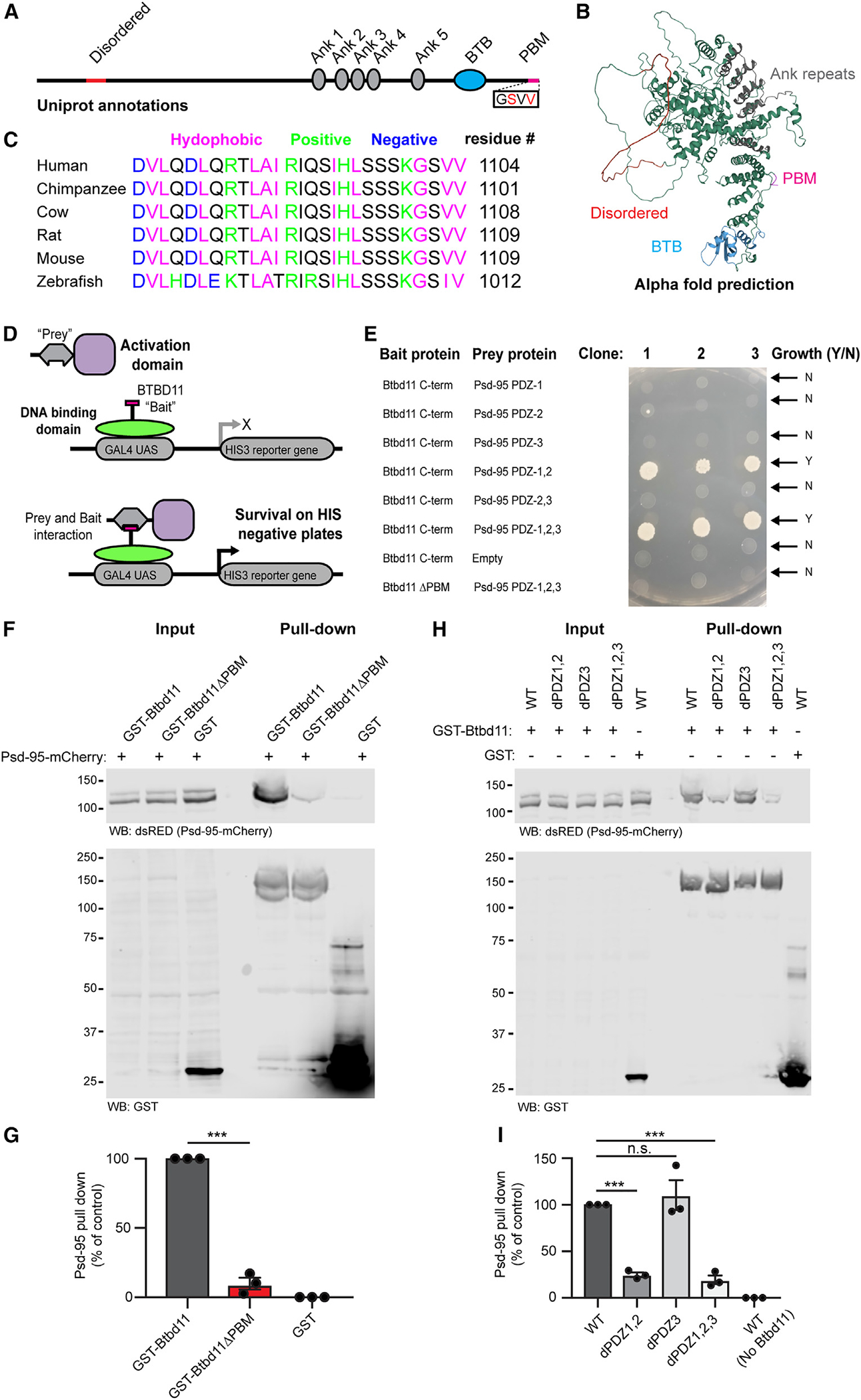
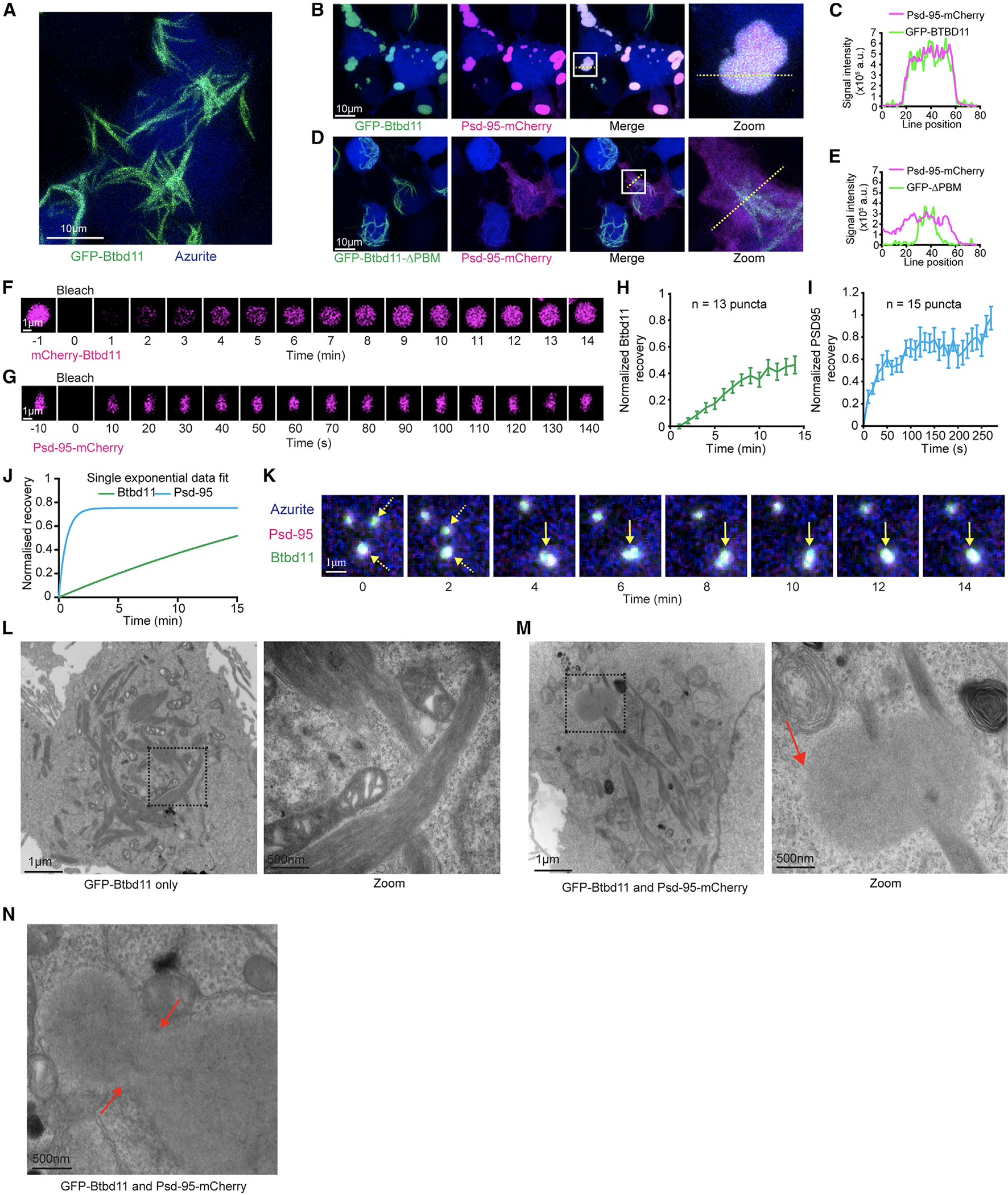
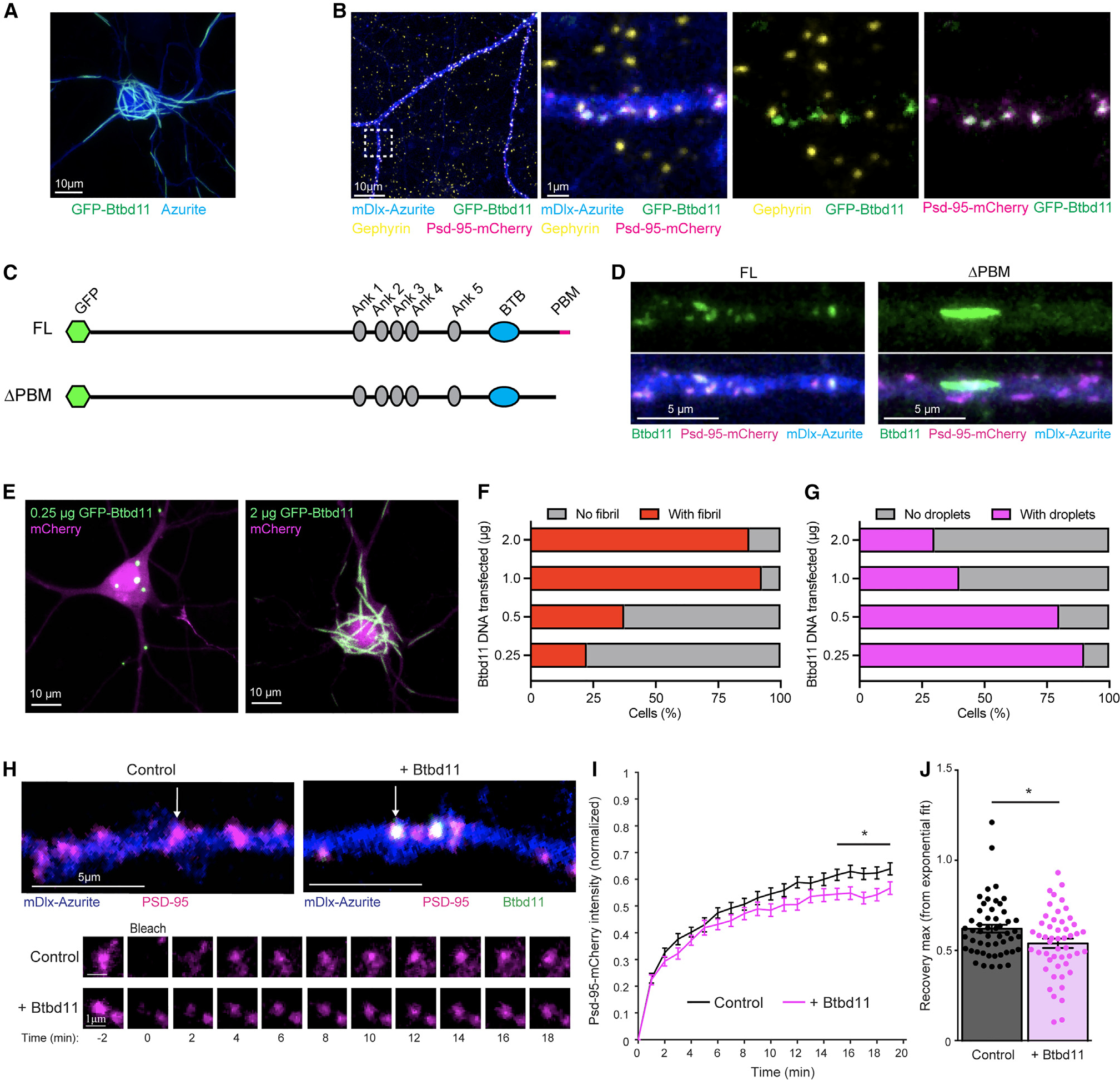
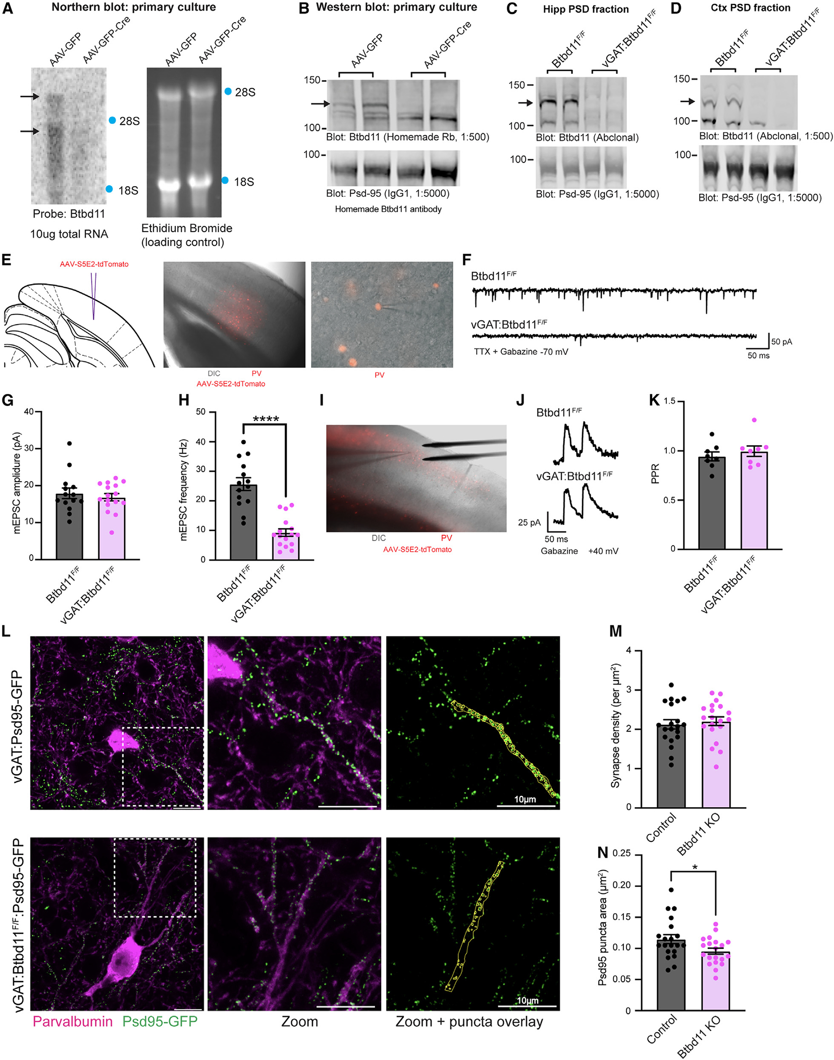
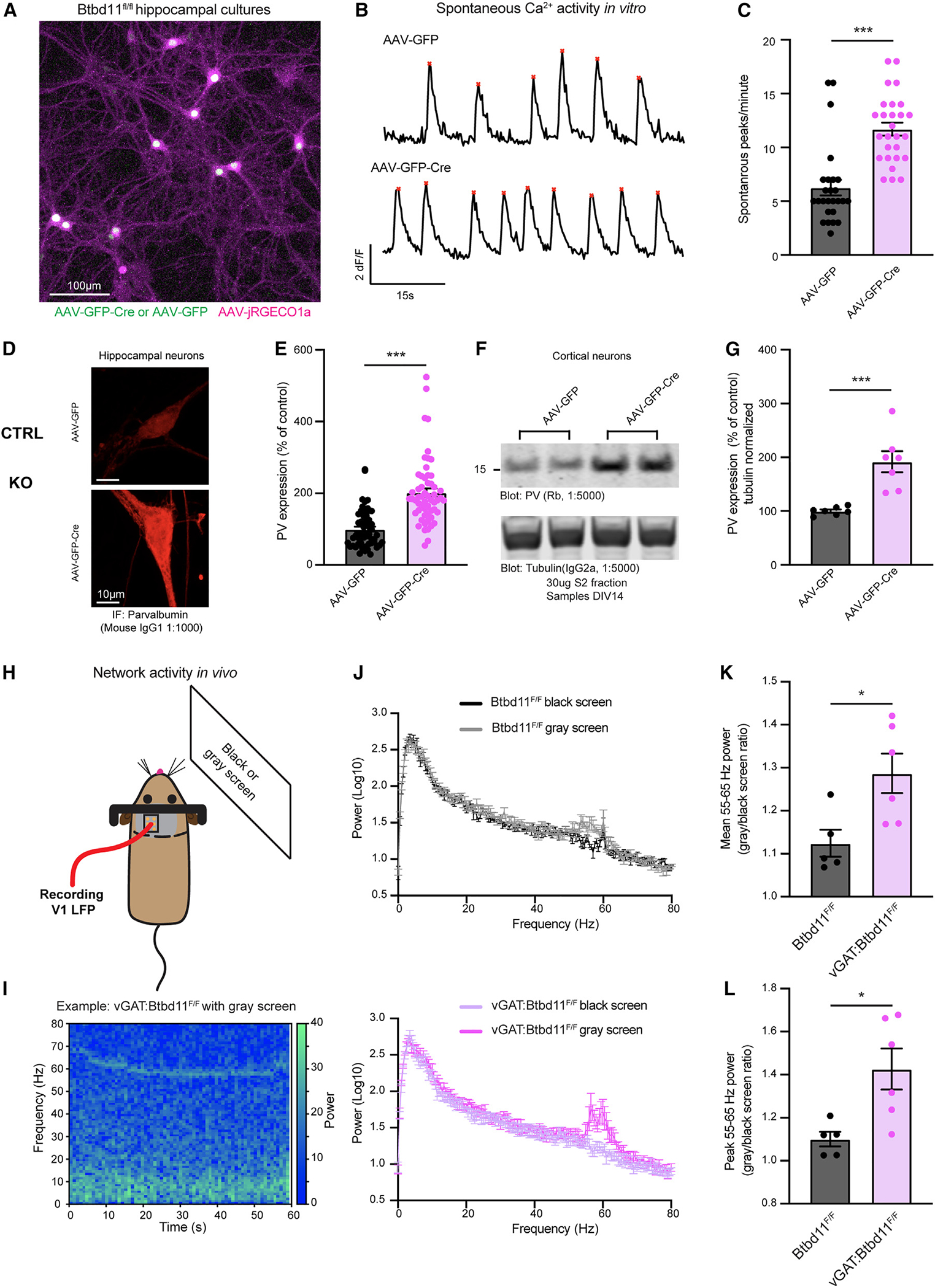
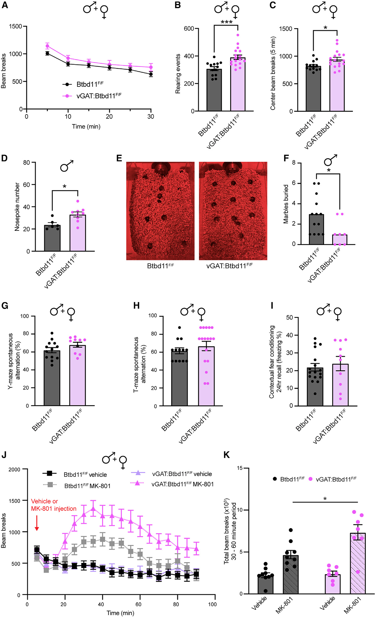
Synapses in the brain exhibit cell-type-specific differences in basal synaptic transmission and plasticity. Here, we evaluated cell-type-specific specializations in the composition of glutamatergic synapses, identifying Btbd11 as an inhibitory interneuron-specific, synapse-enriched protein. Btbd11 is highly conserved across species and binds to core postsynaptic proteins, including Psd-95. Intriguingly, we show that Btbd11 can undergo liquid-liquid phase separation when expressed with Psd-95, supporting the idea that the glutamatergic postsynaptic density in synapses in inhibitory interneurons exists in a phase-separated state. Knockout of Btbd11 decreased glutamatergic signaling onto parvalbumin-positive interneurons. Further, both in vitro and in vivo, Btbd11 knockout disrupts network activity. At the behavioral level, Btbd11 knockout from interneurons alters exploratory behavior, measures of anxiety, and sensitizes mice to pharmacologically induced hyperactivity following NMDA receptor antagonist challenge. Our findings identify a cell-type-specific mechanism that supports glutamatergic synapse function in inhibitory interneurons-with implications for circuit function and animal behavior.

Keywords: Btbd11; CP: Neuroscience; behavior; glutamatergic synapse; inhibitory interneurons; liquid-liquid phase separation; neuronal circuit; parvalbumin; proteomics.


Conflict of interest statement

Declaration of interests R.L.H. is scientific co-founder and SAB member of Neumora Therapeutics and SAB member of MAZE Therapeutics. M.S. is scientific co-founder and SAB member of Neumora Therapeutics and SAB member of Biogen, Cerevel, and Vanqua.

Figures

Figure 1.
Figure 1.. Identification of Btbd11 as an inPSD protein
(A) Schematic of Psd-95-GFP knockin in CaMKII or vGAT positive neurons to label excitatory and inhibitory neurons, respectively. (B) Proteins identified using semi-quantitative mass spectrometry. (C) Psd-95-GFP immuno-isolation with CaMKII:Psd-95-GFP and 4× vGAT:Psd-95-GFP mice (pooled) or a WT animal used for a bead-only control followed by western blots for GFP (detecting Psd-95-GFP, the “bait”) from 10% of the pull-down or Btbd11 (a candidate inPSD) from 30% of the pull-down material. This is a representative example of an experiment conducted 3 times. (D and E) Immunohistochemistry in WT mouse brain slices for Btbd11 (green) or Lhx6 (magenta) in layer 2/3 of the visual cortex (D) and area CA1 (E) of the hippocampus. Note the Btbd11-positive cells are expressing Lhx6, a marker for somatostatin and parvalbumin inhibitory interneurons. (F) Schematic of the CRISPR knockin strategy used to label endogenous Btbd11. (G) Example of GFP-Btbd11 knockin cell from primary cultured rat cortical neurons electroporated with knockin constructs and a mCherry cell fill (magenta). Immunofluorescence was used to boost the GFP-Btbd11 signal (green) and to identify GAD67-positive INs. Yellow arrow indicates a GAD67-positive cell. The area in the yellow dotted box is enlarged to show the GFP-Btbd11 signal. Respective scale bars indicate 10 μm. (H) All 31 GFP-Btbd11 knockin cells were also GAD67 positive. (I and J) Zoom in along the dendrites of GFP-Btbd11 knockin cells with immunofluorescence for Psd-95 or gephyrin, respectively. Yellow arrows indicate a subset of Btbd11 puncta. Scale bars indicate 2 μm. (K) Co-localization of GFP-Btbd11 puncta (identified with CRISPR knockin [KI]) with Psd-95 and gephyrin (visualized with immunofluorescence) using the threshold overlap score (TOS). Bars indicate the mean, and error bars show the SEM. ***p < 0.001. (L) Cumulative frequency plots indicating the puncta-to-puncta distance of Btbd11 with Psd-95 (magenta) or gephyrin (blue). (M) Schematic of the fractionation protocol used to isolate the postsynaptic density (PSD) from mouse cortex. P1, crude nuclear; S1 and S2, supernatant; P2, crude synaptosomal pellet; Syn, synaptosomes; PSD, postsynaptic density. (N) Western blot analysis of Btbd11 levels (top blot, 12 μg protein loaded) in different biochemical fractions of WT animals (1 male and 1 female, 3 months). Note the enrichment of Btbd11 in the PSD fraction. The band corresponding to Btbd11 is indicated with a black arrow (predicted weight 121.6 kDa). The fractionation efficiency was demonstrated with blots for Psd-95 (middle blot, enrichment in PSD) and synaptophysin (bottom blot, enrichment in SYN) with 2 μg protein loaded for each. See Table S2 for full statistical information on all tests. For further details, see Figure S1 and Table S1.
Figure 2.
Figure 2.. Btbd11 contains a PDZ binding motif that interacts with PDZ1,2 of Psd-95
(A) Schematic depiction of Btbd11 with annotations from Uniprot. Red: disordered, gray: ankyrin repeats (Ank), blue: BTB domain (BTB), magenta: PDZ binding motif (PDM). (B) Predicted structure of Btbd11 from AlphaFold with domains shaded as in (A). (C) C-terminal region of Btbd11 in different species showing the conservation of the PBM. (D) Schematic of targeted yeast 2-hybrid experiment to assess binding of Btbd11 with different PDZ domains of Psd-95. (E) Results from targeted yeast 2-hybrid experiment with growth indicating an interaction between Btbd11 and Psd-95. (F) GST pull-down experiments evaluating the ability of GST-Btbd11 or a mutant lacking the PBM (GST-Btbd11ΔPBM) to interact with Psd-95-mCherry in HEK cells. GST only was included as a negative control. (G) Qualification of Psd-95-mCherry pulled down by GST-Btbd11 or GST-Btbd11ΔPBM. (H) GST pull-down experiments in HEK cells evaluating the ability of GST-Btbd11 to interact with Psd-95-mCherry point mutants designed to disrupt PDZ domain binding. (I) Quantification of Psd-95-mCherry mutants pulled down by GST-Btbd11. Bars display the mean, and error bars display SEM. ***p < 0.001. For further details, see Figure S2 and Video S1.
Figure 3.
Figure 3.. Liquid-liquid phase separation of Btbd11 when expressed with Psd-95
(A) Expression of GFP-Btbd11 (green) and an azurite cell fill (blue) in HEK cells led to the formation of large fibril-like assemblies. Scale bar: 10 μm. (B and C) Co-expression of GFP-Btbd11 (green) with Psd-95-mCherry (magenta) and an azurite cell fill (blue). The region within the white box is enlarged on the right (zoom), and the yellow dotted line indicates where a line scan (C) is shown for Psd-95-mCherry and GFP-Btbd11. (D and E) Co-expression of GFP-Btbd11ΔPBM (green) with Psd-95-mCherry (magenta) and an azurite cell fill (blue). The region within the white box is enlarged on the right (zoom), and the yellow dotted line indicates where a line scan (E) is shown for Psd-95-mCherry and GFP-Btbd11ΔPBM. (F and G) Fluorescence recovery after photobleaching (FRAP) of mCherry-Btbd11 and Psd-95-mCherry co-expressed with Psd-95-GFP and GFP-Btbd11, respectively. Note the difference in timescales for Btbd11 and Psd-95. Scale bar: 1 μm. (H and I) Quantification of fluorescence recovery for Btbd11 (n = 13) and Psd-95 (n = 15), respectively. Error bars indicate SEM. (J) Plot of the exponential fit of FRAP data for Btbd11 (green) and Psd-95 (blue). (K) Longitudinal confocal imaging of a HEK cell transfected with GFP-Btbd11 (green), Psd-95-mCherry (magenta), and an azurite cell fill (blue). Yellow dotted arrows indicate two puncta that come together and form a single droplet (solid yellow arrow). Scale bar: 1 μm. (L and M) Electron microscope images of HEK cells transfected with GFP-Btbd11 or GFP-Btbd11 and Psd-95-mCherry, respectively. The black dotted box is enlarged on the right of each panel. Respective scale bars are indicted at the bottom left of each image. In (M), the red arrow indicates a putative droplet observed with Btbd11 and Psd-95 co-expression but not with Btbd11 expression alone. (N) An example of a putative Btbd11 and Psd-95 droplet that looks to be in the process of fusion or fission with red arrows indicating the neck. Respective scale bars are indicated at the bottom left of each image. For further details, see Figure S3 and Video S2.
Figure 4.
Figure 4.. Exogenous expression of Btbd11 stabilizes Psd-95 at glutamatergic synapses
(A) Confocal image from a primary cultured rat hippocampal neuron transfected with GFP-Btbd11 (green) and azurite as a cell fill (blue). Scale bar: 10 μm. (B) Confocal image from a primary cultured putative IN transfected with GFP-Btbd11 (green), Psd-95-mCherry (magenta), and mDlx-azurite (blue) with immunohistochemistry for gephyrin (yellow). The region within the white dotted box is enlarged on the right with different combinations of channels. Scale bars are in the bottom left of the images. (C) Schematic of GFP-Btbd11 and GFP-Btbd11ΔPBM constructs. (D) Confocal image of putative primary cultured hippocampal interneurons, identified with an azurite cell fill (blue) under the control of the mDlx enhancer to drive expression in INs. Full-length (FL) GFP-Btbd11 (left) and GFP-Btbd11ΔPBM (ΔPBM, right) shown in green were co-expressed with Psd-95-mCherry (magenta). Scale bar: 5 μm. (E) Confocal image from transfected primary cultured rat hippocampal neurons with varying amounts of GFP-Btbd11 (green) and mCherry as a cell fill (magenta). Scale bar: 10 μm. (F and G) Quantification of the proportion of cells with fibrils (red bars) or droplets (magenta bars), respectively, when GFP-Btbd11 is transfected in different quantities. (H) Live-cell confocal imaging and FRAP experiments in putative INs (identified with mDlx-azurite) in which Psd-95-mCherry (magenta) is bleached when expressed alone or in the presence of GFP-Btbd11 (green). The bottom panels show FRAP of the individual puncta labeled in the top panels with a white arrow. (I) Quantification of Psd-95-mCherry FRAP under control conditions (black) or with overexpression of Btbd11 (magenta). *p < 0.05. (J) Quantification of the estimated recovery maximum from an exponential fit of the FRAP data for each bleached punctum. Lines or bars show the mean, and error bars display SEM. For further details, see Figure S4 and Video S3.
Figure 5.
Figure 5.. Btbd11 KO reduces glutamatergic signaling in PV-INs
(A) Northern blot to evaluate levels of Btbd11 mRNA in Btbd11F/F cultures transduced with AAV-GFP (left lane) or AAV-GFP-Cre (right lane) and harvested at DIV14. 10 μg RNA was loaded onto a gel, and ethidium bromide staining was used to confirm equal loading of RNA. Locations of 18S and 28S ribosomal RNA are indicated with blue dots. (B) Western blots characterizing the levels of Btbd11 (homemade antibody, top blot) and Psd-95 (bottom blot, as a loading control) in primary cortical Btbd11F/F cultures transduced with AAV-GFP (left lanes) or AAV-GFP-Cre (right lanes). 12 μg protein was loaded from the PSD fraction. Cells harvested at DIV12 and DIV14 (one sample per condition per time point). A black arrow indicates the band corresponding to Btbd11. (C and D) Western blots evaluating the levels of Btbd11 (Abclonal antibody, top blot) and Psd-95 (bottom blot, as a loading control) from the (C) hippocampal and (D) cortical PSD fractions of female Btbd11F/F or vGAT:Btbd11F/F mice aged 3 months. 20 μg protein was loaded. Note the hippocampal PSD material was not generated with a sucrose gradient and so serves as a proxy for the PSD fraction. A black arrow indicates the band corresponding to Btbd11. (E) Schematic depicting the site of AAV-S5E2-tdTomato injection, used to visualize PV-INs in the V1 (left), with zoomed-out (middle) and zoomed-in (right) merged images of differential interference contrast (DIC) and mCherry fluorescence. (F) Example mEPSC traces recorded from V1 PV-INs in Btbd11F/F or vGAT:Btbd11F/F mice recorded at −70 mV in the presence of TTX and gabazine. (G and H) The mEPSC amplitude and frequency, respectively, of mEPSCs recorded from PV-INs from Btbd11F/F (black) animals or vGAT:Btbd11F/F (magenta) mice. ****p < 0.0001. (I) DIC image showing the placement of the electrical stimulating electrode for recordings in the V1. (J) Example traces used to calculate the PPR in Btbd11F/F or vGAT:Btbd11F/F mice with cells held at +40 mV (to allow for the AMPA/NMDA ratio to also be calculated; see Figure S5G). (K) Summary PPR data recorded from PV-INs from Btbd11F/F (black) or vGAT:Btbd11F/F (magenta) mice. (L) Representative confocal images (maximum intensity projections) from the V1 of vGAT:Psd-95-GFP (control) or vGAT:Btbd11F/F:Psd-95-GFP (Btbd11 KO) slices. Endogenous Psd-95-GFP (green) shows putative glutamatergic synapses and is visualized alongside PV detected with immunohistochemistry (magenta). Psd-95-GFP puncta in PV neuron dendrites were manually identified (yellow regions of interest [ROIs], right panel). Scale bar: 10 μm. (M) Glutamatergic synapse density, calculated by the density of Psd-95-GFP puncta, in PV-INs in control and Btbd11 KO animals. (N) Median Psd-95-GFP puncta area per dendritic region in PV-INs of control and Btbd11 KO animals. *p < 0.05. Bars display the mean and error bars show S.E.M. For further details, see Figure S5.
Figure 6.
Figure 6.. Loss of Btbd11 impacts circuit function in vitro and in vivo
(A) Confocal live-cell imaging of primary cultured hippocampal neurons from Btbd11F/F mice transduced with AAV-jRGECO1a (magenta) and AAV-GFP or AAV-GFP-Cre (green). The example image is with AAV-GFP-Cre. Scale bar: 100 μm. (B) Example traces for control (AAV-GFP, top) and KO (AAV-GFP-Cre, bottom) cultures showing the average Ca2+ activity across multiple neurons in one field of view over a 60 s period. Red stars indicate automatically identified peaks. (C) Quantification of large Ca2+ transients across multiple ROIs and coverslips from 3 independent batches of neurons. Control (AAV-GFP) data are shown in black and KO (AAV-GFP-Cre) data in magenta. ***p < 0.001. (D) Confocal image showing immunofluorescence of PV in DIV14 primary culture hippocampal Btbd11F/F neurons transduced with AAV-GFP (control, top) or AAV-GFP-Cre (KO, bottom). Scale bar: 10 μm. (E) Quantification of PV immunofluorescence data with control data in black and KO data in magenta. (F) Western blot from the cytosolic S2 fraction of DIV14 primary cultured cortical Btbd11F/F neurons transduced with AAV-GFP (control) or AAV-GFP-Cre (KO). Top blot shows levels of PV, and the bottom blot shows alpha-tubulin used as a loading control. 30 μg lysate was run. (G) Quantification of western blot data evaluating levels of PV (normalized to alpha-tubulin levels) with control data in black and KO data in magenta. (H) Schematic of in vivo setup used to assess narrowband gamma oscillations in the V1 with presentation of a gray screen. (I) Example spectrogram showing the power over time in the 0–80 Hz range from the V1 of a vGAT:Btbd11F/F mouse presented with a gray screen. (J) Power spectra for Btbd11F/F mice (n = 5, top; dark gray: dark screen, light gray: gray screen) and vGAT:Btbd11F/F mice (n = 6, bottom; light magenta: dark screen, dark magenta gray: gray screen). (K and L) Quantification of the mean or peak 55–65 Hz activity, respectively, presented as a ratio of gray screen/black screen. *p < 0.05. Bars display mean, and error bars show SEM. For further details, see Figure S6 and Videos S4 andS5.
Figure 7.
Figure 7.. Btbd11 KO mice display altered exploratory behavior, reduced anxiety, and sensitization to a NMDA receptor antagonist
(A) Locomotor activity of male and female mice exploring a novel environment. (B) Rearing events during 30 min of open field exploration in male and female animals. ***p < 0.001. (C) Beam breaks in the center of the open field arena during the first 5 min of exploration in male and female animals. *p < 0.05. (D) Number of nose pokes made by male animals. (E) Representative photos showing the number of marbles buried by Btbd11F/F or vGAT:Btbd11F/F mice. (F) Quantification of marble burying data. Bars indicate the median. (G) Spontaneous alternation of male and female animals in a Y-maze test of short-term spatial memory. (H) T-maze spontaneous alternation performance in male and female animals. (I) Contextual fear conditioning recall data 24 h after conditioning. Freezing was used as a proxy for memory performance and compared between male and female animals. (J) Locomotor activity of mice in an open field arena following injection with either saline or MK-801 (0.2 mg/kg). (K) Quantification of the total infrared beam breaks in the 30–60 min period after injection. Unless specified, line or bars display mean, and error bars show SEM. Btbd11F/F animals displayed in black and vGAT:Btbd11F/F displayed in magenta throughout. For further details, see Figure S7.

References

    1. Hu H, Gan J, and Jonas P (2014). Interneurons. Fast-spiking, parvalbumin+ GABAergic interneurons: from cellular design to microcircuit function. Science 345, 1255263. 10.1126/science.1255263. - DOI - PubMed
    1. Marín O (2012). Interneuron dysfunction in psychiatric disorders. Nat. Rev. Neurosci. 13, 107–120. 10.1038/nrn3155. - DOI - PubMed
    1. Lisman JE, Coyle JT, Green RW, Javitt DC, Benes FM, Heckers S, and Grace AA (2008). Circuit-based framework for understanding neurotransmitter and risk gene interactions in schizophrenia. Trends Neurosci. 31, 234–242. 10.1016/j.tins.2008.02.005. - DOI - PMC - PubMed
    1. Lewis DA, Hashimoto T, and Volk DW (2005). Cortical inhibitory neurons and schizophrenia. Nat. Rev. Neurosci. 6, 312–324. 10.1038/nrn1648. - DOI - PubMed
    1. Sohal VS, Zhang F, Yizhar O, and Deisseroth K (2009). Parvalbumin neurons and gamma rhythms enhance cortical circuit performance. Nature 459, 698–702. 10.1038/nature07991. - DOI - PMC - PubMed

Publication types

MeSH terms

Substances