

Predicting vasospasm risk using first presentation aneurysmal subarachnoid hemorrhage volume: A semi-automated CT image segmentation analysis using ITK-SNAP
- PMID: 37262041
- PMCID: PMC10234558
- DOI: 10.1371/journal.pone.0286485
Predicting vasospasm risk using first presentation aneurysmal subarachnoid hemorrhage volume: A semi-automated CT image segmentation analysis using ITK-SNAP
Abstract
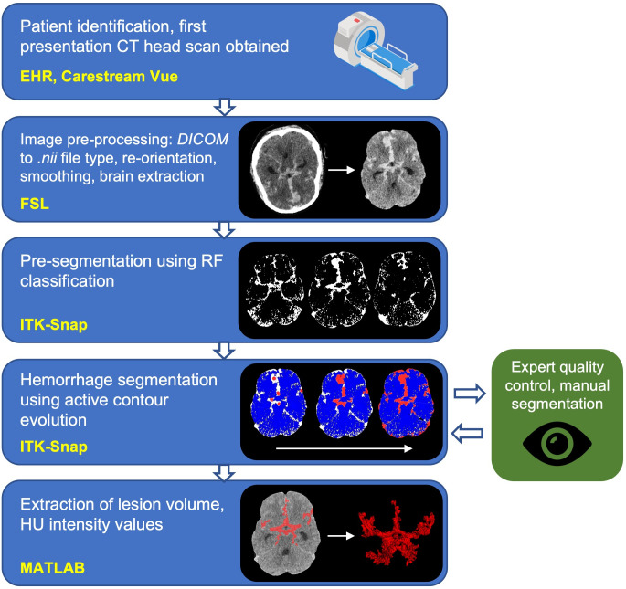
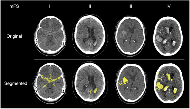
Purpose: Cerebral vasospasm following aneurysmal subarachnoid hemorrhage (aSAH) is a significant complication associated with poor neurological outcomes. We present a novel, semi-automated pipeline, implemented in the open-source medical imaging analysis software ITK-SNAP, to segment subarachnoid blood volume from initial CT head (CTH) scans and use this to predict future radiological vasospasm.
Methods: 42 patients were admitted between February 2020 and December 2021 to our tertiary neurosciences center, and whose initial referral CTH scan was used for this retrospective cohort study. Blood load was segmented using a semi-automated random forest classifier and active contour evolution implemented in ITK-SNAP. Clinical data were extracted from electronic healthcare records in order to fit models aimed at predicting radiological vasospasm risk.
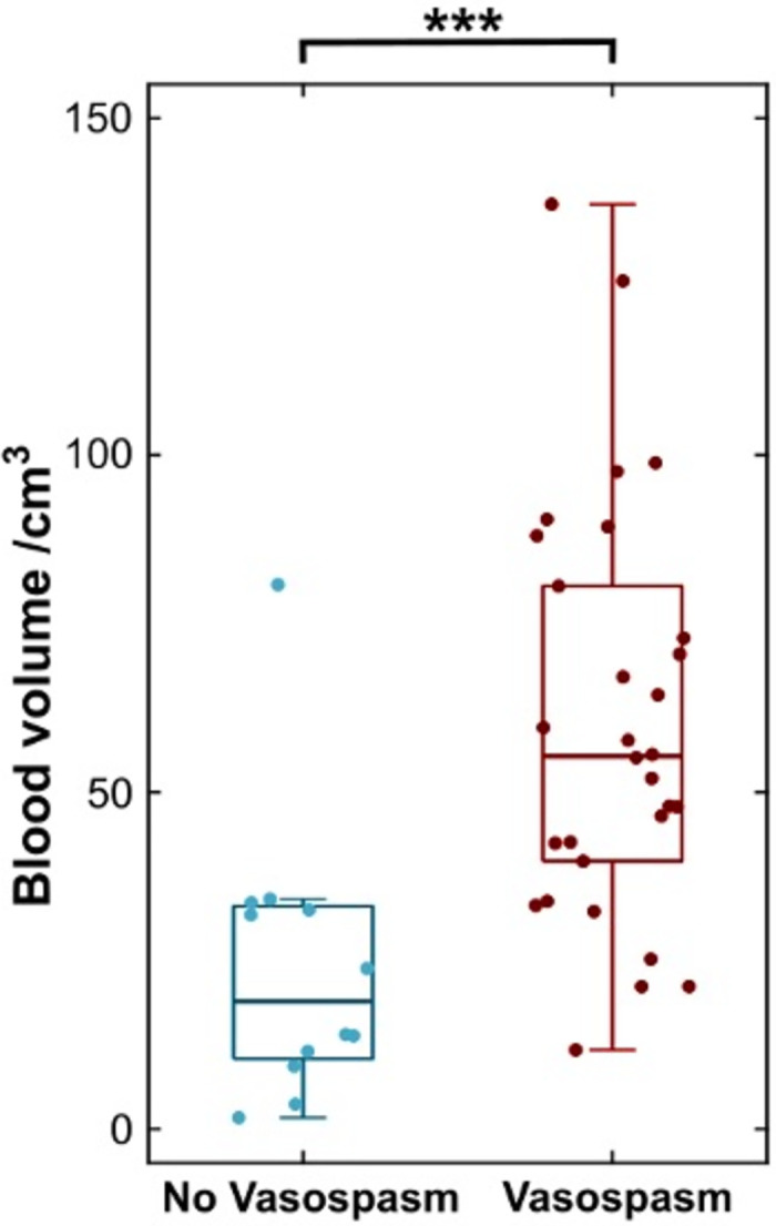
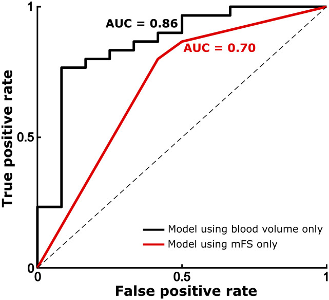
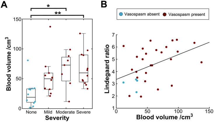
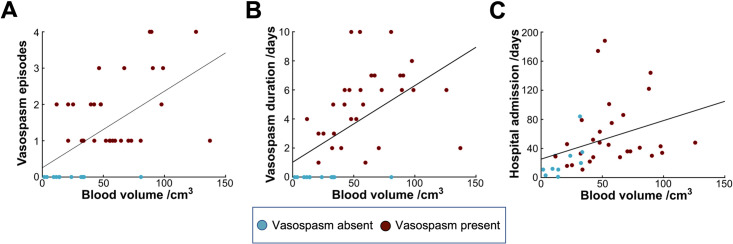
Results: Semi-automated segmentations demonstrated excellent agreement with manual, expert-derived volumes (mean Dice coefficient = 0.92). Total normalized blood volume, extracted from CTH images at first presentation, was significantly associated with greater odds of later radiological vasospasm, increasing by approximately 7% for each additional cm3 of blood (OR = 1.069, 95% CI: 1.021-1.120; p < .005). Greater blood volume was also significantly associated with vasospasm of a higher Lindegaard ratio, of longer duration, and a greater number of discrete episodes. Total blood volume predicted radiological vasospasm with a greater accuracy as compared to the modified Fisher scale (AUC = 0.86 vs 0.70), and was of independent predictive value.
Conclusion: Semi-automated methods provide a plausible pipeline for the segmentation of blood from CT head images in aSAH, and total blood volume is a robust, extendable predictor of radiological vasospasm, outperforming the modified Fisher scale. Greater subarachnoid blood volume significantly increases the odds of subsequent vasospasm, its time course and its severity.
Copyright: © 2023 Street et al. This is an open access article distributed under the terms of the Creative Commons Attribution License, which permits unrestricted use, distribution, and reproduction in any medium, provided the original author and source are credited.
Conflict of interest statement
The authors have declared that no competing interests exist.
Figures
References
Publication types
MeSH terms
LinkOut - more resources
Full Text Sources
