. 2023 Aug 2;31(8):2391-2407.
doi: 10.1016/j.ymthe.2023.05.004. Epub 2023 May 26.

A non-transmissible live attenuated SARS-CoV-2 vaccine

Affiliations

A non-transmissible live attenuated SARS-CoV-2 vaccine

Julia M Adler et al. Mol Ther. .

Abstract

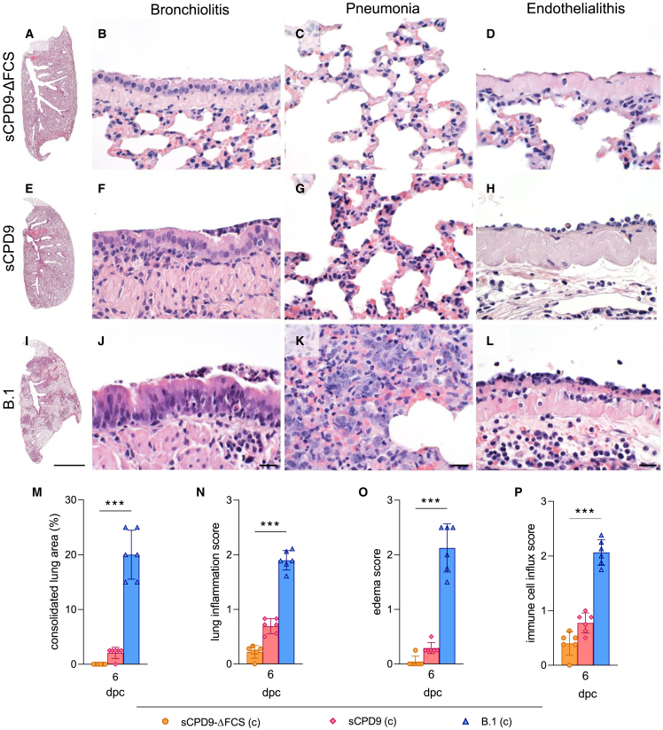
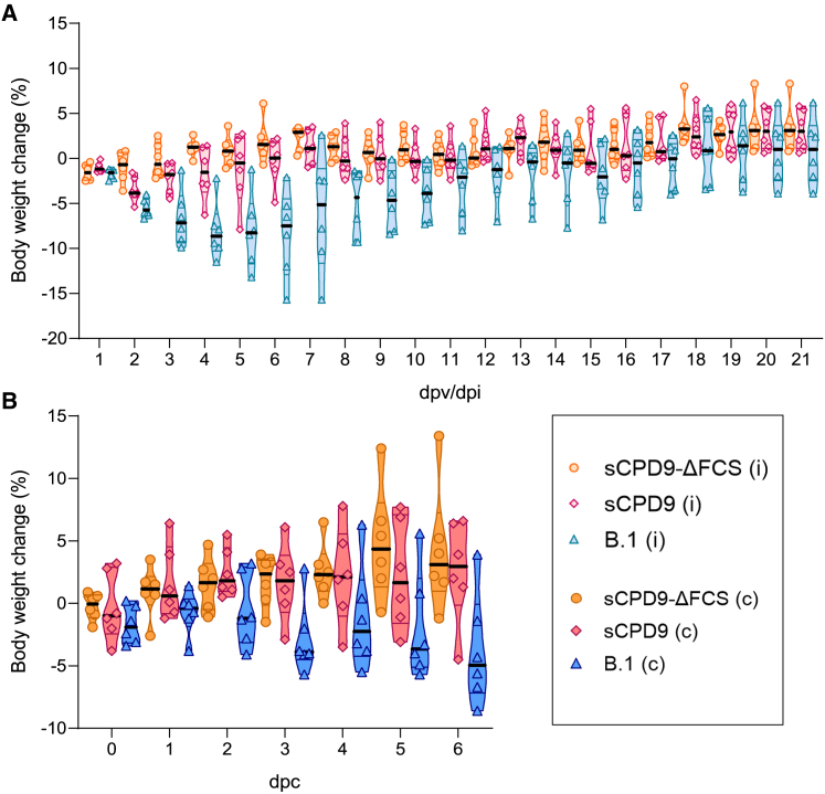
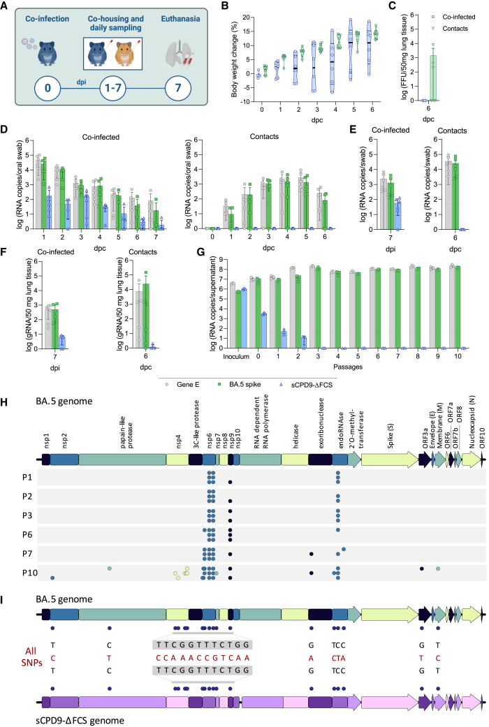
Live attenuated vaccines (LAVs) administered via the mucosal route may offer better control of the COVID-19 pandemic than non-replicating vaccines injected intramuscularly. Conceptionally, LAVs have several advantages, including presentation of the entire antigenic repertoire of the virus, and the induction of strong mucosal immunity. Thus, immunity induced by LAV could offer superior protection against future surges of COVID-19 cases caused by emerging SARS-CoV-2 variants. However, LAVs carry the risk of unintentional transmission. To address this issue, we investigated whether transmission of a SARS-CoV-2 LAV candidate can be blocked by removing the furin cleavage site (FCS) from the spike protein. The level of protection and immunity induced by the attenuated virus with the intact FCS was virtually identical to the one induced by the attenuated virus lacking the FCS. Most importantly, removal of the FCS completely abolished horizontal transmission of vaccine virus between cohoused hamsters. Furthermore, the vaccine was safe in immunosuppressed animals and showed no tendency to recombine in vitro or in vivo with a SARS-CoV-2 field strain. These results indicate that removal of the FCS from SARS-CoV-2 LAV is a promising strategy to increase vaccine safety and prevent vaccine transmission without compromising vaccine efficacy.

Keywords: COVID-19; SARS-CoV-2; live attenuated virus; mucosal vaccination; pneumonia; vaccine; virus transmission.


Conflict of interest statement

Declaration of interests Related to this work, Freie Universität Berlin has filed a patent application for the use of sCPD9 and sCPD9-ΔFCS as vaccine. In this application, J.T., N.O., and D.K. are named as inventors of sCPD9. Freie Universität Berlin is collaborating with RocketVax AG for further development of sCPD9-ΔFCS as vaccine and receives funding for research.

Figures

None
Graphical abstract
Figure 1
Figure 1
Multi-step growth kinetics and plaque sizes (A, B) Multi-step growth kinetics of sCPD9-ΔFCS, sCPD9, B.1-ΔFCS or B.1 viruses on Vero E6 (A) and Vero E6-TMPRSS2 (B) cells. Confluent cells grown in T25 flasks were infected with 100 ffu of the indicated virus and viral titers were determined 24, 48, 72, and 96 h post infection (hpi). The results are shown as means ± SD of duplicates. (C, D) Plaque size diameter of sCPD9-ΔFCS, sCPD9, B.1-ΔFCS, or B.1 viruses on Vero E6 (C) and Vero E6-TMPRSS2 (D) cells. The box-plots relative plaque diameters of 50 plaques normalized against the average plaque diameter of the sCPD9-ΔFCS virus. Shown are the mean and 25th to 75th percentiles with whiskers (min to max).
Figure 2
Figure 2
Virological findings in contact hamsters (A) Schematic overview of the experimental setup. Syrian hamsters were vaccinated either with sCPD9-ΔFCS or sCPD9, or infected with B.1 (wild type) on day 0. On day 1 after the vaccination/infection (dpv/dpi) naive contact hamsters (c) were placed in cohabitation with infected hamsters (i). Contact animals were sampled daily and euthanized after 6 days of cohabitation (dpc). The remaining infected hamsters were challenge-infected with SARS-CoV-2 variant Delta 21 days after vaccination/infection and euthanized on days 23 and 26. (B) Viral genomic RNA (gRNA) copies detected in daily oral swabs from contact hamsters n = 6 (1–6 dpc). Error bars show SD. Statistical analysis was performed using two-way ANOVA and Tukey’s multiple comparison tests. ∗p < 0.05, ∗∗p < 0.01, ∗∗∗p < 0.001, and ∗∗∗∗p < 0.0001. (C) Replication-competent virus particles in lung tissue. (D) gRNA copies detected in oropharyngeal swabs and homogenized lung tissue. (E) SARS-CoV-2 neutralization titers of sera collected at dpc 6 (upper detection limit = 1:1,024). (C–E). Error bars show SD. Dotted lines represent the limit of detection of each assay. Statistical analysis and p values were calculated using the Kruskal-Wallis test and Dunn’s multiple comparison test. ∗p < 0.05, ∗∗p < 0.01, and ∗∗∗p < 0.001.
Figure 3
Figure 3
Histopathological findings in contact hamsters (A–L) Hematoxylin and eosin-stained sections of lungs of naive hamsters that were in contact with the sCPD9-ΔFCS-vaccinated (A)–(D), sCPD9-vaccinated (E)–(H), or B.1-infected hamsters (I)–(L) at day 6 post contact. The lungs of hamsters that were in contact with sCPD9-ΔFCS-vaccinated hamsters showed no signs of inflammation in whole lung scan of left lung lobes (A), major airways (B), peripheral lung tissue (C), or blood vessels (D) at dpi 6. The lungs of hamsters that were in contact with sCPD9-vaccinated hamsters developed hardly any consolidation in left lung lobe scans (E) and only mild bronchiolitis (F), interstitial pneumonia (G), and vascular endothelialitis (H). In contrast, the lungs of hamsters that had contact to B.1-infected hamsters had multifocal patchy consolidation of their lungs (I), marked suppurative and necrotizing to proliferative bronchiolitis (J), marked diffuse alveolar damage with hyperplastic alveolar epithelial cells (K), and strong vascular endothelialitis (L) at 6 dpc. Size bars, 1 cm (A, E, I) or 30 μm (all others). (M)–(P) Histopathological scoring of lung parameters (n = 6). (M) Consolidated lung area in percentage per group on 6 dpc. (N) Inflammatory damage of the lungs is semi-quantitatively assessed in the lung inflammation score including severity of pneumonia; influx of neutrophils, lymphocytes, and macrophages; bronchial epithelial necrosis; bronchitis; alveolar epithelial necrosis; perivascular lymphocyte cuffs; and pneumocyte type II hyperplasia. (O) Edema score accounts for perivascular and alveolar edema and (P) immune cell influx score includes infiltration of lymphocytes, neutrophils, and macrophages as well as perivascular lymphocyte cuffs. (M)–(P) Results are displayed in mean ± SD with symbols indicating individual values. Statistical analysis was done with Kruskal-Wallis and Dunn’s multiple comparisons tests. ∗p < 0.05, ∗∗p < 0.01, and ∗∗∗p < 0.001.
Figure 4
Figure 4
Change in body weight of experimental animals (A) Body weight of infected/vaccinated hamsters (i) during the first 21 days after infection/vaccination. (B) Body weight of contact animals (c) during the cohousing period. (A) and (B) Violin plots (truncated) show weights of individual animals (n = 6), group medians, and quartiles.
Figure 5
Figure 5
Clinical, virological, and serological results in hamsters after challenge infection with SARS-CoV-2 variant Delta (A) Change in body weight of animals after challenge infection. Violin plots (truncated) show weights of individual animals (n = 6), group medians, and quartiles. (B) Viral gRNA in oropharyngeal swabs and lung tissue. (C) Infectious virus particles in homogenized lung tissue (n = 3). (D) Neutralizing activity of hamster sera collected on day 0 (before challenge, n = 6), and on days 2 and 5 post challenge (dpch, n = 3) against SARS-CoV-2 variants B.1, Delta, and BA.1 and BA.5 (upper detection limit = 1:1,024). (B)–(D) Error bars display SD. Dotted lines show the lower limits of detection. The statistical analysis was performed using two-way ANOVA and Tukey’s multiple comparison tests. ∗p < 0.05, ∗∗p < 0.01, ∗∗∗p < 0.001, and ∗∗∗∗p < 0.0001.
Figure 6
Figure 6
Pathological findings in hamsters after challenge infection with SARS-CoV-2 variant Delta The lungs of hamsters vaccinated with sCPD9-ΔFCS (A–E) or sCPD9 (F–J) had no or only minimal signs of pulmonary inflammation or other changes, including consolidation as seen in whole scans of left lung lobes (A, F), bronchiolitis (B, G), interstitial pneumonia (C, H), vascular endothelialitis (D, I), or lambertosis-like epithelial hyperplasia (E, J). Slightly stronger lesions were seen in hamsters that had been infected with B.1, including mild patchy consolidation of pulmonary parenchyma (K), moderate necrotizing and proliferative bronchiolitis (L), interstitial pneumonia (M), mild vascular endothelialitis (N), and marked lambertosis-like epithelial proliferation (O) 26 days after infection. In contrast to all three vaccinated/infected groups, unvaccinated (mock) hamsters developed strong histopathology lesions at 5 days after challenge, including marked patchy to confluent parenchymal consolidation (P), suppurative to necrotizing bronchiolitis (Q), marked diffuse alveolar damage (R), and strong vascular endothelialitis (S) but no lambertosis-like epithelial hyperplasia (T). (A) to (T), hematoxylin and eosin-stained sections of lesions representative of each group. Size bars, 1 cm (A, F, K, P), 50 μm (E, J, O, T), or 30 μm (all others). (U) Lung pathology scores with percentage of lung area consolidated by SARS-CoV-2 infection. (V) Lung inflammation score including influx of neutrophils, lymphocytes, and macrophages; bronchial epithelial necrosis; bronchitis; alveolar epithelial necrosis; and perivascular lymphocyte cuffs, as well as pneumocyte type II hyperplasia. (W) Pulmonary edema score including perivascular and alveolar edema and (X) immune cell influx score accounting for infiltration of lymphocytes, neutrophils, and macrophages, as well as perivascular lymphocyte cuffs (n = 3). (U–X) Results are shown in mean ± SD with symbols representing individual values. Data were analyzed using two-way ANOVA and Tukey’s multiple comparisons tests. ∗p < 0.05, ∗∗p < 0.01, and ∗∗∗p < 0.001.
Figure 7
Figure 7
In vivo and in vitro co-infection with sCPD9-ΔFCS and BA.5 (A) Experimental design. Syrian hamsters were co-infected with sCPD9-ΔFCS and the Omicron variant BA.5. One day post infection (dpi), naive contact hamsters were placed in cohabitation with infected hamsters. Oral swabs were collected daily. All animals were euthanized after 6 days of cohousing. (B) Change in body weight of animals after co-infection or contact to co-infected animals. Violin plots (truncated) show weights of individual animals (n = 6), group medians, and quartiles. (C) Infectious virus particles in homogenized lung tissue. Viral gRNA copies detected in oral swabs (D), oropharyngeal swabs (E), and lung tissue (F) from co-infected and contact hamsters (n = 6), using assays targeting the SARS-CoV-2 E gene (envelope), which is present in both viruses, or sequences that are uniquely present in one of the two different viruses—the spike gene of Omicron BA.5 virus, or the recoded sCPD9 region of sCPD9-ΔFCS virus. (G) Replication of Omicron BA.5 and sCPD9-ΔFCS in CaLu-3 cells. CaLu-3 cells were infected with 1,000 ffu of sCPD9-ΔFCS and 100 ffu of BA.5 and after 72 h of incubation, 1% of the supernatant was used as an inoculum for the next virus passage (n = 3). (C)–(G) Error bars show SD with symbols indicating individual results. Dotted lines represent the limits of detection. (H, I) CaLu-3 cells were infected with 1,000 ffu of sCPD9-ΔFCS and 100 ffu of BA.5 and after 72 h of incubation, either 1% or 5% of the supernatant was used as an inoculum for the next virus passage (n = 3). (H) SNPs identified in passages 1, 2, 3, 6, 7, and 10 of the co-infection experiments and their respective location within the BA.5 reference genome. The panel shows the SNPs identified in the three replicates that contained the most SNPs in each passage, irrespective of the passaging condition. Only SNPs that were identified in >10% of sequence reads are depicted. (I) All unique SNPs that emerged during the co-infection experiments (with >10% read support) in comparison with both BA.5 and sCPD9-ΔFCS genome reference sequences.
Figure 8
Figure 8
Effect of immunosuppression on safety, efficacy, and transmission of sCPD9-ΔFCS (A) Schematic overview of the experimental setup. Syrian hamsters were immunosuppressed by daily subcutaneous injections of dexamethasone (Dex) at a dose of 2 mg/kg, starting 3 days prior to vaccination or contact. After 3 days of treatment, the hamsters were vaccinated with sCPD9-ΔFCS and subsequently cohoused with naive and immunosuppressed contact animals 24 h after vaccination. Oral swabs were collected daily from all hamsters. Contacts were euthanized 6 days post contact (dpc), while vaccinated hamsters were euthanized 21 days post vaccination (dpv). (B) Change in body weight of immunosuppressed animals after vaccination or contact with vaccinated hamsters. Violin plots (truncated) show weights of individual animals (n = 6), group medians, and quartiles. (C) Viral gRNA copies in oral swabs. (D) gRNA copies in oropharyngeal swabs and lung tissue. (E) Replication-competent virus in lung tissue. (F) Neutralizing activity of sera collected from immunocompetent (IC) and immunosuppressed and sCPD9-ΔFCS-vaccinated animals 21 days post vaccination (dpv) against SARS-CoV-2 variant B.1 (upper detection limit = 1:1,024). A Mann-Whitney test failed to identify siginficant (p < 0.05) differences. (C)–(J) Error bars show SD with symbols representing individual values (n = 6). Dotted lines indicate the limits of detection.

References

    1. Pons-Salort M., Burns C.C., Lyons H., Blake I.M., Jafari H., Oberste M.S., Kew O.M., Grassly N.C. Preventing vaccine-derived poliovirus emergence during the polio endgame. PLoS Pathog. 2016;12:e1005728. doi: 10.1371/journal.ppat.1005728. - DOI - PMC - PubMed
    1. Bull J.J., Smithson M.W., Nuismer S.L. Transmissible viral vaccines. Trends Microbiol. 2018;26:6–15. doi: 10.1016/j.tim.2017.09.007. - DOI - PMC - PubMed
    1. Layman N.C., Tuschhoff B.M., Nuismer S.L. Designing transmissible viral vaccines for evolutionary robustness and maximum efficiency. Virus Evol. 2021;7:veab002. doi: 10.1093/ve/veab002. - DOI - PMC - PubMed
    1. Nuismer S.L., May R., Basinski A., Remien C.H. Controlling epidemics with transmissible vaccines. PLoS One. 2018;13:e0196978. doi: 10.1371/journal.pone.0196978. - DOI - PMC - PubMed
    1. Smithson M.W., Basinki A.J., Nuismer S.L., Bull J.J. Transmissible vaccines whose dissemination rates vary through time, with applications to wildlife. Vaccine. 2019;37:1153–1159. doi: 10.1016/j.vaccine.2019.01.018. - DOI - PMC - PubMed

Publication types

Supplementary concepts