. 2023 Oct;165(4):874-890.e10.
doi: 10.1053/j.gastro.2023.05.038. Epub 2023 May 30.

Transforming Growth Factor-β Blockade in Pancreatic Cancer Enhances Sensitivity to Combination Chemotherapy

Affiliations

Transforming Growth Factor-β Blockade in Pancreatic Cancer Enhances Sensitivity to Combination Chemotherapy

Li Qiang et al. Gastroenterology. 2023 Oct.

Abstract

Background & aims: Transforming growth factor-b (TGFb) plays pleiotropic roles in pancreatic cancer, including promoting metastasis, attenuating CD8 T-cell activation, and enhancing myofibroblast differentiation and deposition of extracellular matrix. However, single-agent TGFb inhibition has shown limited efficacy against pancreatic cancer in mice or humans.

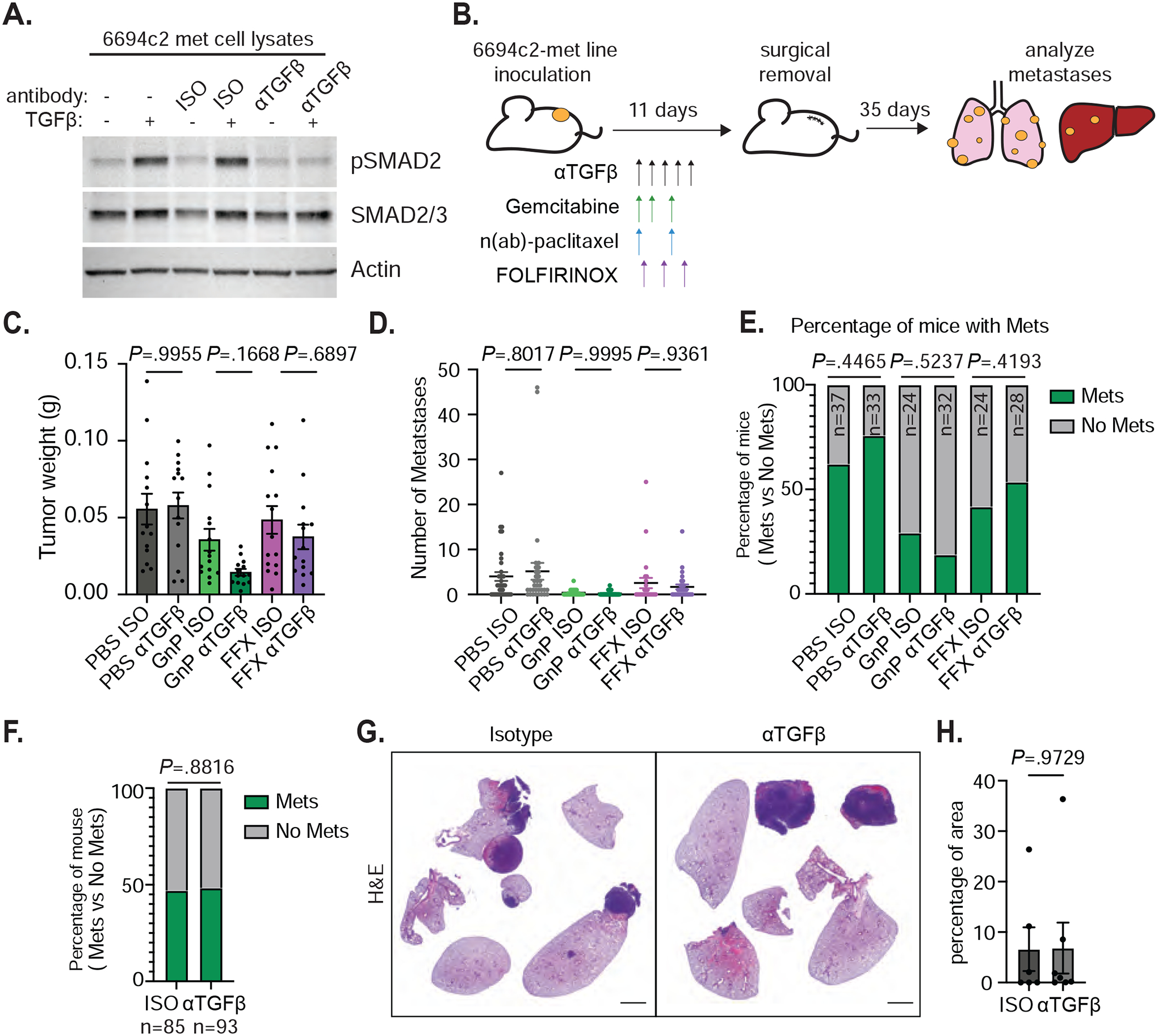
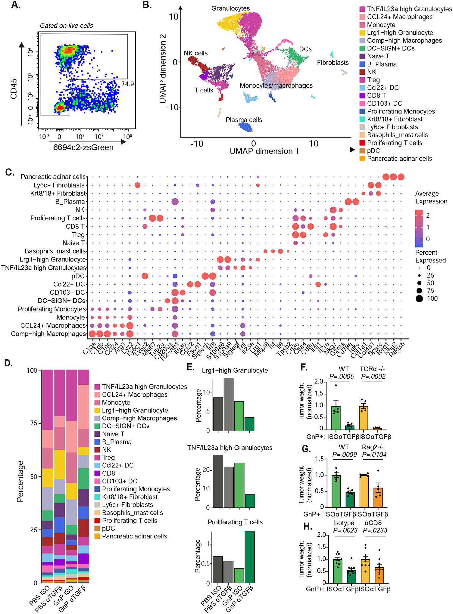
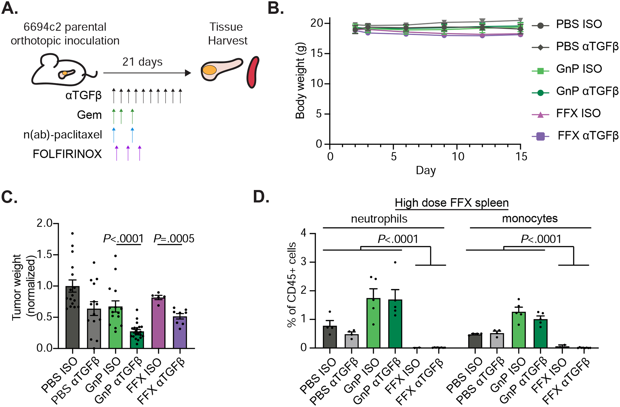
Methods: We evaluated the TGFβ-blocking antibody NIS793 in combination with gemcitabine/nanoparticle (albumin-bound)-paclitaxel or FOLFIRINOX (folinic acid [FOL], 5-fluorouracil [F], irinotecan [IRI] and oxaliplatin [OX]) in orthotopic pancreatic cancer models. Single-cell RNA sequencing and immunofluorescence were used to evaluate changes in tumor cell state and the tumor microenvironment.

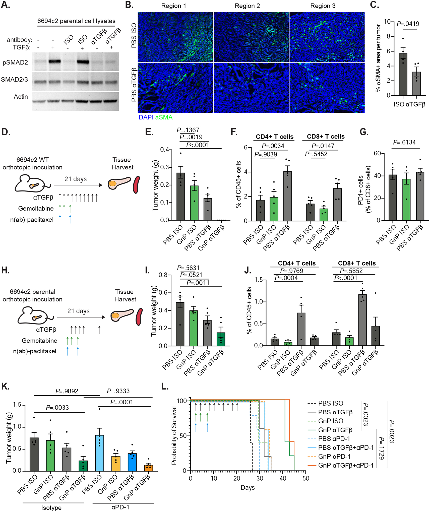
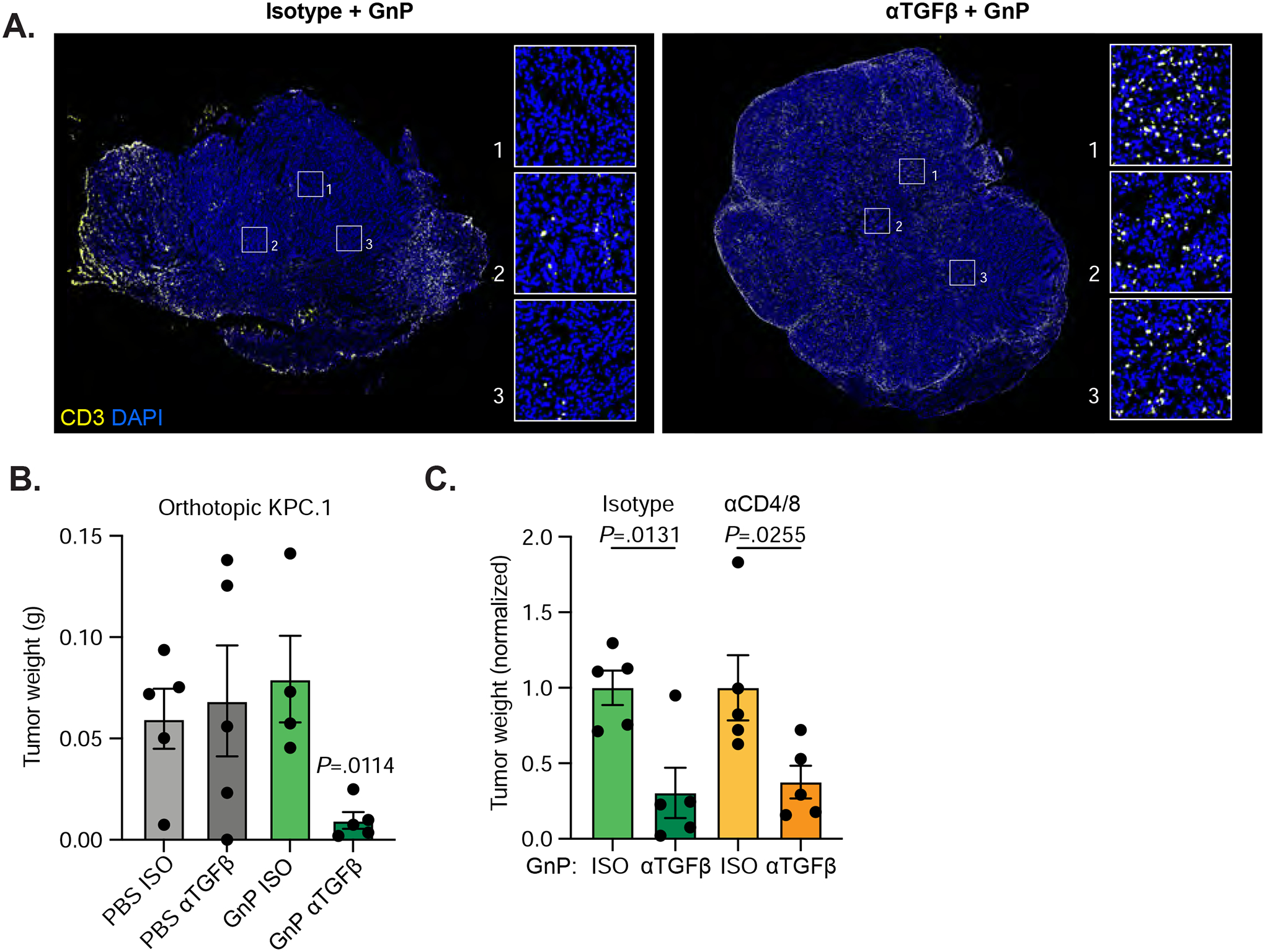
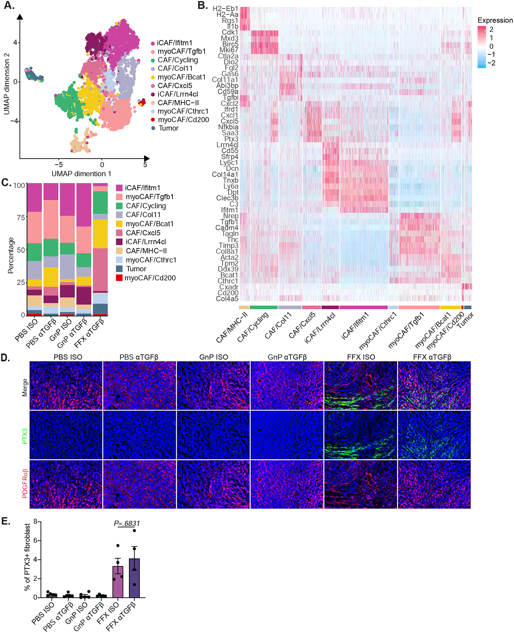
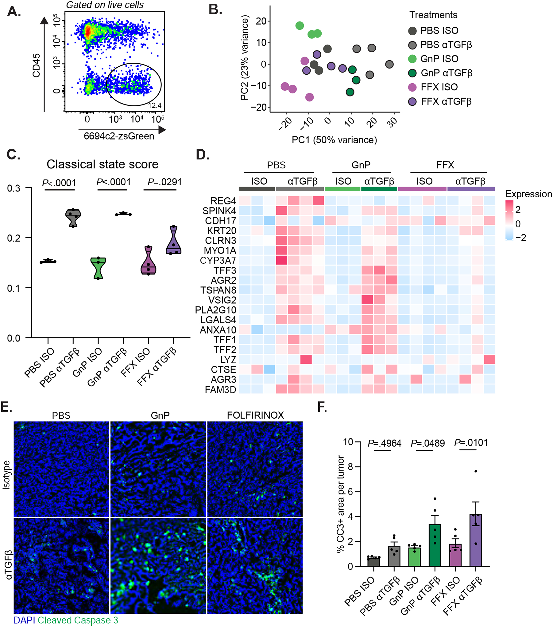
Results: Blockade of TGFβ with chemotherapy reduced tumor burden in poorly immunogenic pancreatic cancer, without affecting the metastatic rate of cancer cells. Efficacy of combination therapy was not dependent on CD8 T cells, because response to TGFβ blockade was preserved in CD8-depleted or recombination activating gene 2 (RAG2-/-) mice. TGFβ blockade decreased total α-smooth muscle actin-positive fibroblasts but had minimal effect on fibroblast heterogeneity. Bulk RNA sequencing on tumor cells sorted ex vivo revealed that tumor cells treated with TGFβ blockade adopted a classical lineage consistent with enhanced chemosensitivity, and immunofluorescence for cleaved caspase 3 confirmed that TGFβ blockade increased chemotherapy-induced cell death in vivo.

Conclusions: TGFβ regulates pancreatic cancer cell plasticity between classical and basal cell states. TGFβ blockade in orthotropic models of pancreatic cancer enhances sensitivity to chemotherapy by promoting a classical malignant cell state. This study provides scientific rationale for evaluation of NIS793 with FOLFIRINOX or gemcitabine/nanoparticle (albumin-bound) paclitaxel chemotherapy backbone in the clinical setting and supports the concept of manipulating cancer cell plasticity to increase the efficacy of combination therapy regimens.

Keywords: Chemotherapy; Immunotherapy; Pancreatic cancer; TGF-β; Tumor microenvironment.


Conflict of interest statement

SKD received research funding for this project from Novartis Institute for Biomedical Research. VC and MP are employees of Novartis Institute for Biomedical Research. SKD received research funding unrelated to this project from Eli Lilly and Company, Genocea, and Bristol-Myers Squibb and is a founder, science advisory board member (SAB) and equity holder in Kojin. MD has research funding unrelated to this project from Eli Lilly; he has received consulting fees from Genentech, ORIC Pharmaceuticals, Partner Therapeutics, SQZ Biotech, AzurRx, Eli Lilly, Mallinckrodt Pharmaceuticals, Aditum, Foghorn Therapeutics, Palleon, and Moderna; and he is a member of the Scientific Advisory Board for Neoleukin Therapeutics, Veravas, and Cerberus Therapeutics. SR holds equity in Amgen. AJA has consulted for Anji Pharmaceuticals, Arrakis Therapeutics, AstraZeneca, Boehringer Ingelheim, Oncorus, Inc., Merck & Co., Inc., Mirati Therapeutics, Nimbus Therapeutics, Plexium, Revolution Medicines, Reactive Biosciences, Riva Therapeutics, Servier Pharmaceuticals, Syros Pharmaceuticals, Tknife Therapeutics. AJA holds equity in Riva Therapeutics. AJA has research funding from Bristol Myers Squibb, Deerfield, Inc., Eli Lilly, Mirati Therapeutics, Novartis, Novo Ventures, Revolution Medicines, and Syros Pharmaceuticals.

Figures

Figure 1:
Figure 1:. TGFβ blockade does not affect metastasis formation in vivo.
A) 6694c2-met cells were cultured with 5ng/ml TGFβ, and/or 20ug/ml TGFβ blocking or isotype control antibody for 24h. Protein lysates were collected and analyzed by immunoblot. B) Diagram of experimental metastasis model. 180,000 6694c2-met cells were implanted subcutaneously in the lower back of C57BL/6 mice. Mice were treated with αTGFβ or isotype control i.p. every 2 days starting on day 2 for 5 doses. Gemcitabine was given on days 2, 4 and 7; nab-paclitaxel was dosed on days 2 and 7. FOLFIRINOX (5mg/kg oxaliplatin, 50mg/kg irinotecan, 75mg/kg leucovorin, and 75mg/kg 5-FU) was dosed on day 3. Primary tumors grew for 11 days before surgical removal. Metastases in the lung, liver and lymph node were analyzed at day 46. C) Primary tumor weight upon surgical removal. Data are representative of 3 independent experiments. D) Total metastases visually counted in different treatment groups. E) The percentage of mice containing metastases. F) The percentage of mice containing metastases from all isotype-treated or all αTGFβ-treated groups pooled from 3 independent experiments. G) Representative H&E image of metastases in the lungs from isotype and αTGFβ single agent treated mice. H) Quantification was performed on the H&E image for the percentage of area of metastases in the lungs from isotype and αTGFβ single agent treated mice. Scale bars = 2000px. Error bars are SEM throughout. Mann-Whitney t-test was used for comparison between two groups.
Figure 2:
Figure 2:. Anti-TGFβ GnP combination treatment is highly effective in poorly immunogenic mouse tumors.
A) 6694c2 parental cells were cultured with 5ng/ml TGFβ, and/or 20ug/ml TGFβ blocking or isotype control antibody for 24h. Protein lysates were collected and analyzed by immunoblot. B) 6694c2 tumors were inoculated into C57BL/6 mice, treated with TGFβ blocking antibody or isotype control and harvested at the midstage of growth on day 15 and frozen in OCT. Representative images from different regions of tumors stained with an antibody against αSMA and DAPI nuclear counterstain are shown. Scale bars = 100μm. C) Quantification of αSMA+ area per tumor. D) Diagram of experimental protocol for orthotopic 6694c2 mouse model for Figure 2E–G. 50,000 6694c2 parental cells were inoculated orthotopically into the pancreas of C57BL/6 mice. E). Tumor weights were measured on day 21. F) Tumor infiltrating CD4 and CD8 T cells were analyzed by flow cytometry of day 21 tumors. G) Percentage of PD1+ tumor infiltrating CD8 T cells. H) Diagram of experimental protocol Figure 2I–J. TGFβ blockade or isotype were dosed i.p. on day 4, day 6, day 8, day 10 and day 20; gemcitabine was given i.p. on day 4, day 7 and day 10; nab-paclitaxel was dosed i.v. on day 4 and day 10. I) Tumor weights were measured on day 21. J) Tumor infiltrating CD4 T cells and CD8 T cells were analyzed by flow cytometry. K-L) 6694c2 tumors were inoculated into mice, treated with TGFβ blockade (every 2 days), PD-1 blockade (twice a week) or corresponding isotype controls until day 20, along with gemcitabine and nab-paclitaxel treatments. K) Tumor weights were measured on day 21; L) Survival of a separate cohort was monitored. N=5 per group. Error bars are SEM throughout.
Figure 3.
Figure 3.. Anti-TGFβ GnP combination treatment reverses the immunosuppressive environment, yet is not dependent on T cells.
A) 6694c2-zsGreen cells were inoculated orthotopically into C57BL/6 mice, treated with αTGFβ or Isotype +/− GnP as illustrated in Figure 2D. Tumors were harvested on day 15, digested, and live single cells except zsGreen+ tumor cells were sorted by FACS for single-cell transcriptional profiling. n = 5 tumors pooled per group. Representative plot and gating strategy are shown. B) UMAP plot of single-cell RNAseq data. C) Bubble plot outlining the expression of canonical lineage cell markers used for cell cluster annotation. D) Cluster representation per sample. E) Fractional representation of indicated clusters across different treatment groups. F-G) 6694c2 cells were inoculated orthotopically in C57BL/6 or TCRα−/− (F), or RAG2−/− (G) mice. Mice were treated with αTGFβ or Isotype +/− GnP as illustrated in Figure 2D. Tumors were harvested at 21 days after inoculation and weighed. H) 6694c2 cells were inoculated orthotopically in C57BL/6 treated with αTGFβ or Isotype +/− GnP as illustrated in Figure 2D. Mice are also treated with depleting antibodies against to CD4 and CD8 (150μg each antibody per mouse) starting day 0, every 3 days. Control mice received isotype treatment. Tumors were harvested at day 21. Error bars are SEM throughout.
Figure 4.
Figure 4.. TGFβ blockade significantly increases tumor infiltrating T cells in highly immunogenic pancreatic cancer.
A) KPC.1 cells were inoculated orthotopically into C57BL/6 mice treated with αTGFβ or isotype +/− GnP as in Figure 2D. Tumors were harvested on day 18. Representative images from tumors treated with isotype+GnP or αTGFβ+GnP and stained with anti-CD3. Scale bars = 1000μm. B) Tumors weights at harvest. n = 5 per group. C) C57BL/6 mice inoculated orthotopically with KPC.1 cells were treated with αTGFβ or Isotype +/− GnP as in Figure 2D. Mice were also treated with depleting antibodies against CD4 and CD8 (150μg each antibody per mouse) or isotype control antibodies every 3 days starting at day 0. Tumors were harvested at day 21.
Figure 5.
Figure 5.. Anti-TGFβ FOLFIRINOX combination treatment effectively reduces tumor burden in poorly immunogenic mouse tumors.
A) Diagram of experimental protocol for orthotopic 6694c2 mouse model with different treatments. TGFβ blocking antibody and GnP were given as in Figure 2D. FOLFIRINOX (5mg/kg oxaliplatin, 75mg/kg leucovorin, 50mg/kg irinotecan, and 15mg/kg 5-FU) were dosed i.v. at days 3, 6, and 9. B) Mouse body weight was measured. C) Tumor weights at day 15. Results are combined from three independent experiments. D) Mice inoculated with 6694c2 cells were treated with TGFβ blocking antibody or isotype control, +/−GnP, or +/− high dose FOLFIRINOX (5mg/kg oxaliplatin, 75mg/kg leucovorin, 50mg/kg irinotecan, and 75mg/kg 5-FU; dosed i.v. at day 3 and day 6). Spleens were harvested 15 days after inoculation. Spleen neutrophils (CD11b+, Gr-1high, SSChigh) and monocytes (CD11b+, Gr-1mid, SSCmid) were analyzed by flow cytometry.
Figure 6.
Figure 6.. FOLFIRINOX treated mice have a distinct population of cancer associated fibroblasts.
A) 6694c2-zsGreen cells were inoculated orthotopically into C57BL/6 mice treated with αTGFβ or Isotype, +/− GnP as illustrated in Figure 2D, or +/− FOLFIRINOX (5mg/kg oxaliplatin, 75mg/kg leucovorin, 50mg/kg irinotecan, and 75mg/kg 5-FU; dosed by i.v. at day 3 and day 6). Tumors were digested and enriched for fibroblasts using CD31 and CD90 positive selection with magnetic beads followed by single-cell transcriptional analysis. n ≥ 5 tumors pooled per group. Combined UMAP plot of single-cell RNAseq data for fibroblast populations. B) Expression heatmap of representative genes differently expressed. C) Cluster representation per sample. D) C57BL/6 mice inoculated orthotopically with 6694c2 WT cells were treated with αTGFβ or isotype control, +/− GnP as illustrated in Figure 2D, or +/− FOLFIRINOX (5mg/kg oxaliplatin, 75mg/kg leucovorin, 50mg/kg irinotecan, and 15mg/kg 5-FU; dosed i.v. at day 3, day 6 and day 9). Tumors were cryopreserved at day 15, sectioned and stained with DAPI and antibodies against PDGFRαβ and PTX3. Representative images are shown. Scale bars = 100μm. E) Quantification of PTX3+ PDGFRαβ+ area per tumor.
Figure 7.
Figure 7.. TGFβ blockade enhances tumor cell susceptibility to chemotherapy by polarizing tumor cells to a classical state.
A) 6694c2-zsGreen cells were inoculated orthotopically into C57BL/6 mice, treated with αTGFβ or Isotype, +/− GnP as illustrated in Figure 2D, or +/− FOLFIRINOX (5mg/kg oxaliplatin, 75mg/kg leucovorin, 50mg/kg irinotecan, and 15mg/kg 5-FU; dosed by i.v. at day 3, day 6 and day 9) as in Figure 5A. Tumors were harvested on day 15, digested, and stained with antibody against CD45. Live zsGreen+CD45− cells were sorted by FACS for limited input bulk transcriptional profiling. B) PCA plot with each dot representing an individual sample from different treatment groups. C) Violin plots depicting the classical state score across different treatment groups. D) Expression heatmap of genes associated with the classical state across different treatment groups. E) 6694c2 WT cells were inoculated orthotopically into C57BL/6 mice; treated with αTGFβ or isotype every 2 days starting at day 2 post-inoculation; +/− gemcitabine treatment on days 3, 6, and 9, paclitaxel treatment on days 3 and 9; +/− FOLFIRINOX treatment (5mg/kg oxaliplatin, 75mg/kg leucovorin, 50mg/kg irinotecan, and 15mg/kg 5-Fu) on days 3, 6, and 9. Tumors were harvested at day 12 and stained with DAPI and anti-cleaved caspase 3. Representative images are shown. Scale = 100μm. F) Percentage area of cleaved caspase 3 per tumor. 10 random tumor regions were imaged and quantified per tumor excluding the edge of the tumors or the necrotic centers. Each dot represents the average CC3+ area of the 10 random tumor regions from the same tumor sample. N=5 tumors per group.

References

    1. Rahib L, Smith BD, Aizenberg R, et al. Projecting cancer incidence and deaths to 2030: the unexpected burden of thyroid, liver, and pancreas cancers in the United States. Cancer Res 2014;74:2913–21. - PubMed
    1. Hosein AN, Dougan SK, Aguirre AJ, et al. Translational advances in pancreatic ductal adenocarcinoma therapy. Nat Cancer 2022;3:272–286. - PubMed
    1. Principe DR, DeCant B, Mascarinas E, et al. TGFbeta Signaling in the Pancreatic Tumor Microenvironment Promotes Fibrosis and Immune Evasion to Facilitate Tumorigenesis. Cancer Res 2016;76:2525–39. - PMC - PubMed
    1. Zhong Y, Macgregor-Das A, Saunders T, et al. Mutant p53 Together with TGFbeta Signaling Influence Organ-Specific Hematogenous Colonization Patterns of Pancreatic Cancer. Clin Cancer Res 2017;23:1607–1620. - PMC - PubMed
    1. Whittle MC, Izeradjene K, Rani PG, et al. RUNX3 Controls a Metastatic Switch in Pancreatic Ductal Adenocarcinoma. Cell 2015;161:1345–60. - PMC - PubMed

Publication types

MeSH terms