. 2023 Sep;53(10):531-543.
doi: 10.1016/j.ijpara.2023.02.009. Epub 2023 May 30.

Exo-erythrocytic development of two Haemoproteus species (Haemosporida, Haemoproteidae), with description of Haemoproteus dumbbellus, a new blood parasite of bunting birds (Emberizidae)

Affiliations

Exo-erythrocytic development of two Haemoproteus species (Haemosporida, Haemoproteidae), with description of Haemoproteus dumbbellus, a new blood parasite of bunting birds (Emberizidae)

Mélanie Duc et al. Int J Parasitol. 2023 Sep.

Abstract

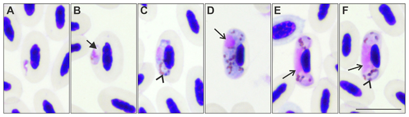
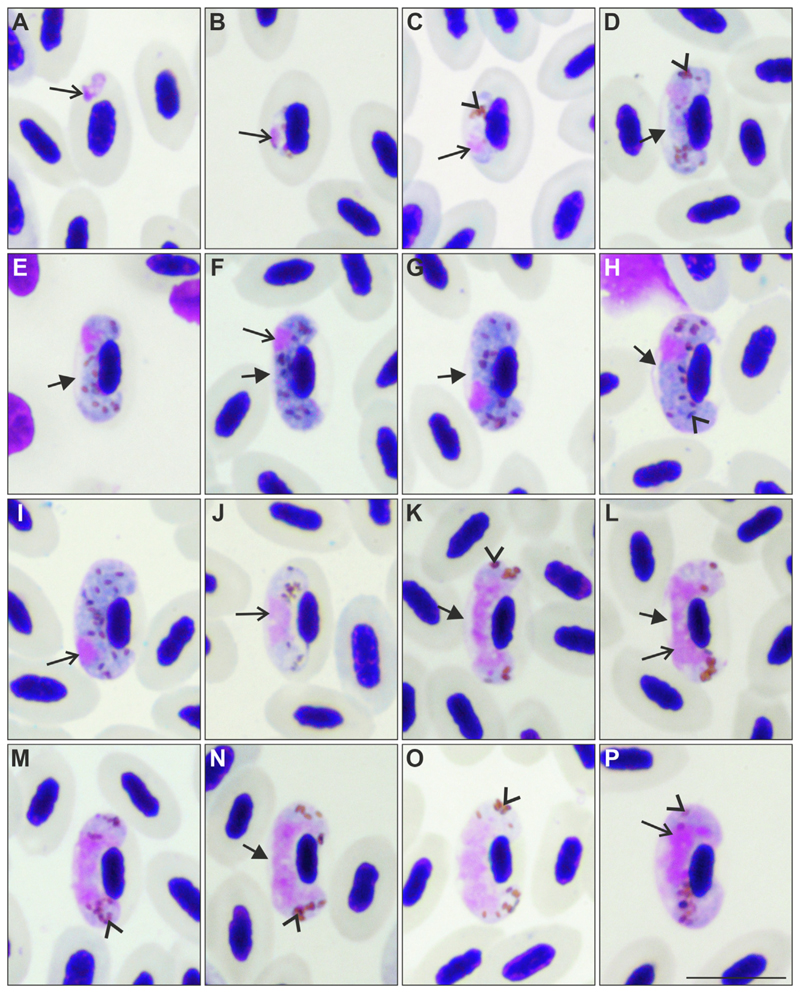
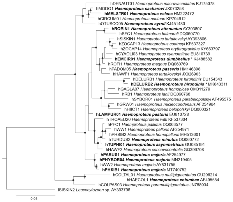
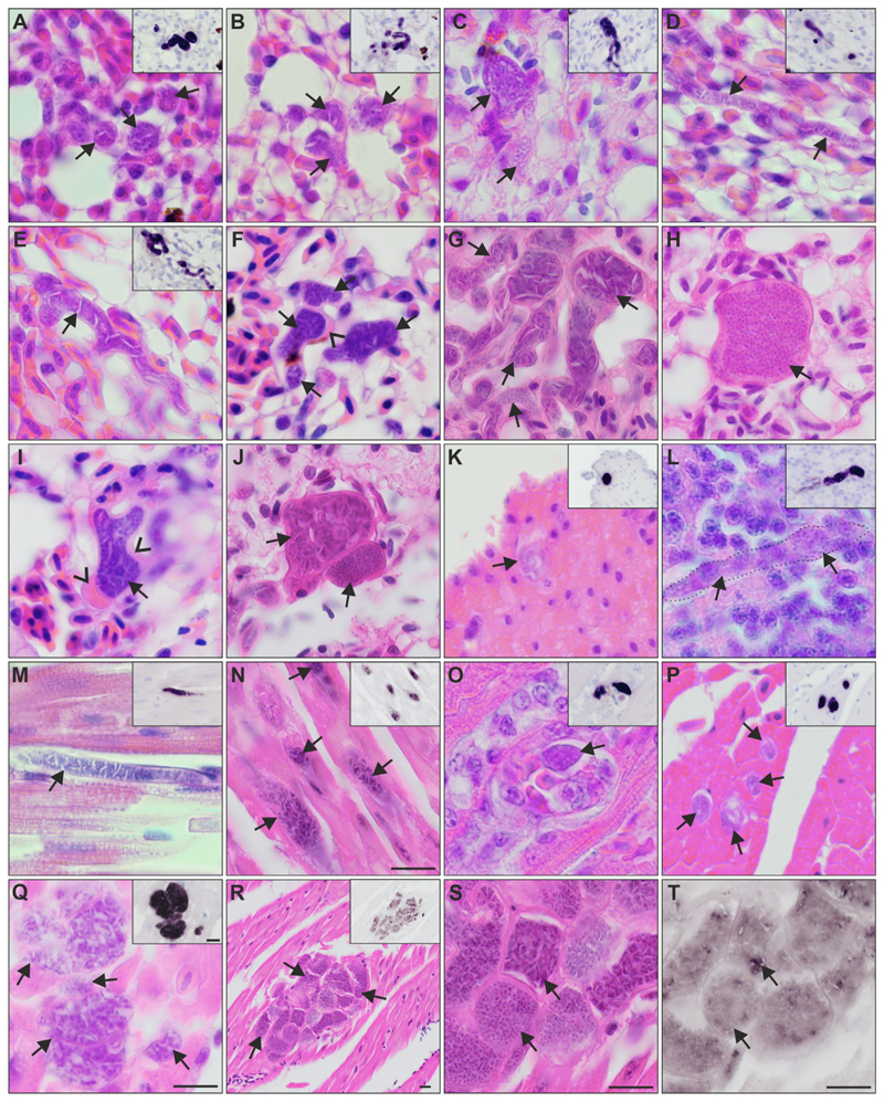
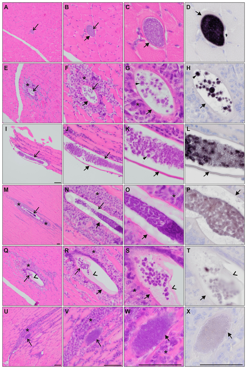
Avian haemosporidians are widespread parasites categorized into four families of the order Haemosporida (Apicomplexa). Species of the subgenus Parahaemoproteus (genus Haemoproteus) belong to the Haemoproteidae and are transmitted by Culicoides biting midges. Reports of death due to tissue damage during haemoproteosis in non-adapted birds have raised concerns about these pathogens, especially as their exo-erythrocytic development is known for only a few Haemoproteus spp. More research is needed to better understand the patterns of the parasites' development in tissues and their impact on avian hosts. Yellowhammers Emberiza citrinella (Emberizidae) and common house martins Delichon urbicum (Hirundinidae) were screened for Haemoproteus parasites by microscopic examination of blood films and PCR-based testing. Individuals with single infection were selected for histological investigations. H & E-stained sections were screened for detection and characterization of the exo-erythrocytic stages, while chromogenic in situ hybridization (CISH) and phylogenetic analysis were performed to confirm the Haemoproteus origin and their phylogenetic relationships. Haemoproteus dumbbellus n. sp. was discovered in Emberiza citrinella single-infected with the lineage hEMCIR01. Meronts of H. dumbbellus n. sp. developed in various organs of five of six tested individuals, a pattern which was reported in other Haemoproteus species clustering in the same clade, suggesting this could be a phylogenetic trait. By contrast, in Delichon urbicum infected with the Haemoproteus lineage hDELURB2, which was linked to the more distantly related parasite Haemoproteus hirundinis, only megalomeronts were found in the pectoral muscles of two of six infected individuals. All exo-erythrocytic stages were confirmed to be Haemoproteus parasites by CISH using a Haemoproteus genus-specific probe. While the development of meronts seems to be typical for species of the clade containing H. dumbbellus, further investigations and data from more species are needed to explore whether a phylogenetic pattern occurs in meront or megalomeront formation.

Keywords: Avian haemosporidians; Chromogenic in situ hybridization; Haemoproteus; Haemoproteus dumbbellus n. sp.; Megalomeronts; Meronts.


Figures

Fig. 1
Fig. 1
Gametocytes of Haemoproteus hirundinis (lineage hDELURB2) from the blood of a common house martin Delichon urbicum. Developmental stages are (A, B) young gametocytes (Note the presence of a vacuole in young gametocytes in (B) as indicated by the arrow); (C) growing, and (D) fully grown macrogametocytes; (E) growing, and (F) fully grown microgametocytes. Parasite nuclei in D-F are indicated by arrows. The arrowheads indicate pigment granules. Scale-bar = 10 μm.
Fig. 2
Fig. 2
Gametocytes of Haemoproteus dumbbellus n. sp. (lineage hEMCIR01) from the blood of a yellowhammer Emberiza citrinella. Developmental stages are (A-C) young gametocytes; (D-I) macrogametocytes; (J-P) microgametocytes. The following forms can be distinguished among them: (D, E) growing macrogametocytes, (F, G) advanced macrogametocytes, (H, I) fully grown macrogametocytes, (J, K) growing microgametoccytes, (L-M) advanced microgametocytes, and (N-P) fully grown microgametocytes. Note that early gametocytes (A) do not adhere to the erythrocyte nuclei, but all other blood stages (B-P) adhere to them. The long arrow indicates the parasite nucleus and the arrowheads indicate pigment granules. An unfilled space (indicated by the short arrow in D-H, K, L, N) is present between gametocytes and the envelope of erythrocytes from the stage of developing gametocytes to the stage of fully grown gametocytes. This gives the parasite a dumbbell-like shape at most stages of growth, which is a characteristic feature of this parasite species. Note that this space often maintains in fully grown gametocytes (H, N), a rare character in Haemoproteus species. Fully grown gametocytes fill erythrocytes till the poles; they enclose the nuclei of erythrocytes with their ends but do not encircle them completely (H, I, O, P). The macrogametocyte nucleus is subterminal in position; it does not adhere to the erythrocyte nucleus (F-I). Scale-bar = 10 μm.
Fig. 3
Fig. 3
Bayesian phylogenetic tree of partial cytochrome b sequences of 37 Haemoproteus spp. lineages and one Leucocytozoon sp. lineage as outgroup. Parasites were represented by MalAvi lineage names (Bensch et al., 2009), followed by their species names and GenBank sequences accession numbers. Bold font indicates species (and lineage when known) for which exo-erythrocytic stages (meronts or megalomeronts) have been described. Parasites from this study indicated with an asterisk. Posterior probabilities (PP) are provided with symbols: triangles, PP 0.7–0.8; squares, PP 0.8–0.9; and circles, PP 0.9–1.
Fig. 4
Fig. 4
Meronts of Haemoproteus dumbbellus (lineage hEMCIR01) found in (A-J) the lungs, (K) gizzard, (L) brain, and (M-T) heart of two yellow hammers Emberiza citrinella. The meront generic origin in H & E-stained preparations was confirmed by chromogenic in situ hybridization (CISH) using a Haemoproteus genus-specific probe indicated by a purple signal in the insets of panels A-E and K-R. Independently of their maturation stage, meronts (indicated by arrows) differed greatly in shape, being round (A, B, K), oval (O, P) or elongate (D, L-N), and ranging in size from less than 10 μm (P) to more than 50 μm (D, E, M, Q). Elongate meronts in the heart appear to follow the shape of the muscle cells (M, N), whereas meronts in the lung tissue (E) were often of capillary shape. Meronts were surrounded by a thin eosinophilic wall, occasionally with bulges of unclear origin located at the periphery of nearly mature parasites (F, I). Note the development of angular-shaped cytomeres separated by clefts, which is a characteristic feature of meront maturation in this parasite species. Such clefts are particularly well visible between cytomeres before merozoite formation (E, G, J-upper arrow), and they disappear in mature meronts, which are overfilled with discrete merozoites (H, J-lower arrow). R-T show the same group of meronts at different magnifications. Note the difference in maturation among meronts (R-T), with mature, roundish merozoites, characterized by weak CISH signals (S, T, lower arrow), or the still developing cytomeres and stronger CISH signals (S, T, upper arrow). Arrowheads indicate the eosinophilic wall. Scale-bar = 10 μm.
Fig. 5
Fig. 5
Megalomeronts of Haemoproteus hirundinis (lineage hDELURB2) found in the (A-X) pectoral muscles of two common house martins Delichon urbicum. The megalomeront generic origin in H & E-stained preparations was confirmed by chromogenic in situ hybridization (CISH) using a Haemoproteus genus-specific probe (purple signal) on a subsequent section (D, H, L, P, T, X). Megalomeronts (indicated by the longer arrows) were elongate, following the muscles cells. In transverse sections, they appeared as roundish or oval bodies (A-D). Megalomeronts were covered by a capsular-like wall of host origin (indicated by the shorter arrows). Note the presence of predominantly small roundish cytomeres, with merozoites budding-off at their periphery, giving maturing cytomeres various star-like appearances (indicated by the triangles in G, H, K). Note the differences in probe signal intensity among megalomeronts of different maturation, varying from strong signal in young megalomeronts (D, H) over less intense signal in maturing megalomeronts (L) to almost no visible signal in fully mature megalomeronts (P, T, X). Inflammatory host cell infiltration (indicated by the asterisks) was observed around megalomeronts (E-G, I-K, M-O, Q-S, U-W). Note the presence of a thin membrane partially detached from the inner wall of the megalomeront (indicated by the arrowheads in Q-T), and still covering the parasite. Mature megalomeronts were overfilled with discrete, roundish merozoites (W). Scale-bar = 50 μm.

Similar articles

Cited by

References

    1. Atkinson CT, Greiner EC, Forrester DJ. Pre-erythrocytic development and associated host responses to Haemoproteus meleagridis (Haemosporina: Haemoproteidae) in experimentally infected domestic turkeys. J Protozool. 1986;33:375–381. - PubMed
    1. Atkinson CT, Forrester DJ, Greiner EC. Pathogenicity of Haemoproteus meleagridis (Haemosporina: Haemoproteidae) in experimentally infected domestic turkeys. J Parasitol. 1988;74:228. doi: 10.2307/3282448. - DOI - PubMed
    1. Beadell JS, Gering E, Austin J, Dumbacher JP, Peirce MA, Pratt TK, Atkinson CT, Fleischer RC. Prevalence and differential host-specificity of two avian blood parasite genera in the Australo-Papuan region. Mol Ecol. 2004;13:3829–3844. doi: 10.1111/j.1365-294X.2004.02363.x. - DOI - PubMed
    1. Bennett GF, Campbell AG. Avian haemoproteidae. I. Description of Haemoproteus fallisi n. sp. and a review of the haemoproteids of the family Turdidae. Can J Zool. 1972;50:1269–1275. doi: 10.1139/z72-172. - DOI - PubMed
    1. Bennett GF, Peirce MA, Ashford RW. Avian haematozoa: Mortality and pathogenicity. J Nat Hist. 1993;27:993–1001. doi: 10.1080/00222939300770621. - DOI

Publication types

LinkOut - more resources